1. Introduction
Unilateral cleft lip repair aims to restore a symmetrical lip and nose, and whichever technique is used, from the rotation-advancement of Millard (1958) to the anatomical subunit approximation of Fisher (2005), the surgeon is trying to match the cleft side to the other. Symmetry is therefore the natural yardstick of the result. In practice it is seldom measured. Most audits rely on panels who rate cropped photographs on an ordinal scale, the best known being that of Asher-McDade et al. (1991).
Panel rating has real merits, since it reflects how the face is perceived, but its weaknesses are well documented. A systematic review of aesthetic outcome assessment in cleft care found a wide variety of methods, none of them accepted as a standard (Sharma et al., 2012). The international standard set of cleft outcomes includes appearance but leaves the means of objective measurement open (Allori et al., 2017).
Three-dimensional surface imaging could fill that gap. This article proposes a workflow by which a cleft team could use it to quantify nasolabial symmetry. No images were analysed for this article, and the workflow has yet to be tested.
2. Background
Two strands of earlier work shape the design: the performance of the imaging systems, and the ways in which asymmetry of a surface can be expressed as a number.
2.1. Surface imaging systems
Stereophotogrammetry reconstructs a surface from synchronised photographs taken from different angles. Laser scanning and structured light achieve the same end by other means. For infants and young children the decisive property is capture time, because a system that acquires the face in milliseconds tolerates movement that would ruin a scan lasting several seconds. Validation studies of commercial stereophotogrammetry systems have found measurement error well below a millimetre on inanimate objects and on faces (Aldridge et al., 2005; Weinberg et al., 2006). A practical guide to acquisition is given by Heike et al. (2010).
2.2. Ways of expressing asymmetry
The classical approach measures distances and angles between landmarks, following the definitions of Farkas (1994), and compares the cleft with the non-cleft side. It is transparent but uses only a handful of points. The alternative treats the whole surface. The face is reflected to produce its mirror image, the two are superimposed, and the distance between them at each point is the local asymmetry. The statistical basis for analysing bilateral shapes in this way was set out by Klingenberg et al. (2002), and Claes et al. (2011) extended it to dense surface data. The range of three-dimensional methods applied to patients with clefts has been catalogued in a systematic review by Kuijpers et al. (2014).
3. The Proposed Workflow
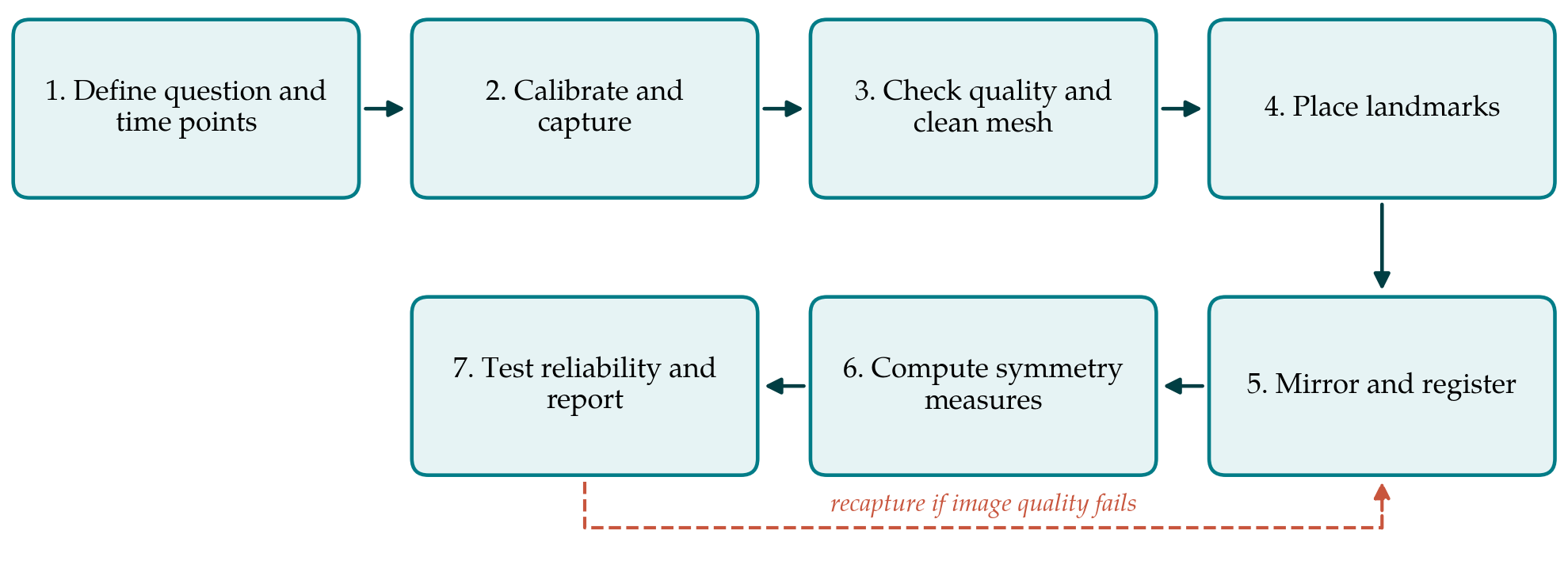
The workflow has seven steps, shown in order in Figure 1. They are designed for a clinic with one imaging system and no dedicated engineer, and each ends with a product that can be checked before the next begins.
3.1. Step 1: Define the question and the time points
A unit should first decide what the measurement is for. Comparing an operative technique before and after a change of practice, tracking individual patients through growth, and contributing to a multi-centre audit make different demands. The decision fixes the ages at which images are taken, which should coincide with existing clinic visits, and the region of interest. For lip repair the region is the nasolabial area bounded by the nasal dorsum, the alar bases and cheeks, and the lower border of the upper lip. Whether lip and nose are reported separately should also be settled now, since a symmetrical lip beneath an asymmetrical nose is a common result that a single figure would conceal.
3.2. Step 2: Calibrate and capture
The system is calibrated according to the manufacturer's procedure at the start of each session and the calibration logged. The subject sits upright, or on a carer's lap, with the head in a natural position, hair held back and the face at rest with lips gently closed. Expression is the main threat to validity: a smile or a cry changes nasolabial form far more than any surgical variable. Several captures are taken, and the operator selects the one closest to a neutral face at the time, while the child is still present. The published acquisition guide gives detailed advice on positioning and on working with young children (Heike et al., 2010).
3.3. Step 3: Check quality and prepare the mesh
Each image is inspected against a short written list: complete coverage of the nostrils and alar bases, no holes or spikes in the region of interest, no motion artefact and a neutral expression. An image that fails is retaken, which is why the check belongs in the clinic and not at a later date. The accepted mesh is then cropped to the face, stray fragments are removed, and the file is stored in an open format under a study code with no identifying name. Facial images are identifiable by nature, so storage and consent must meet the standards applied to clinical photographs.
3.4. Step 4: Place landmarks
A small set of landmarks is placed on the textured surface: the inner canthi and nasion as stable midline and upper-face references, pronasale, subnasale, the alare and subalare points on each side, the peaks of the Cupid's bow, the midpoint of the vermilion border and the corners of the mouth. Definitions are written down with illustrations and follow Farkas (1994) wherever a definition exists. Some of these points are located far less reproducibly than others on three-dimensional scans (Gwilliam et al., 2006), and in a repaired cleft the scar may displace or blur the very landmarks of interest. Each operator therefore marks a training set twice before contributing data.
3.5. Step 5: Mirror and register
The mesh is reflected across an arbitrary plane to create its mirror image. Original and mirror are then superimposed in two stages. A first alignment uses the landmarks, with paired left and right points exchanged, by Procrustes fitting (Gower, 1975). A second refines it by the iterative closest point algorithm of Besl and McKay (1992). The region used for this refinement is a matter of judgement that affects every later result. If the whole face is used, the asymmetric nose and lip pull the registration toward themselves and their asymmetry is understated. The workflow therefore proposes registering on the forehead, orbits and upper nasal dorsum, which the cleft affects least, and then measuring in the nasolabial region.
3.6. Step 6: Compute symmetry measures
No single number describes asymmetry adequately, and the workflow reports a small panel of measures whose definitions and weaknesses are summarised in Table 1. The principal surface measure is the root mean square distance between the original and mirrored meshes within the region of interest, reported for the lip and the nose separately. It is accompanied by a colour-coded distance map, which shows where the deviation lies and is the output most useful to the operating surgeon. Landmark measures are expressed as ratios of the cleft to the non-cleft side so that they are independent of facial size and can be compared across ages.
| Measure | Definition | What it captures | Main weakness |
|---|---|---|---|
| Root mean square surface distance | Mean deviation between original and mirrored mesh in a region | Overall asymmetry of lip or nose | Depends on registration region and mesh density |
| Distance map | Point-by-point deviation shown in colour on the face | Location and direction of asymmetry | Qualitative; hard to summarise across patients |
| Lip height ratio | Subnasale to Cupid's bow peak, cleft over non-cleft side | Vertical lip length match | Landmark blurred by scar |
| Alar base width ratio | Subnasale to alare, cleft over non-cleft side | Lateral position of the alar base | Sensitive to head pose in sparse data |
| Nostril axis and sill height | Angle and vertical offset of paired nostril landmarks | Residual nasal deformity | Nostril interior poorly captured |
| Landmark asymmetry score | Procrustes distance between landmark set and its relabelled mirror | Whole-configuration asymmetry | Limited to chosen landmarks |
3.7. Step 7: Test reliability and report
Before any measure is used, the unit establishes its own measurement error. A subset of subjects is imaged twice in one visit, and each image is processed twice by each operator. Agreement is examined with the limits-of-agreement method of Bland and Altman (1986), reliability with the intraclass correlation coefficient (Shrout and Fleiss, 1979) and landmark error with the formula of Dahlberg (1940). Repeat capture matters most, because it includes variation in expression and pose that repeated processing of one image cannot reveal. Reports should state the system and software versions, the age at imaging, the registration region, the landmarks, and the error estimates alongside the results.
4. Fitting the Workflow to a Cleft Service
The workflow was drafted with three practical limits in mind. Clinic time is short, so capture and the quality check are meant to fit within an ordinary appointment, with landmarking and analysis done later in batches. Expertise is uneven, so the steps that need judgement, namely the choice of registration region and the landmark definitions, are fixed once in a written protocol and not left to each operator. Software changes, so raw meshes are kept in an open format and every analysis records the version used, allowing older images to be reprocessed when methods improve.
Objective measures should sit beside subjective ones, not replace them. A face can be measurably asymmetric and still look acceptable, and the reverse can occur where scar colour or texture draws the eye. The workflow therefore recommends that a panel rating and a patient-reported or parent-reported measure be collected at the same visits. Reading the three together is more informative than any one alone: agreement between them gives confidence in a result, and disagreement points to features, such as scar quality or lip movement, that a resting surface scan does not record.
5. Limitations and What Remains to Be Shown
The workflow is a proposal built from published components. It has not been validated here, and several of its choices rest on reasoning and not on comparison. The first matter to be shown is repeatability in the intended population: published accuracy figures come largely from adults and mannequins, and error in a moving infant will be larger. The second is the effect of the registration region, which could be examined by processing the same images with alternative regions. The third is meaning: whether the surface measure tracks panel ratings, and what size of difference observers and patients actually perceive, is not established. A threshold separating acceptable from unacceptable asymmetry cannot be stated at present.
Further limits are inherent. A static image at rest says nothing about symmetry in movement, which matters greatly for the lip. Surface imaging records skin only and gives no information on the dental arch or skeletal base that influence nasolabial form during growth. Reference data for normal facial asymmetry by age and population are sparse, so a patient's score is hard to place in context. The measures support comparison within a unit over time more securely than comparison between units using different systems.
6. Conclusion
Surface imaging has made objective assessment of cleft lip repair technically feasible in an ordinary clinic. The seven-step workflow proposed here links capture, quality control, landmarking, registration on unaffected regions and a small panel of symmetry measures, and it requires every unit to quantify its own error before drawing conclusions. Its usefulness depends on validation work that has yet to be done, and the article has set out what that work should include.
Conflict of interest
To be declared by the author(s) before publication.
Funding
To be declared by the author(s) before publication.