1. Introduction
Coronary computed tomography angiography (CCTA) has moved from a specialist examination to the recommended first test for many patients with stable chest pain. European and North American guidelines both give it a class I recommendation as an initial investigation in appropriately selected patients (Knuuti et al., 2020; Gulati et al., 2021), and the SCOT-HEART trial showed that a strategy including CCTA was followed at five years by fewer deaths from coronary heart disease or non-fatal myocardial infarctions than standard care alone (SCOT-HEART Investigators, 2018). The strength of the test lies in its negative predictive value: a normal scan rules out obstructive disease with great reliability.
Its weakness has been known for as long. Dense coronary calcification appears larger on the image than it is, a phenomenon usually called blooming, and the artefact encroaches on the lumen. In the multicentre accuracy studies carried out with 64-detector scanners, specificity fell in patients with high calcium scores (Budoff et al., 2008), and one of the principal trials excluded patients with an Agatston score above 600 (Miller et al., 2008). The patients affected are older, more often diabetic or in renal failure, and more likely to have the obstructive disease the test is meant to detect. A calcified segment that cannot be graded sends the patient on to invasive angiography or functional testing, which is the outcome CCTA was meant to avoid.
Photon-counting detector CT (PCCT) entered clinical use in 2021, when the first whole-body system received regulatory clearance (Rajendran et al., 2022). Several of its properties address the calcium problem directly. This review considers how far the published evidence supports the expectation that PCCT can characterise plaque, and not only grade stenosis, in heavily calcified coronary arteries.
2. Scope and Approach
This is a narrative review of the published literature. It draws on the technical reviews that describe photon-counting detectors, on the early phantom, ex vivo and clinical reports of coronary imaging with PCCT, and on the older body of work that defines what plaque characterisation by CT means and why calcium interferes with it. No formal search protocol or quantitative synthesis was attempted, because the clinical literature is young, heterogeneous in scanner type and reconstruction settings, and largely made up of single-centre feasibility studies. The emphasis is on mechanisms and on the kind of evidence available, so that the reader can judge new reports as they appear. The phrase "heavily calcified" is used in the loose clinical sense of vessels in which calcification limits interpretation on a conventional scanner, without a fixed score threshold.
3. Why Calcium Limits Conventional Coronary CT
Conventional scanners use energy-integrating detectors. Each element consists of a scintillator that converts X-rays to light and a photodiode that converts the light to current, and the elements are separated by reflective septa that stop light spreading to neighbours. The septa cannot be made much thinner, so shrinking the elements reduces the active area and wastes dose. The practical limit on detector pitch sets the in-plane resolution of coronary CT at a size that is a meaningful fraction of the lumen of a distal or diseased coronary segment (Willemink et al., 2018; Flohr et al., 2020).
Blooming follows from this. A voxel that is partly filled with calcium takes on a high attenuation value throughout, so the plaque is drawn larger than it is and the lumen smaller. Smooth reconstruction kernels, chosen to control noise, spread the effect further. Beam hardening adds dark bands beside dense calcium that can imitate low-attenuation plaque, and residual cardiac motion smears a bright object across more voxels than a faint one (Kalisz et al., 2016). The combined result is overestimation of stenosis and, just as damaging for plaque characterisation, loss of the tissue immediately around the calcium. Table 1 sets these problems beside the detector properties discussed in the next section.
| Problem in calcified vessels | Origin in energy-integrating CT | Relevant photon-counting property | State of the evidence |
|---|---|---|---|
| Blooming of calcified plaque | Partial-volume averaging in large voxels; smooth kernels | Smaller detector elements, sharper kernels | Consistent across phantom, ex vivo and clinical reports (Flohr et al., 2020) |
| Overestimated stenosis | Blooming encroaches on the lumen | Ultra-high-resolution acquisition | High accuracy in one high-risk cohort (Hagar et al., 2023) |
| Dark bands beside calcium | Beam hardening of a polychromatic beam | High-energy monoenergetic images | Principle established with dual-energy CT (McCollough et al., 2015) |
| Hidden non-calcified plaque | Artefact covers adjacent soft tissue | Resolution with higher soft-tissue contrast | Qualitative support; quantitative thresholds not recalibrated |
| Noise at thin slices | Electronic noise added to weak signals | Counting threshold set above the noise floor | Well documented technically (Willemink et al., 2018) |
| Lumen obscured entirely | No routine spectral information | Material decomposition and calcium subtraction | Phantom data; clinical accuracy unproven |
The loss matters because the features that carry risk are mostly not calcified. Pathological studies locate the precursor of plaque rupture in the thin-cap fibroatheroma, a lesion with a large necrotic core and often only speckled calcification (Virmani et al., 2000). On CT its correlates are low attenuation, positive remodelling, spotty calcification and the napkin-ring sign (Motoyama et al., 2009; Maurovich-Horvat et al., 2014), and in SCOT-HEART the burden of low-attenuation plaque predicted myocardial infarction more strongly than calcium score or stenosis severity (Williams et al., 2020). High-risk plaque features are now reporting modifiers in CAD-RADS 2.0 (Cury et al., 2022). In a heavily calcified artery they are the first things to disappear behind artefact.
4. Photon-Counting Detectors and the Coronary Arteries
A photon-counting detector is a slab of semiconductor, cadmium telluride or cadmium zinc telluride in most designs and silicon in one, across which a high voltage is applied. An absorbed X-ray photon releases a cloud of charge that is swept to pixelated anodes and produces a pulse whose height is proportional to the energy of the photon. Electronics compare each pulse with a set of thresholds and count it in the corresponding energy bin (Taguchi and Iwanczyk, 2013; Danielsson et al., 2021). There is no light, and so no need for septa. Figure 1 links the resulting properties to their consequences for calcified vessels.
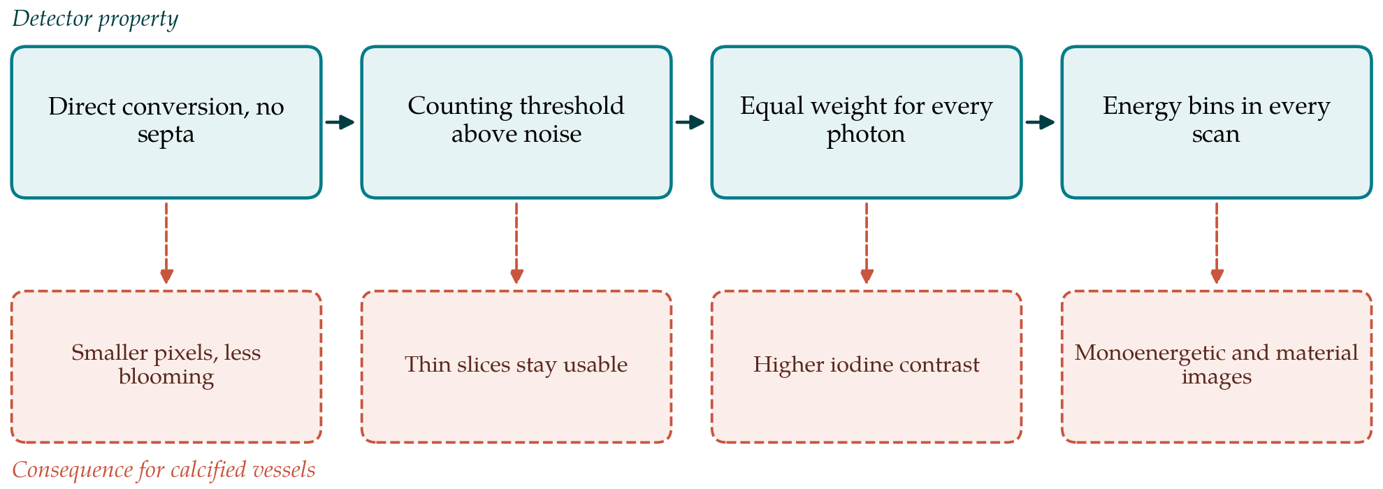
Four consequences follow. First, the pixels can be made much smaller without sacrificing geometric efficiency; the first clinical system offers an ultra-high-resolution mode with a slice thickness of 0.2 mm (Rajendran et al., 2022). Second, the lowest threshold is set above the level of electronic noise, which is therefore excluded from the count instead of added to the signal. Third, every photon contributes one count whatever its energy, whereas an energy-integrating detector gives more weight to high-energy photons that carry little contrast information; the contrast of iodine against soft tissue rises as a result. Fourth, energy-resolved data are acquired in every examination, at full temporal resolution and without the need to select a dual-energy mode in advance (Leng et al., 2019).
The spectral data support the same reconstructions as dual-energy CT, which rest on the observation that attenuation in the diagnostic range can be expressed as a combination of two basis functions (Alvarez and Macovski, 1976). Virtual monoenergetic images at low energies raise iodine contrast; at high energies they suppress blooming and beam hardening at the price of a fainter lumen. Material decomposition yields iodine maps and virtual non-contrast images, and can in principle subtract calcium (McCollough et al., 2015). The detectors are not ideal. Charge shared between neighbouring pixels and fluorescence escape distort the recorded spectrum, and at high flux pulses pile up and are miscounted, so spectral separation is good but falls short of what the bin structure alone would suggest (Willemink et al., 2018).
5. Evidence in Calcified Coronary Disease
The published evidence can be grouped under three headings: the grading of stenosis, the description of plaque composition, and the use of spectral reconstructions for calcium scoring and calcium removal. The strength of the evidence declines in that order.
5.1. Stenosis grading
The clearest evidence concerns blooming and lumen visibility. Phantom and ex vivo studies summarised in the technical reviews consistently show that calcified plaque occupies a smaller apparent volume, with sharper edges, on photon-counting images than on energy-integrating images of the same object, and that the gain grows as slices become thinner and kernels sharper (Flohr et al., 2020; Leng et al., 2019). The first human coronary examinations on a prototype system reported better subjective image quality and better conspicuity of calcifications, stents and non-calcified plaque than a dual-layer scanner in the same patients (Si-Mohamed et al., 2022).
Feasibility work with the clinical dual-source system showed that ultra-high-resolution coronary angiography can be acquired with electrocardiographic gating and that sharp vascular kernels improve the delineation of plaque and lumen, with noise rising steeply as the kernel sharpens (Mergen et al., 2022). The comparison with invasive angiography that matters most so far was made in patients assessed before transcatheter aortic valve replacement, a group with a high prevalence of dense coronary calcium and of previous stenting; ultra-high-resolution PCCT showed high accuracy for obstructive disease in that setting (Hagar et al., 2023). The finding is encouraging because it comes from the population in which conventional CCTA is weakest. It is a single-centre result, and the reference standard was angiographic stenosis and not a measure of flow limitation.
5.2. Plaque composition
Evidence on composition is thinner and more indirect. Higher resolution should, and in ex vivo comparisons does, reveal small calcifications, thin fibrous rims and lipid-rich cores that are blurred together on standard images. The qualitative high-risk features described on older scanners are therefore likely to be seen more often and with more confidence (Maurovich-Horvat et al., 2014). Quantification is another matter. Plaque volumes are obtained by assigning voxels to calcified, fibrous or low-attenuation classes according to fixed attenuation thresholds, such as the 30 HU boundary used for low-attenuation plaque (Motoyama et al., 2009). When blooming shrinks, voxels formerly counted as calcium are reassigned, and when the energy of a monoenergetic reconstruction changes, every attenuation value shifts.
It follows that plaque burdens measured on PCCT cannot be assumed to be interchangeable with those from the energy-integrating scanners on which the prognostic thresholds were derived (Williams et al., 2020). A smaller calcified volume and a different non-calcified volume in the same patient is the expected result of better resolution and not a sign of error, but it means that risk categories would need to be recalibrated before they are applied. In a heavily calcified vessel the direction of the change favours interpretation, since tissue once hidden becomes measurable. Whether newly visible low-attenuation plaque beside dense calcium carries the same prognostic meaning as low-attenuation plaque in a lightly calcified vessel has not been tested.
5.3. Calcium scoring and spectral reconstructions
The Agatston score was defined with a threshold of 130 HU under specific acquisition conditions (Agatston et al., 1990), and decades of risk data depend on that definition. Photon-counting systems reproduce conventional conditions through monoenergetic reconstruction at a matched energy, and phantom work indicates that scores comparable to conventional values can be obtained in this way (Flohr et al., 2020). Virtual non-contrast images raise the possibility of deriving a calcium score from the angiographic acquisition alone. Agreement with true unenhanced scoring is imperfect in the dual-energy literature, because iodine and calcium are not perfectly separated (McCollough et al., 2015), and the same caution applies here.
The reverse operation, removing calcium to leave a clean lumen, is the most direct spectral answer to the calcified vessel. Calcium-subtraction algorithms based on photon-counting data have been described and tested in phantoms, and they are attractive for vessels with circumferential calcification. Their difficulty is the one that affects all material decomposition near a partial-volume boundary: a voxel containing both calcium and iodine may be assigned wrongly, and motion makes the error worse. High-energy monoenergetic images are a simpler and more robust option that reduces blooming without any classification step, though they lower lumen contrast at the same time (Leng et al., 2019). No spectral reconstruction has yet been shown to improve diagnostic accuracy over the ultra-high-resolution images themselves.
6. Limitations of the Technology and of the Evidence
Resolution is bought with noise. Halving the voxel dimensions leaves far fewer photons in each voxel, and sharp kernels amplify what noise there is, so ultra-high-resolution coronary images depend on iterative or learned denoising and, in large patients, on a higher dose (Mergen et al., 2022). Temporal resolution is set by gantry rotation and source geometry and is not improved by the detector. A calcified plaque in a vessel that moves during acquisition will still blur, and at finer spatial resolution a given displacement spans more voxels (Kalisz et al., 2016). Heart rate control therefore remains as necessary as before.
The evidence base has its own limits. Most reports come from a small number of centres with early access to one manufacturer's system, alongside prototypes built on different sensor materials, and results from one design may not transfer to another (Danielsson et al., 2021). Study populations are modest and often selected. Reconstruction settings vary between reports, which makes plaque measurements hard to compare. Reference standards are usually invasive angiography for stenosis and, for composition, either nothing or an older CT scan; histological or intravascular validation in living patients is scarce. Above all, there are no trials showing that a PCCT-guided strategy changes downstream testing, treatment or events in patients with heavy calcification, which is the standard by which CCTA itself was eventually judged (SCOT-HEART Investigators, 2018).
7. Gaps and Future Work
Three questions seem most pressing. The first is diagnostic: multicentre accuracy studies in patients with high calcium scores, using invasive physiological measurement as the reference, would establish whether patients now sent directly to the catheter laboratory can safely be imaged first. The second is metrological: thresholds, kernels and monoenergetic levels for plaque quantification need to be standardised, and the relation between photon-counting and conventional plaque volumes characterised, before reporting systems such as CAD-RADS can incorporate the new measurements (Cury et al., 2022). The third is prognostic, and can be answered only by follow-up of cohorts scanned with the new detectors.
Further technical development is likely to help. More energy bins and better correction for charge sharing would sharpen material separation, and K-edge imaging of contrast agents other than iodine has been demonstrated experimentally as a way of separating lumen from calcium entirely (Willemink et al., 2018). These remain research directions. For the present, the practical gain for calcified vessels comes mainly from spatial resolution.
8. Conclusion
Photon-counting CT attacks the calcium problem at its source. Smaller detector elements reduce the partial-volume effect that produces blooming, the exclusion of electronic noise and the more favourable weighting of low-energy photons make the smaller voxels usable, and spectral data offer further means of suppressing or subtracting calcium. Early clinical reports indicate that stenosis can be graded accurately in patients who would have been poor candidates for CCTA a few years ago, and that plaque beside dense calcification can be seen where it was previously hidden. The measurements that describe that plaque are not yet standardised, and their prognostic meaning has not been established on the new scanners. Until multicentre accuracy and outcome data are available, PCCT in heavily calcified vessels is best regarded as a substantial technical advance whose clinical value is probable but unproven.
Conflict of interest
To be declared by the author(s) before publication.
Funding
To be declared by the author(s) before publication.