1. Introduction
The recurrent laryngeal nerve supplies every intrinsic muscle of the larynx except cricothyroid, and it carries sensation from the mucosa below the vocal folds. A lesion on one side leaves a fold lying paramedian, with a weak, breathy voice and sometimes aspiration. A lesion on both sides can close the airway. Because the nerve runs within a few millimetres of the back of the thyroid lobe, any operation on the gland puts it at risk, and the rate of nerve palsy has become the usual yardstick of a thyroid surgeon's work (Hermann et al., 2002).
For the first decades of thyroid surgery the common advice was to stay away from the nerve and never to look for it. That advice was overturned by Lahey (1938), who dissected and showed the nerve in every operation, and later by Riddell (1970), whose long personal series made identification the accepted standard. Identification, however, presupposes that the surgeon knows where the nerve may be found, and the difficulty is that it varies: in its course, in the way it divides, and in its relations to the structures used as landmarks.
This review describes those variations and what each implies for operative technique. It is written for surgeons in training and for anatomists who teach the neck, and it tries to separate what is firmly known from what rests on a handful of dissection series with differing definitions.
2. Scope and Approach
This is a narrative review of the published literature. It draws on standard anatomical texts, cadaveric and operative series, pooled analyses of anatomical prevalence, and the consensus statements of the International Neural Monitoring Study Group. No formal search protocol was applied and no new data are presented. Where sources disagree, the disagreement is reported and, where possible, explained by differences in method. Frequencies are given in words unless a figure is consistently reported across large series.
3. Development and Usual Course
The asymmetry of the two nerves is a product of the embryonic aortic arches. Each nerve at first passes to the sixth pharyngeal arch below the sixth arch artery. As the heart descends, the nerves are drawn down and hook around whatever arterial arch persists caudal to them. On the left the distal sixth arch survives as the ductus arteriosus, so the nerve loops beneath the arch of the aorta beside the ligamentum arteriosum. On the right the distal sixth arch and the fifth arch disappear, and the nerve rises to loop under the fourth arch derivative, the subclavian artery (Standring, 2016).
The consequence in the neck is a difference of obliquity. The left nerve has a long, nearly vertical ascent and usually lies in or close to the tracheo-oesophageal groove. The right nerve leaves the root of the neck more laterally and crosses obliquely towards the larynx, so that low in the neck it may lie well away from the trachea. Both end by passing deep to the lower border of the inferior constrictor, behind the cricothyroid joint. That entry point is the one constant in an otherwise variable course, and the inferior cornu of the thyroid cartilage, which can be felt at operation, marks it reliably.
4. Variations in Course
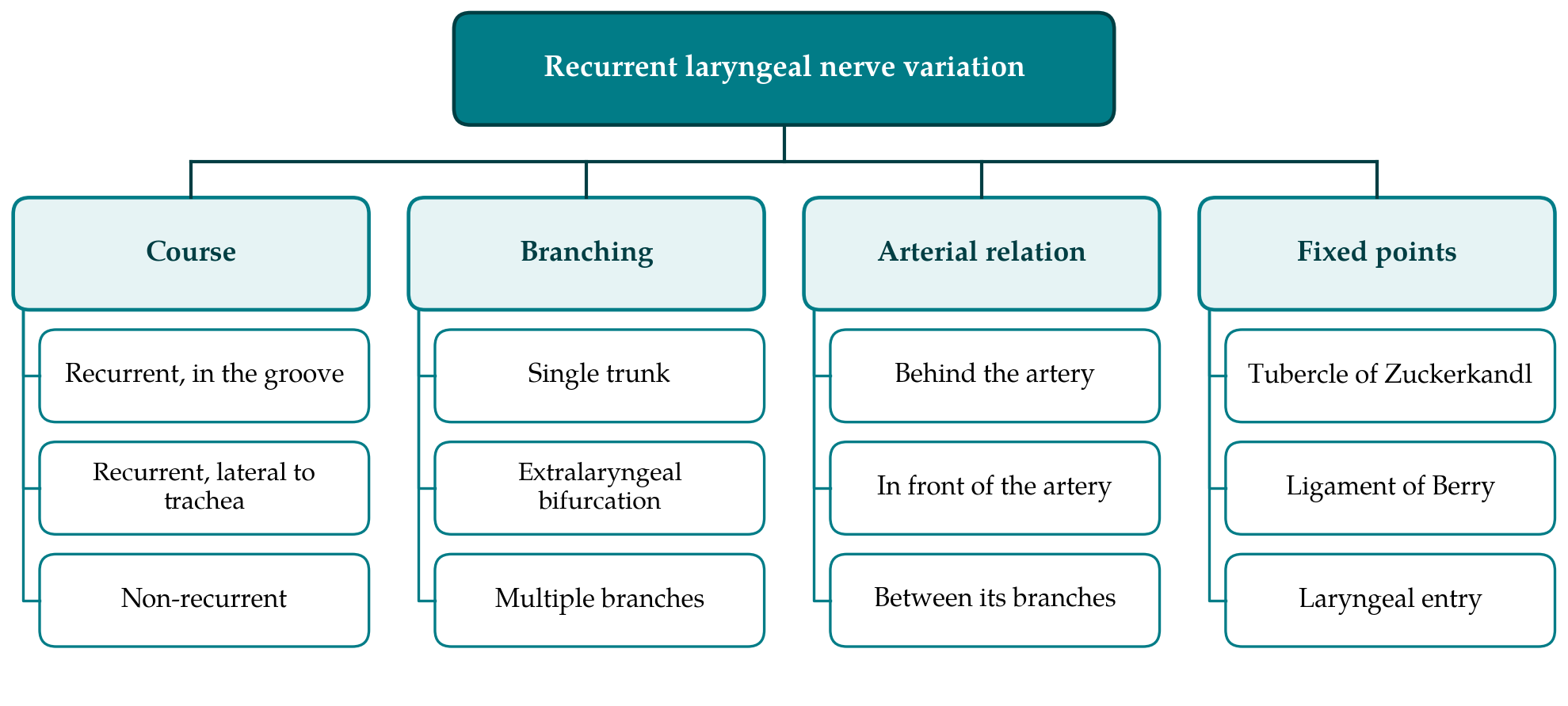
Even the ordinary recurrent nerve does not keep to the groove as often as is supposed. Dissection studies have found a fair proportion of nerves lying lateral to the trachea, and a few lying anterolateral to it, where they are exposed to injury as the lobe is drawn forwards (Steinberg et al., 1986). Enlargement of the gland changes the picture further. A large or retrosternal goitre can stretch the nerve over its surface or displace it forwards, and a nerve that is fixed to the capsule of a nodule will not be where the surgeon expects. The main variations discussed in this review are grouped in Figure 1.
4.1. The non-recurrent nerve
The most striking variation is the nerve that does not recur at all. It has been known to anatomists since the early nineteenth century and is explained by the same embryology that produces the normal asymmetry. When the right fourth arch regresses, the right subclavian artery arises as the last branch of the aortic arch and passes behind the oesophagus. Nothing then holds the right nerve down, and it leaves the vagus in the neck to run more or less transversely to the larynx. The anomaly is uncommon: large surgical series and a pooled analysis agree that it is found in well under one in a hundred right sides (Henry et al., 1988; Toniato et al., 2004; Henry et al., 2017). On the left it requires situs inversus or a right-sided arch with an aberrant left subclavian artery and is extremely rare.
A non-recurrent nerve is in danger because it lies where no nerve is expected. It may cross at the level of the upper pole, where it can be taken for a vessel and divided with the superior pedicle, or lower, alongside the inferior thyroid artery. The rule that follows is simple. If the right nerve cannot be found in its usual place, no transverse structure passing from the carotid sheath to the larynx should be cut until a non-recurrent nerve has been excluded. The retro-oesophageal subclavian artery can be recognised on cross-sectional imaging or ultrasound before operation, and its presence predicts the nerve anomaly (Toniato et al., 2004).
5. Extralaryngeal Branching
The nerve often divides before it reaches the larynx. Reported frequencies differ widely, from a minority of nerves in some series to a clear majority in others (Nemiroff and Katz, 1982; Cernea et al., 2009). A meta-analysis that pooled a very large number of nerves concluded that branching is common and that it is recorded more often in cadaveric than in operative studies (Henry et al., 2016). Much of the spread is a matter of definition. Some authors count only a division of the main trunk into laryngeal branches; others include the fine twigs to the trachea, oesophagus and inferior constrictor that almost every nerve gives off. Magnification, the level at which the nerve is first exposed and the patience of the dissector also affect the count.
What matters to the surgeon is where the motor fibres run. Stimulation studies have shown that when the nerve bifurcates, the fibres to both the abductor and the adductor muscles generally travel in the anterior branch, while the posterior branch is largely sensory (Serpell et al., 2009; Kandil et al., 2011). The anterior branch is also the thinner and the more superficial of the two in the region of the ligament of Berry. A surgeon who finds a substantial posterior branch and assumes it to be the whole nerve may therefore divide the part that moves the vocal fold. The practical answer is to identify the nerve low, to follow it upwards to its entry beneath the constrictor, and to treat any structure running forwards from it as nerve until it is proved otherwise.
6. Relation to the Inferior Thyroid Artery
The crossing of the nerve and the inferior thyroid artery was for many years the favoured place to look for the nerve, and it is still useful, but the relation is inconstant. The nerve may pass behind the artery, in front of it, or between its terminal branches, and the arrangement on one side does not predict the other (Reed, 1943). Later series have confirmed all three patterns and have added many sub-types according to how the artery and the nerve each divide (Yalçin, 2006; Makay et al., 2008). There is general agreement that the right nerve is found in front of or between the arterial branches more often than the left, which fits its more oblique and superficial course.
Two points follow for technique. First, the artery should not be ligated as a trunk close to the nerve; its branches are divided individually on the capsule of the gland, which also protects the blood supply of the parathyroid glands. Second, a nerve lying in front of the artery or among its branches is lifted with the lobe when the gland is rotated medially, and traction then falls directly on it. Table 1 summarises this and the other variations together with the precaution each calls for.
| Variation | What is found | Surgical precaution |
|---|---|---|
| Lateral or anterior course | Nerve lies outside the tracheo-oesophageal groove, commoner on the right | Search a wide field low in the neck before dividing tissue |
| Non-recurrent nerve | Nerve leaves the vagus in the neck; aberrant right subclavian artery | Cut no transverse band until the nerve is found |
| Extralaryngeal branching | Trunk divides below the larynx; motor fibres mostly anterior | Trace every branch to the laryngeal entry |
| Arterial relation | Nerve behind, in front of or between branches of the artery | Divide arterial branches singly on the capsule |
| Tubercle of Zuckerkandl | Nerve usually deep to the tubercle, occasionally on its surface | Mobilise the tubercle with the nerve in view |
| Ligament of Berry | Nerve close to, and at times within, the ligament | Avoid traction and diathermy; accept a small remnant if needed |
7. The Tubercle of Zuckerkandl and the Ligament of Berry
The tubercle of Zuckerkandl is a posterolateral projection of the thyroid lobe, present to some degree in most glands. Pelizzo et al. (1998) drew attention to its value as a pointer: the nerve usually runs in the cleft between the tubercle and the trachea, covered by it. When the tubercle is large it hides the nerve and must be turned forwards before the nerve can be seen. Less often the nerve runs across its lateral surface, where it is easily stretched or cut as the lobe is delivered. A prominent tubercle is a signal to slow down.
Just above lies the ligament of Berry, the condensation of fascia that binds the gland to the cricoid cartilage and the upper tracheal rings. Here the nerve, or its anterior branch, is within a few millimetres of the gland and is tethered, so it cannot slide away from traction. An autopsy study found the nerve dorsolateral to the ligament in the specimens examined and not running through it (Sasou et al., 1998), although surgeons regularly report nerves that appear embedded in its fibres or in thyroid tissue caught within it. The final centimetre before the laryngeal entry is, by common consent, where most injuries happen.
Neuromonitoring studies support that view and add something about mechanism. When the point of injury can be located along a nerve that is visibly intact, it is usually near the ligament, and the commonest cause is traction during medial rotation of the lobe, ahead of clamping, ligature and heat (Chiang et al., 2008). The nerve that stops conducting has, in most cases, never been cut. This is why a nerve that looks perfect at the end of the operation is no guarantee of a moving cord.
8. Implications for Operative Practice
The case for finding the nerve in every operation rests on decades of consistent observation. In a very large multicentre audit, palsy was less frequent when the nerve had been dissected and displayed than when it had not, and the individual surgeon's results varied more than any other factor (Hermann et al., 2002). Knowledge of variation is what makes identification safe. A surgeon who expects a single trunk in the groove behind the artery will be right often enough to become confident and wrong often enough to do harm.
Intraoperative neuromonitoring, in which the nerve is stimulated and the response recorded from electrodes on the tracheal tube, has been standardised by an international guideline (Randolph et al., 2011). Whether it lowers the rate of permanent palsy is unsettled. A large prospective multicentre study and a randomised trial both showed lower rates of transient palsy with monitoring in some groups, notably high-risk operations, without a clear difference in permanent injury (Dralle et al., 2004; Barczyński et al., 2009), and a meta-analysis of comparative studies found no significant advantage over visualisation alone (Pisanu et al., 2014). Permanent palsy is infrequent enough that very large trials would be needed to show a difference.
Monitoring nevertheless helps with variant anatomy in ways that palsy rates do not capture. It allows the anterior and posterior divisions of a branched nerve to be told apart by their response. Stimulation of the vagus low in the neck that gives no response, with a response higher up, reveals a non-recurrent nerve before it is seen. And a loss of signal on the first side gives the surgeon grounds to stop and stage the second side, which is the surest protection against bilateral palsy (Schneider et al., 2018). Monitoring is an addition to anatomical knowledge and identification by sight, not a replacement for either.
One further implication concerns audit. Reported palsy rates depend heavily on whether and when the larynx is examined after operation; units that examine every patient find more palsies than those that examine only patients with a changed voice (Jeannon et al., 2009). Comparisons between series, including comparisons of anatomical risk factors, mean little unless the method of laryngeal assessment is the same.
9. Gaps and Future Work
The anatomical literature has three weaknesses. Definitions of branching differ so much that pooled prevalence figures are hard to interpret, and an agreed classification that separates laryngeal from extralaryngeal twigs would help. Most large series come from a small number of centres, and it is not known how far differences between populations are real and how far they reflect technique. Finally, almost all operative data describe diseased glands, in which the nerve may have been displaced, so the normal range is known mainly from cadavers, whose tissues differ from those of the living. High-resolution ultrasound and nerve mapping during monitored operations offer ways of recording the anatomy in life, and prospective registries with uniform definitions would do more than further small dissection series.
10. Conclusion
The recurrent laryngeal nerve varies in its course, its branching and its relations, and each variation has a predictable consequence at operation. The non-recurrent nerve is rare but is cut if it is not thought of. Branching is common, and the branch that matters is usually the anterior one. The relation to the inferior thyroid artery cannot be relied on, and the region of the tubercle of Zuckerkandl and the ligament of Berry is where a fixed nerve meets traction. Routine identification, followed to the laryngeal entry, is well supported. Neuromonitoring is a useful aid whose effect on permanent injury is still unproven. Safe thyroid surgery continues to depend on a surgeon who knows how variable this nerve is.
Conflict of interest
To be declared by the author(s) before publication.
Funding
To be declared by the author(s) before publication.