1. Introduction
Laser in situ keratomileusis (LASIK) leaves a lamellar plane in the cornea that persists for life. In most eyes it is invisible and of no consequence. Under particular conditions fluid collects in it, separating the flap from the stromal bed, and the result is known as interface fluid syndrome. The first reports appeared within a few years of LASIK becoming common. Lyle and Jin (1999) described interface fluid in association with lamellar keratitis and epithelial ingrowth, Rehany et al. (2000) and Fogla et al. (2001) drew attention to the misleadingly low pressure readings that accompany it, and Hamilton et al. (2002) documented glaucomatous damage in eyes whose steroid-induced pressure rise had gone unrecognised behind a fluid cleft.
Those early accounts concerned the weeks after surgery, when topical steroids are in use. The same mechanism can operate much later. Anything that raises intraocular pressure or weakens the endothelial pump in an eye with an old flap can drive fluid into the interface, and published reports describe the syndrome after intraocular surgery, during uveitis and with endothelial disease, long after the refractive procedure (McLeod et al., 2005; Ortega-Usobiaga et al., 2012; Luceri et al., 2016). As the large population treated with LASIK since the 1990s reaches the age of cataract, glaucoma and endothelial dystrophy, such presentations can be expected to become more frequent.
This article sets out a structured pathway for recognising and working up late onset interface fluid syndrome. It is written for general ophthalmologists, cataract and glaucoma surgeons and emergency eye services as much as for refractive surgeons, because the eye is unlikely to return to the clinic where the flap was made. The pathway is proposed on the basis of the literature. It reports no cases and has not been tested in practice by the present work.
2. Background
Three points from the literature shape the pathway: why the interface remains vulnerable, the two routes by which fluid reaches it, and the reasons the diagnosis is missed.
2.1. A wound that does not fully heal
Histological and mechanical study of human corneas after LASIK showed that the central and paracentral interface heals with a hypocellular primitive scar of very low cohesive strength, while only the flap margin forms a firmer hypercellular scar (Schmack et al., 2005). The central lamellar plane is therefore a potential space indefinitely. This is the anatomical basis for flap dislocation long after surgery, and it explains why interface fluid is not confined to the postoperative period.
2.2. Two mechanisms
Stromal hydration is set by the balance between the swelling pressure of the stroma, the intraocular pressure pushing fluid forward and the endothelial pump removing it. Dawson et al. (2007) reproduced interface fluid in eye bank corneas with LASIK flaps by raising pressure and, separately, by damaging the endothelium, and proposed a staged description running from diffuse interface haze to a visible fluid pocket. The clinical literature follows the same division. In the pressure-driven form the commonest cause in the early period is a steroid response, a phenomenon reviewed in general terms by Kersey and Broadway (2006); later causes include any secondary glaucoma. In the endothelial form the pump fails through dystrophy, surgical trauma, inflammation or graft failure, and pressure may be normal. The two can coexist, as in uveitis with raised pressure.
2.3. Why the diagnosis is missed
Early interface fluid produces a diffuse haze that resembles diffuse lamellar keratitis, the sterile interface inflammation described by Smith and Maloney (1998) and managed with intensive steroid (Linebarger et al., 2000). When the haze is in fact pressure-induced, more steroid raises the pressure further. Belin et al. (2002) named this presentation pressure-induced interlamellar stromal keratitis, and Nordlund et al. (2004) showed that it can arise late. The second trap is tonometry. A Goldmann probe applied centrally measures the pressure in the fluid pocket and reads low or normal while the true pressure is high (Senthil et al., 2010). The third is history: years after surgery the flap edge may be faint and the operation unmentioned.
3. The Proposed Pathway
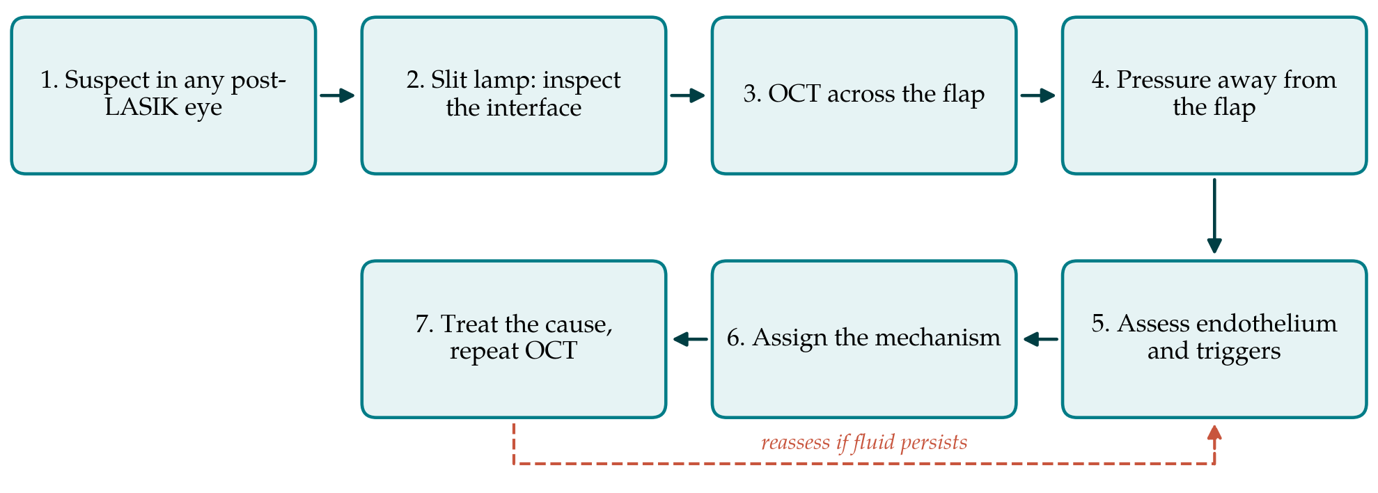
The pathway has seven steps, shown in Figure 1. They are ordered so that the cheapest observations come first and so that no treatment is started before the mechanism has been considered. The steps are described below with the reasoning for each.
3.1. Step 1: Hold the suspicion
The pathway begins with a rule of thumb: in any eye with a history of LASIK that develops blurred vision, a myopic or hyperopic shift, or corneal haze after a new event, interface fluid should be on the list. Relevant events are a course of topical, periocular, intravitreal or systemic steroid, intraocular surgery, uveitis, trauma and any new diagnosis of raised pressure or endothelial disease. The refractive history should be asked for directly and the flap edge looked for, since patients may not volunteer an operation performed long ago.
3.2. Step 2: Examine the interface at the slit lamp
A narrow, bright, oblique slit beam is used to look for an optically empty cleft between flap and bed, most easily seen centrally. In early disease there may be only a fine diffuse haze confined to the interface with a clear overlying flap. Microcystic epithelial oedema suggests high pressure or established endothelial failure. Cells in the interface, focal infiltrates, an epithelial defect or a nest of epithelium at the flap margin point to other diagnoses, which Table 1 sets beside the two forms of interface fluid.
| Condition | Interface appearance | True pressure | Effect of more steroid |
|---|---|---|---|
| Pressure-driven interface fluid | Diffuse haze or clear cleft; no cells | Raised | Worsens if a steroid responder |
| Endothelial interface fluid | Cleft with stromal and epithelial oedema | Normal or raised | Little direct effect on fluid |
| Diffuse lamellar keratitis Smith and Maloney, 1998 | Granular cellular infiltrate, often peripheral at first | Normal | Improves |
| Epithelial ingrowth | Pearls or sheets extending from the flap edge | Normal | No effect |
| Infectious keratitis | Focal infiltrates, may cross lamellae | Variable | May worsen; treat infection |
3.3. Step 3: Image the interface
Anterior segment optical coherence tomography shows interface fluid as a hyporeflective band or pocket between flap and bed and can detect it before a cleft is visible at the slit lamp (Wirbelauer and Pham, 2005). The pathway asks for a scan through the corneal centre in at least two meridians, stored so that later scans can be compared with it. Where the instrument reports flap and total thickness, these are recorded as well. Imaging also documents the flap itself, which settles any doubt about previous surgery.
3.4. Step 4: Measure pressure away from the fluid
No central applanation reading is accepted as the pressure in an eye with suspected interface fluid. The literature recommends measurement over the peripheral cornea beyond the flap, where a small-tipped or portable device can be applied, and comparison with the central reading; a peripheral value clearly higher than the central one supports the diagnosis (Randleman and Shah, 2012). Dynamic contour tonometry is less dependent on corneal thickness after LASIK than applanation (Kaufmann et al., 2003), but it has not been shown to be reliable over a fluid cleft and should be treated with the same caution. Digital palpation is crude and still worth doing. The optic disc is examined and, if the view allows, a visual field and nerve fibre layer scan are obtained, because damage may already have occurred by the time the syndrome is recognised (Hamilton et al., 2002).
3.5. Step 5: Assess the endothelium and look for triggers
The fellow eye is examined for guttae, and specular microscopy is attempted in both eyes, accepting that the affected cornea may be too hazy for a count. The anterior chamber is checked for cells, flare and keratic precipitates. The drug history is taken again with specific attention to steroid in any form. Recent intraocular surgery is noted with its date, since endothelial trauma and postoperative steroid act together in that setting.
3.6. Step 6: Assign the mechanism
The findings are then sorted into one of three working categories: pressure-driven, where true pressure is raised and the endothelium appears healthy; endothelial, where pressure is normal and there is evidence of pump failure; or mixed. Writing the category in the record is deliberate. It forces the clinician to state which variable treatment is aimed at and makes a later change of mind visible.
3.7. Step 7: Treat the cause and confirm resolution
Management follows from the mechanism and is described in the reviews cited here; the pathway adds no new treatment. In the pressure-driven form, published practice is to withdraw or reduce steroid where it is the cause and to lower pressure medically, with surgical control of glaucoma if that fails (Randleman and Shah, 2012). In the endothelial form the options reported are those for corneal decompensation generally, including endothelial keratoplasty, after which interface fluid has been reported to clear (Luceri et al., 2016). Uveitis is treated on its merits with close attention to pressure. In every case the scan from Step 3 is repeated, and resolution of the hyporeflective space is taken as the sign that the mechanism was correctly identified. Fluid that persists returns the clinician to Step 4.
4. Fitting the Pathway to Practice
Few clinics outside refractive centres have every instrument the pathway mentions. The essential elements are a slit lamp, a means of measuring pressure peripherally and the habit of asking about LASIK. Optical coherence tomography of the anterior segment is now widely available on retinal instruments with an adaptor lens, and where it is absent the slit lamp findings and the central to peripheral pressure difference carry the diagnosis. Services that perform cataract or vitreoretinal surgery on eyes with old flaps could reasonably add a line to the preoperative record noting the flap and a line to postoperative instructions about haze and falsely low pressure.
The pathway also has a place in teaching. Trainees learn diffuse lamellar keratitis as the cause of interface haze and steroid as its treatment, and the pressure-induced mimic is the exception that causes harm. Presenting the two side by side, as in Table 1, with tonometry technique as the discriminating step, addresses the specific error that the early reports described.
5. Limitations and What Remains to Be Shown
The pathway rests on case reports, small series, one laboratory model and narrative reviews. There are no controlled studies of diagnostic strategy in this condition, and its rarity makes them unlikely. The discriminating features in Table 1 are those the literature describes, and their sensitivity and specificity have not been measured. The value of peripheral tonometry is supported by reasoning and by reported cases, not by comparison with manometry in affected eyes. The staged description of Dawson et al. (2007) was derived from donor corneas and has not been tested for agreement between clinical observers.
Three things would strengthen the proposal. A registry of late presentations with standard fields for trigger, interval since surgery, tonometry by site and imaging would show how often each mechanism occurs. An agreement study using stored scans and slit lamp photographs would show whether clinicians classify the same eye in the same way. An audit of time to correct diagnosis before and after the pathway is introduced in a service would show whether it changes anything. Until such work is done the pathway should be read as an organised summary of published advice and not as a validated protocol.
6. Conclusion
A LASIK flap leaves a permanent potential space, and interface fluid can fill it at any later time when pressure rises or the endothelium fails. Late cases are missed for reasons that are well described: the surgery is forgotten, the haze is taken for inflammation and the central pressure reading is falsely reassuring. The seven-step pathway proposed here orders the examination so that those errors are checked for in turn, and it ties treatment to a stated mechanism confirmed by repeat imaging. It is offered as a proposal for discussion and testing.
Conflict of interest
To be declared by the author(s) before publication.
Funding
To be declared by the author(s) before publication.