1. Introduction
A CRISPR experiment in a crop begins with a choice of guide RNA, and that choice is made from sequence alone. Design tools rank candidates using rules learned mostly from mammalian screens and from a few model plants (Doench et al., 2016; Concordet and Haeussler, 2018; Liu et al., 2017). The rankings are useful, but a guide that scores well can still cut poorly at its target in a particular genome, for reasons that include chromatin state, local sequence context and the presence of a polymorphism in the target site of the genotype actually being transformed.
In a species where stable transformation takes a few weeks, trying three guides and keeping the best is an acceptable way to proceed. In the grain legumes it is not. Soybean transformation is restricted to a narrow set of responsive genotypes and takes months (Jacobs et al., 2015; Michno et al., 2015), and chickpea, pigeonpea, cowpea and groundnut are harder still, with regeneration rather than gene transfer forming the bottleneck. A guide that turns out to be inactive after a full transformation cycle has cost most of a season.
Protoplasts provide a faster answer. Mesophyll cells stripped of their walls take up nucleic acids and proteins readily when treated with polyethylene glycol, and a transient assay in the species of interest reports whether a guide cuts its own target in its own genomic context (Yoo et al., 2007; Sheen, 2001). The assay has been used for this purpose since the first demonstrations of CRISPR in plants (Li et al., 2013; Shan et al., 2013).
What is missing is a single account written for legume laboratories, where the tissue is less cooperative than Arabidopsis rosette leaf and where the available protocols have to be reconciled. This article proposes one. The procedure is assembled from published methods and from the standard practice of protoplast work; it is a proposal and has not been validated with data reported here.
2. Background
Plant protoplasts have been a workhorse of cell biology since enzymatic wall digestion became routine, and their uses and limits are well documented (Davey et al., 2005). The transient expression system developed in Arabidopsis mesophyll (Yoo et al., 2007) set the pattern that most later protocols follow: digest leaf strips in a cellulase and macerozyme mixture buffered with mannitol, purify the released cells, transfect with polyethylene glycol and calcium, and assay after incubation in the dark.
Two developments make the system particularly suitable for guide screening. The first is delivery of the editor as a preassembled ribonucleoprotein rather than as a plasmid. Protein and guide RNA act immediately and are degraded within the cell, which gives a cleaner readout of intrinsic guide activity and introduces no DNA (Woo et al., 2015; Svitashev et al., 2016). The second is the replacement of mismatch-cleavage assays by amplicon sequencing, which resolves the actual spectrum of insertions and deletions instead of giving a single crude estimate (Clement et al., 2019). Where sequencing capacity is limited, decomposition of Sanger traces offers a usable approximation (Brinkman et al., 2014).
The limits should be stated at the outset. A protoplast is a wounded, wall-less cell under osmotic stress, and nothing about its chromatin or its repair activity can be assumed to match that of a dividing embryogenic cell in culture. The assay is a filter for guides, not a prediction of editing outcome in a regenerated plant, and it says nothing about transformation competence of the genotype. Earlier work on targeted mutagenesis in legumes made the same point in a different context (Curtin et al., 2011).
3. The Proposed Procedure
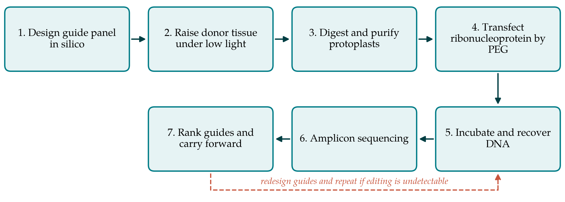
The assay runs in seven steps and is designed to be completed within about a week of having guide sequences in hand. Table 1 lists the steps with what each produces and the control that makes it interpretable, and Figure 1 shows how they connect, including the loop back to design when no guide in the panel performs. The steps are then described in turn.
| Step | What is done | Output and control |
|---|---|---|
| 1. Design | Select six to eight guides across the target, check against the genotype assembly | Guide panel; targets confirmed by resequencing |
| 2. Donor tissue | Grow seedlings at low light, use young fully expanded leaflets | Uniform tissue; same age across replicates |
| 3. Digestion | Cellulase and macerozyme in mannitol buffer, gentle vacuum infiltration | Protoplast suspension; viability stain |
| 4. Transfection | Polyethylene glycol with preassembled ribonucleoprotein | Treated cells; no-guide and no-protein controls |
| 5. Recovery | Incubate in the dark, then extract genomic DNA | DNA of adequate quality; untreated reference sample |
| 6. Sequencing | Amplify the target, sequence the amplicon, call indels Clement et al., 2019 | Indel spectrum; background from untreated DNA |
| 7. Ranking | Order guides by editing frequency and by indel profile | Ranked panel; two guides carried forward |
3.1. Step 1: Design the guide panel
Six to eight guides spread across the target region give a reasonable chance that at least two will be active. Candidates are selected with a design tool (Concordet and Haeussler, 2018; Liu et al., 2017) and filtered for predicted off-target sites in the species assembly, which matters in legumes because of the widespread gene duplication that follows polyploidy. The step most often skipped is the one that matters most: the target site should be amplified and sequenced from the exact accession to be used, since a single polymorphism in the seed region or in the protospacer-adjacent motif will make an otherwise good guide inactive. Where the species carries duplicated copies of the target gene, the panel should also be checked against each copy, since a guide that matches two of three homoeologues gives a result that is difficult to interpret later.
3.2. Step 2: Prepare donor tissue
Protoplast yield depends more on the source tissue than on anything done afterwards. Seedlings raised under moderate light and high humidity produce leaves with thin cuticles and loose mesophyll. Young fully expanded leaflets, taken before the tissue toughens, digest best. Plants that have been stressed or allowed to dry give low yields and fragile cells, and because the assay compares guides against each other, tissue of uniform age should be used across all treatments in a run.
3.3. Step 3: Digest and purify
The digestion mixture of the standard Arabidopsis protocol, cellulase and macerozyme in a mannitol osmoticum buffered near pH 5.7 with added calcium (Yoo et al., 2007), is the sensible starting point. Legume leaves generally need longer digestion, and the enzyme concentration and incubation time should be adjusted once for each species and then fixed, since changing them between runs makes guide comparisons unreliable. Leaf strips cut with a fresh blade, or lower leaf surfaces abraded gently, release cells faster than whole leaflets. Brief vacuum infiltration helps the enzyme enter the tissue. Released protoplasts are filtered, washed and counted, and viability is checked with a vital stain before the suspension is used.
3.4. Step 4: Transfect the ribonucleoprotein
Cas9 protein and the synthetic guide are combined and allowed to assemble before contact with cells. The complex is mixed with the protoplast suspension and polyethylene glycol with calcium, held briefly, then diluted out and the cells washed. Two controls are needed in every run: protoplasts treated with polyethylene glycol and no complex, which establishes the background indel rate of the sequencing method, and protoplasts treated with Cas9 protein and no guide, which shows that the protein alone does nothing. A positive control guide known to work in the species, once one is available, turns the assay from a relative comparison into an absolute one.
3.5. Step 5: Incubate and recover DNA
Transfected cells are incubated in the dark in the osmotic buffer. A period of roughly one to two days allows cutting and repair while keeping losses acceptable, and protoplasts of most legume species do not survive much longer without a regeneration medium. Genomic DNA is then extracted from the whole population. Yields are small, so a method suited to low input is preferable, and the sample should be kept as a single pool rather than divided, since the editing frequency is a property of the population.
3.6. Step 6: Sequence and call edits
The target region is amplified with primers placed outside any predicted cut site, barcoded, and sequenced. Analysis with a dedicated tool reports the proportion of reads carrying insertions or deletions at the expected position and the composition of that spectrum (Clement et al., 2019). Where short-read sequencing is not available, trace decomposition from Sanger reads gives a workable estimate for guides that are clearly active (Brinkman et al., 2014), though it resolves low frequencies poorly. The background measured in the no-complex control is subtracted before guides are compared.
3.7. Step 7: Rank and carry forward
Guides are ordered by editing frequency, and the two highest are taken into stable transformation. Frequency is not the only criterion. A guide producing a high proportion of single-base insertions or of deletions that shift the reading frame is preferable when a knockout is wanted, while a guide producing mostly in-frame deletions may be the better choice when the aim is to alter a domain rather than remove the protein. If no guide in the panel edits above background, the design is revisited rather than the protocol: the usual cause is a target sequence that differs from the reference assembly.
4. Fitting the Assay to an Ordinary Laboratory
The assay was put together with modest facilities in mind. It needs a growth room, a laminar flow bench, a bench centrifuge with a swing-out rotor, a haemocytometer and an inverted microscope. Commercial Cas9 protein and synthetic guides remove the need for in vitro transcription. The only step that may have to be sent out is sequencing, and a small amplicon pool is inexpensive to have read commercially. Because no DNA is introduced and the cells are destroyed at the end of the run, the biosafety position is simpler than for stable transformation in many jurisdictions, though local rules should be checked.
Two practical habits make the difference between a working assay and a frustrating one. Fix the digestion conditions per species and record them, so that results from different weeks can be compared. And run the whole panel in one batch, since variation between protoplast preparations is larger than many of the differences between guides that the assay is meant to detect.
5. Limitations and What Remains to Be Shown
This article proposes a procedure and does not report its application. Three things would need to be established before it could be recommended without qualification. The first is predictive value: whether the ranking obtained in protoplasts agrees with editing frequencies measured in stably transformed plants of the same species, across enough targets for the agreement to be meaningful. The second is reproducibility: whether a given guide ranks consistently across independent protoplast preparations and between laboratories. The third is transferability: whether the digestion conditions that work for one legume can be adapted to another without a fresh round of optimisation.
Several limits will remain whatever those studies show. The assay cannot test guides for a target expressed only in a tissue that protoplasts do not represent, and it cannot report on homology-directed repair, which is rare in somatic plant cells and would need a different readout. It gives no information about regeneration, which in the hardest legume species is the real obstacle. And a guide that performs well in a wall-less mesophyll cell may behave differently in embryogenic callus, where chromatin and cell cycle differ. The assay narrows the field; it does not settle it.
6. Conclusion
Guide screening is worth doing wherever transformation is expensive, and in the grain legumes it is expensive enough that a week spent in protoplasts is cheap insurance. The procedure set out here brings the established protoplast and ribonucleoprotein methods into one sequence written for legume tissue, names the controls that make the readout interpretable, and states where it is likely to fail. It remains a proposal. Laboratories that adopt it are encouraged to report the correspondence between their protoplast rankings and their eventual transformation outcomes, since that comparison, accumulated across species and targets, is what would turn a reasonable procedure into a validated one.
Conflict of interest
To be declared by the author(s) before publication.
Funding
To be declared by the author(s) before publication.