1. Introduction
Acute ureteric obstruction by a stone is one of the commonest urological emergencies, and its management is covered in detail by international guidelines (Türk et al., 2016a; Assimos et al., 2016). Those guidelines say little about one complication: the escape of urine from the collecting system into the renal sinus and perinephric space. Wider use of computed tomography has shown that this is not as unusual as was once thought, and radiology reports now mention perinephric fluid or contrast extravasation often enough that the on-call urologist needs a considered response.
The difficulty is that the evidence consists of single case reports and retrospective series from individual hospitals, with no agreed terminology and no comparative studies. Some authors report that nearly all patients do well without intervention; others place a stent in every case. A clinician reading this literature cannot easily tell whether the two groups were treating the same condition.
This article proposes a structured pathway for the diagnosis and management of spontaneous rupture of the renal pelvis caused by an obstructing ureteric stone, and by extension of forniceal rupture, from which it must be distinguished. The pathway is assembled from published guidance and from the reasoning in the available series. It does not describe a patient or report outcomes, and it has not been tested in practice.
2. Background
Three matters need to be clear before a pathway can be designed: how the leak arises, what the terms mean, and what is known about its course.
2.1. Mechanism and terminology
When a ureter is suddenly blocked while the kidney continues to filter, pressure in the collecting system rises. The classical work on pyelovenous backflow showed that fluid then leaves the system through the fornices of the calyces, where the wall is thinnest, and tracks into the renal sinus, veins and lymphatics (Hinman and Lee-Brown, 1924). Forniceal rupture is this process made visible: a small tear that decompresses the kidney and usually seals when the obstruction is relieved. In the series analysed by Gershman et al. (2011) ureteric stones were by far the commonest cause, and the responsible stones were typically small and lodged in the distal ureter.
Rupture of the renal pelvis is a different lesion, a breach in the wall of the pelvis itself. It is much less common, tends to produce a larger and more persistent leak, and has been associated with pre-existing weakness of the wall from infection, previous surgery or long-standing obstruction. The two are often reported under the same heading of spontaneous urinary extravasation, which makes the literature hard to interpret. In this article the word spontaneous means that no trauma or instrumentation preceded the leak, and the pathway asks the clinician to record which of the two lesions is present whenever imaging allows.
2.2. What the published series suggest
The largest series of fornix rupture found that most patients were managed successfully and that serious complications were uncommon (Doehn et al., 2010). A later report asked directly whether conservative treatment was safe and concluded that, in selected patients without infection, it was (Morgan et al., 2017). At the other end of the range of practice, primary ureteroscopy with removal of the stone has been described as initial treatment (Kalafatis et al., 2004). These reports are retrospective and concern mainly forniceal leaks. For true pelvic rupture the evidence is limited to isolated cases, and the proposals below lean on first principles and on guidance written for obstruction in general.
3. The Proposed Pathway
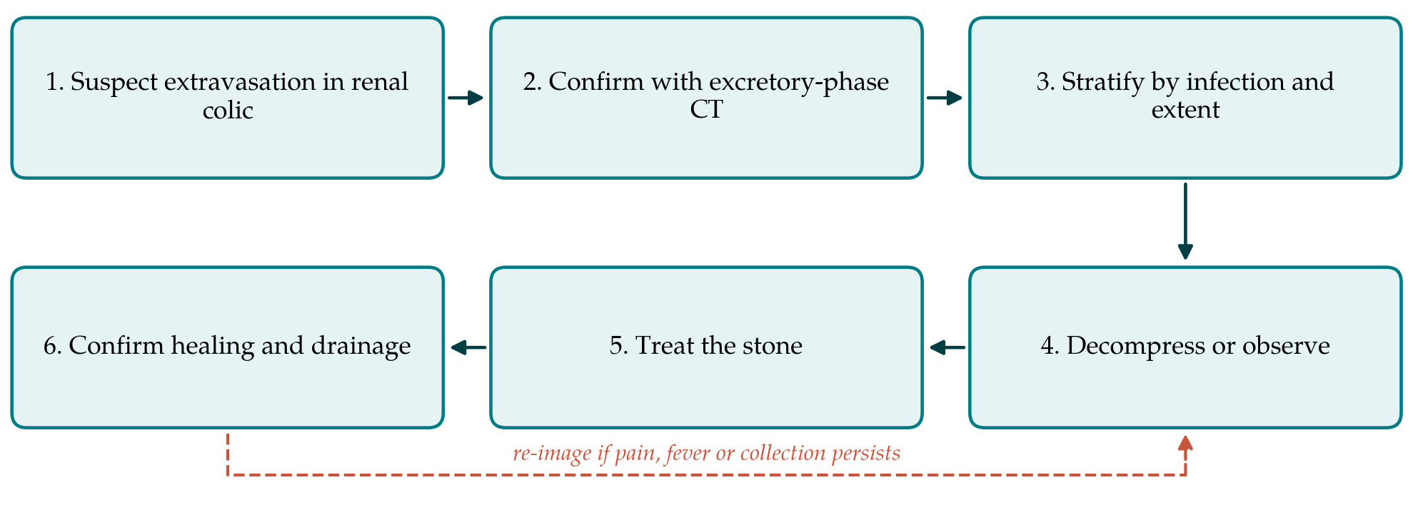
The pathway has six steps, shown in sequence in Figure 1. Each is described below with its purpose and the judgement it requires. The steps are intended to be followed in order, with a return to imaging if the clinical course departs from what is expected.
3.1. Step 1: Suspect the diagnosis
No symptom is specific. Extravasation is usually found in someone being assessed for ordinary renal colic. Features that should raise suspicion are pain that changes character, from colicky to constant or from severe to suddenly eased, flank tenderness with guarding that is out of proportion to simple colic, and fever. Because a sudden fall in pressure can relieve pain, improvement does not exclude a leak. The first step is therefore an attitude: to keep the diagnosis in mind when the clinical picture and the size of the stone do not match.
3.2. Step 2: Confirm with appropriate imaging
Non-contrast computed tomography is the standard first test for suspected ureteric stone (Smith et al., 1995; Coursey et al., 2012), and ultrasonography is an accepted initial alternative (Smith-Bindman et al., 2014). Neither demonstrates a leak directly. Non-contrast images show perinephric stranding and fluid, which are common in obstruction without rupture. The pathway therefore proposes that, when a fluid collection is seen around the kidney or along the ureter and renal function permits, contrast-enhanced computed tomography with a delayed excretory phase be obtained. Opacified urine outside the collecting system confirms extravasation, and the site and volume of the leak help to separate a forniceal tear from a breach of the pelvis. Where contrast cannot be given, the diagnosis rests on the size and behaviour of the collection on repeat imaging.
3.3. Step 3: Stratify the case
Management should follow from risk, not from the radiological label alone. Table 1 lists the features the pathway asks the clinician to assess and the response proposed for each. Infection carries the most weight. An obstructed kidney with signs of infection is a urological emergency under existing guidelines whether or not urine has leaked (Türk et al., 2016a), and sepsis should be recognised with the criteria of the international consensus definitions (Singer et al., 2016). Infected urine in the retroperitoneum adds the risk of abscess.
| Feature assessed | Why it matters | Proposed response |
|---|---|---|
| Fever, raised inflammatory markers or positive urine culture | Infected urine outside the kidney may form an abscess | Urgent decompression and antibiotics in line with guidelines |
| Site of leak: fornix or pelvis | Pelvic breaches are larger and seal less readily | Lower threshold for drainage in pelvic rupture |
| Size of urinoma | Large collections may compress the kidney or become infected | Consider percutaneous drainage of the collection |
| Solitary kidney or impaired renal function | Little reserve if obstruction persists | Decompress early |
| Stone size and position | Determines the chance of spontaneous passage | Observation only if passage is likely |
| Pain not controlled by analgesia | Suggests continuing high pressure or enlarging leak | Decompress and re-image |
3.4. Step 4: Observe or decompress
For a small forniceal leak in someone who is afebrile, with a stone likely to pass, good renal function and two kidneys, the pathway proposes observation: analgesia, with non-steroidal anti-inflammatory drugs as the first choice where they are not contraindicated (Holdgate and Pollock, 2004), clinical review, and a low threshold for repeat imaging. Whether prophylactic antibiotics are needed in the absence of infection is unknown, and the pathway leaves the decision to local policy. Medical expulsive therapy may be considered on the same terms as for any ureteric stone (Hollingsworth et al., 2006).
In all other cases, the pathway proposes decompression. In obstruction with infection, a retrograde ureteric stent and a percutaneous nephrostomy were found to be equally effective in a randomised comparison (Pearle et al., 1998), and observational data link decompression to lower mortality in sepsis with ureteric stones (Borofsky et al., 2013). The choice can be made on availability and on the condition of the patient. A stent has the advantage of splinting the collecting system and lowering pressure across a pelvic tear. A nephrostomy avoids instrumenting a ureter under pressure and allows antegrade study later. A large or infected urinoma may need a separate percutaneous drain. Management of sepsis itself follows established principles of early antibiotics, resuscitation and source control (Wagenlehner et al., 2013).
3.5. Step 5: Treat the stone
Definitive treatment of the stone follows the usual indications and techniques (Türk et al., 2016b). The question specific to this setting is timing. In the presence of infection, guidelines advise that stone removal be deferred until the infection has been treated. Where there is no infection, primary ureteroscopy has been reported (Kalafatis et al., 2004), but pressurised irrigation in a system that has already leaked could in principle enlarge the extravasation. The pathway therefore proposes that definitive treatment normally be delayed until the leak has had time to seal, and that primary ureteroscopy be reserved for small forniceal leaks treated with low irrigation pressure by an experienced operator.
3.6. Step 6: Confirm healing
The final step is to show that the leak has closed, that any collection has resolved and that the kidney drains. For a small forniceal tear with an uneventful course, clinical recovery and a follow-up ultrasound scan may be enough. After pelvic rupture, or where a drain or nephrostomy was placed, the pathway proposes contrast imaging before tubes are removed. Complications should be recorded in a standard form, for which the classification of Dindo et al. (2004) is suitable, so that experience can be pooled between units.
4. Fitting the Pathway to Local Practice
Hospitals differ in what they can offer out of hours. Where interventional radiology is not available at night, retrograde stenting becomes the default method of decompression, and the pathway accommodates this. Where delayed-phase imaging is not routinely added to stone protocols, agreement is needed with radiology on the circumstances in which it will be performed, since a second scan adds radiation and contrast exposure and should not be requested for every patient with perinephric stranding. A short local document that names the features in Table 1, the person responsible for each decision and the interval for review is likely to be more useful than a detailed protocol.
5. Limitations and What Remains to Be Shown
The pathway is a proposal built on guidelines written for obstruction and infection in general and on a small, retrospective literature. It has not been validated. Several of its choices are judgements that others might make differently: the weight given to the site of the leak, the preference for delaying ureteroscopy, and the suggestion that uninfected forniceal tears may be observed. The rarity of true pelvic rupture means that no single centre is likely to accumulate enough cases to test these choices alone.
A prospective multicentre registry would be the realistic means of evaluation. It would need to record the site and size of the leak on excretory-phase imaging, the presence of infection at presentation, the treatment given and its timing, and a small set of outcomes: unplanned intervention, infected collection, length of stay, and renal drainage at follow-up. Such data would show whether the stratification in Table 1 separates patients who do well with observation from those who do not, which is the question on which the usefulness of the pathway depends.
6. Conclusion
Urinary extravasation from a kidney obstructed by a ureteric stone ranges from a trivial forniceal leak to a breach of the renal pelvis with an infected collection. The six-step pathway proposed here asks the clinician to confirm the diagnosis with excretory-phase imaging, to let infection, the site and size of the leak and renal reserve determine whether to observe or decompress, to time stone treatment with the healing of the collecting system in mind, and to document resolution. It offers a common structure for decisions that are at present made case by case, and it awaits prospective testing.
Conflict of interest
To be declared by the author(s) before publication.
Funding
To be declared by the author(s) before publication.