1. Introduction
Diabetic retinopathy is among the leading causes of visual loss in adults of working age, and the number of people affected is projected to keep rising, with the steepest growth in low and middle income regions (Teo et al., 2021). The sight-threatening stages are usually silent until late. Laser photocoagulation and intravitreal therapy preserve vision when applied in time, which makes the condition a textbook candidate for screening under the criteria set out by Wilson and Jungner (1968). The guidelines of the International Council of Ophthalmology describe what screening should contain at each level of resource and when to refer (Wong et al., 2018).
Conventional screening depends on a tabletop fundus camera, mains electricity and a trained photographer. Outreach camps, held for a day in a school, clinic or place of worship, seldom have these. Smartphone-based imaging has been promoted as the answer. This communication looks at that proposal from the point of view of a camp organiser deciding whether, and how, to adopt it.
2. What the Devices Are and What Validation Shows
Three kinds of system are described in the literature. The simplest holds a condensing lens in front of the phone camera and uses the phone's light source in the manner of indirect ophthalmoscopy (Haddock et al., 2013). Clip-on adapters place a small optical module over the camera to give a direct ophthalmoscope view, as in the devices studied by Russo et al. (2015) and Bastawrous et al. (2016). Integrated handheld cameras use the phone as sensor, screen and storage behind dedicated optics with internal fixation, and behave much like a portable fundus camera.
Validation results differ by type. Rajalakshmi et al. (2015) compared an integrated smartphone camera with a conventional camera after dilation in a diabetes clinic and found close agreement for detecting retinopathy and its sight-threatening forms. Smartphone ophthalmoscopy through an adapter agreed well with slit lamp biomicroscopy when performed by an ophthalmologist through a dilated pupil (Russo et al., 2015). In contrast, a comparison in which a phone and condensing lens were set against non-mydriatic and mydriatic photography found the phone method the least sensitive of the three (Ryan et al., 2015). The reasonable summary is that optics and dilation matter more than the fact that a phone is involved.
Two cautions apply before these results are carried into a camp. The studies were done mostly in clinics, by trained staff, on people already under diabetes care, with dim rooms and time to repeat a poor image. A camp offers bright ambient light, queues, older attenders with cataract and small pupils, and operators who may have learnt the device that week. The second caution is the field of view. Phone systems capture a narrower field than tabletop cameras, so several overlapping images are needed to cover the posterior pole, and peripheral disease may be missed.
Practical matters decide much of the image quality. Handheld imaging is a skill, and the published comparisons used operators who had practised it. A camp should allow supervised practice before the day, set aside a darkened corner or a simple hood so that pupils stay larger and the screen can be read, and plan for spare charged batteries and enough storage on the device. Seating the attender and bracing the operator's hand against the forehead steadies the image. Media opacity defeats any camera, so people in whom no view can be obtained because of cataract are referred on that ground, which is often a benefit to them in its own right.
3. Elements of a Camp Pathway
Table 1 sets out the choices a camp has to make. None of them is specific to smartphones, but the portability of the device tempts organisers to skip them. Pupil dilation is the first. Gradable images through undilated pupils are hard to obtain with handheld devices, particularly in older people, and a camp that decides against dilation should expect a large share of ungradable eyes. A protocol that fixes the number and position of fields per eye, typically one centred on the macula and one on the disc, gives graders a consistent set and makes omissions visible.
| Element | Decision to be made | Point to consider |
|---|---|---|
| Pupil | Dilate all, dilate selectively, or not at all | Undilated handheld imaging yields more ungradable eyes |
| Image set | Fields per eye and their centring | Narrow field of view; macula and disc both needed |
| Quality check | Who accepts or repeats an image, and when | Check on site while the person is still present |
| Grading | On site, remote, or automated | Use a recognised scale Wilkinson et al., 2003 |
| Ungradable eyes | Rule for referral | Treat as screen-positive, not as normal |
| Referral | Where, how soon, and how tracked | Treatment must be reachable and affordable |
| Data | Consent, storage, transfer | Images on personal phones need explicit safeguards |
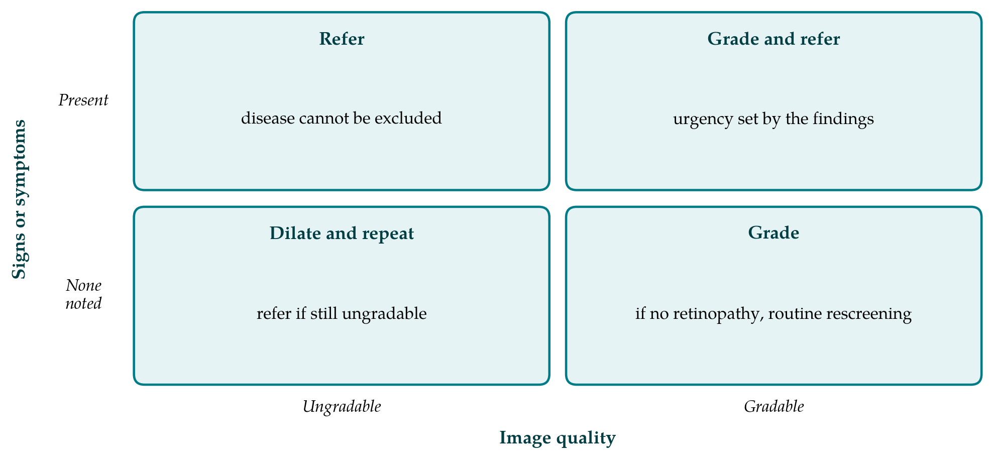
Quality control belongs at the camp and not at the reading centre. An image found to be ungradable a week later means a person who cannot easily be recalled. The operator, or a second person, should confirm before the attender leaves that the macula and disc are in focus and adequately lit. Figure 1 shows a simple decision rule that combines image quality with what else is known. Its main purpose is to make one point explicit: an eye that could not be imaged adequately has not been screened, and after a repeat attempt it is referred.
Grading should follow a published severity scale so that referral thresholds mean the same thing from one camp to the next. The international clinical scale was designed for this purpose and is simple enough for trained non-physician graders (Wilkinson et al., 2003). Visual acuity is measured alongside imaging, as the international guidelines advise, since reduced acuity may signal macular oedema or other disease that a two-field photograph does not show (Wong et al., 2018).
4. Automated Grading
Deep learning systems can detect referable retinopathy in conventional photographs with high accuracy (Gulshan et al., 2016), and the same approach has been applied to smartphone images. Rajalakshmi et al. (2018) reported good performance of an automated system on images from an integrated smartphone camera, and Natarajan et al. (2019) evaluated an algorithm that runs on the phone without an internet connection in a community setting. For a camp the attraction is a result while the person is still present, which makes counselling and referral immediate.
Three limits should temper the enthusiasm. An algorithm is validated on images from particular cameras and populations, and its performance on another device or in eyes with cataract cannot be assumed. Automated systems reject poor images, so the rate of ungradable results may rise, and the rule for those eyes has to be settled in advance. Regulatory status differs between countries, and a camp should use a system only in the way its approval allows. Automated output is best treated as a triage aid with human review of positives and of a sample of negatives.
5. The Referral Problem
Screening does nothing for sight unless those who screen positive are examined and treated. This is the weakest link in outreach work and the one least affected by better cameras. A camp should not begin imaging until it has an agreement with a treating centre, a means of telling each referred person where and when to attend, and a list that allows someone to check afterwards who arrived. Cost and distance are the usual barriers, and where treatment is beyond reach the classical screening criteria are not met (Wilson and Jungner, 1968). Consent and data protection also deserve a written procedure, since retinal images linked to names are health records wherever they are stored.
6. Conclusion
Smartphone-based fundus cameras make retinal imaging possible in places a conventional camera cannot reach, and the better devices, used through a dilated pupil by trained operators, have performed well in published clinic-based comparisons. Evidence from camps themselves is thinner, and adapter-only methods are less reliable. The gain from portability is realised only when a camp also controls image quality on site, refers ungradable eyes, grades to a recognised scale and secures a route to treatment. Studies that report gradability, referral uptake and treatment received under real camp conditions would be more useful to organisers than further accuracy comparisons in clinics.
Conflict of interest
To be declared by the author(s) before publication.
Funding
To be declared by the author(s) before publication.