1. Introduction
Transplanting fat without its blood supply is an old idea with a long record of disappointment. Peer (1950) weighed human fat grafts a year after transfer and found that roughly half their mass had been lost, a figure that discouraged the technique for a generation. Liposuction revived interest by making fat available in quantity, but early attempts at breast injection produced palpable lumps and calcifications. In 1987 a committee of the American Society of Plastic and Reconstructive Surgeons advised against fat injection for breast enlargement on the ground that the resulting changes might hinder the detection of cancer (ASPRS, 1987).
The position changed as technique improved. Coleman (2006) described a method of atraumatic harvest, purification and placement in very small parcels, and Coleman and Saboeiro (2007) reported its use in the breast with durable results. A task force of the American Society of Plastic Surgeons then reviewed the field and concluded that fat grafting could be considered for breast surgery, while stressing that results depended on technique and that the evidence was weak (Gutowski, 2009). Large clinical series followed (Delay et al., 2009).
After mastectomy the stakes differ from those of cosmetic augmentation. The recipient site is scarred, often irradiated, and has been the seat of a cancer. This review considers what the literature says about the persistence of grafted fat in that setting and about its oncological safety.
2. Approach of This Review
This is a narrative review of the published literature. It relies on the experimental studies that describe how fat grafts heal, the clinical series and volumetric studies that report retention in the breast, the cohort studies and meta-analyses that address recurrence, and the statements of professional societies. The sources were selected for their influence on the debate, and the review does not claim to have identified every relevant report. Cosmetic augmentation of the healthy breast is considered only where it supplies evidence that reconstruction lacks.
3. Uses of Fat Grafting After Mastectomy
Three uses can be distinguished. The commonest is as an adjunct: filling the step at the upper pole of an implant, softening the edge of a flap, or correcting a contour irregularity after the main reconstruction is complete. The second is treatment of the irradiated chest wall. Rigotti et al. (2007) reported improvement in tissues damaged by radiotherapy after injection of lipoaspirate and attributed it to the stromal cell content of the graft. The third is reconstruction of the whole breast by fat alone over several sessions, which requires volumes far larger than the scarred mastectomy envelope will readily accept. Whole-breast reconstruction of this kind is practised in few centres, and most of the clinical evidence concerns the first two uses.
4. Graft Retention
Retention is the proportion of injected volume that persists as living tissue once healing is complete. It is the product of biology and technique, and the literature on each is considered in turn.
4.1. How a fat graft survives
Grafted fat depends on diffusion from the recipient bed until new vessels reach it. The experimental work of Eto et al. (2012) showed what this implies. Adipocytes survive only in a thin outer layer of each deposit, within a few hundred micrometres of its surface. Beneath that layer the adipocytes die, but adipose-derived stromal cells, which tolerate ischaemia better, survive and regenerate new adipocytes. In the centre of a large deposit both cell types die. Kato et al. (2014) followed the process over three months and described how dead fat is either replaced by new adipocytes or left as an oil cyst walled off by fibrosis, which may later calcify.
Two consequences are accepted across the field. Fat must be placed in small parcels so that as much of it as possible lies within reach of diffusion, and the volume seen in the early weeks, which includes oedema and dead fat not yet resorbed, overstates the final result. The regenerative role of the stromal cells rests on the demonstration by Zuk et al. (2001) that adipose tissue contains a population of multipotent cells.
4.2. Technique
Each step of the procedure has been examined for its effect on survival, and Figure 1 summarises the factors usually cited. The evidence is less decisive than the confidence of individual authors suggests. Rohrich et al. (2004) found no difference in adipocyte viability among the usual donor sites. The review by Strong et al. (2015) concluded that no method of harvest or processing, whether centrifugation, filtration or washing, had been shown to be clearly superior in clinical use. What the literature agrees on is the principle: handle the tissue gently, remove oil and fluid, and distribute the graft widely.
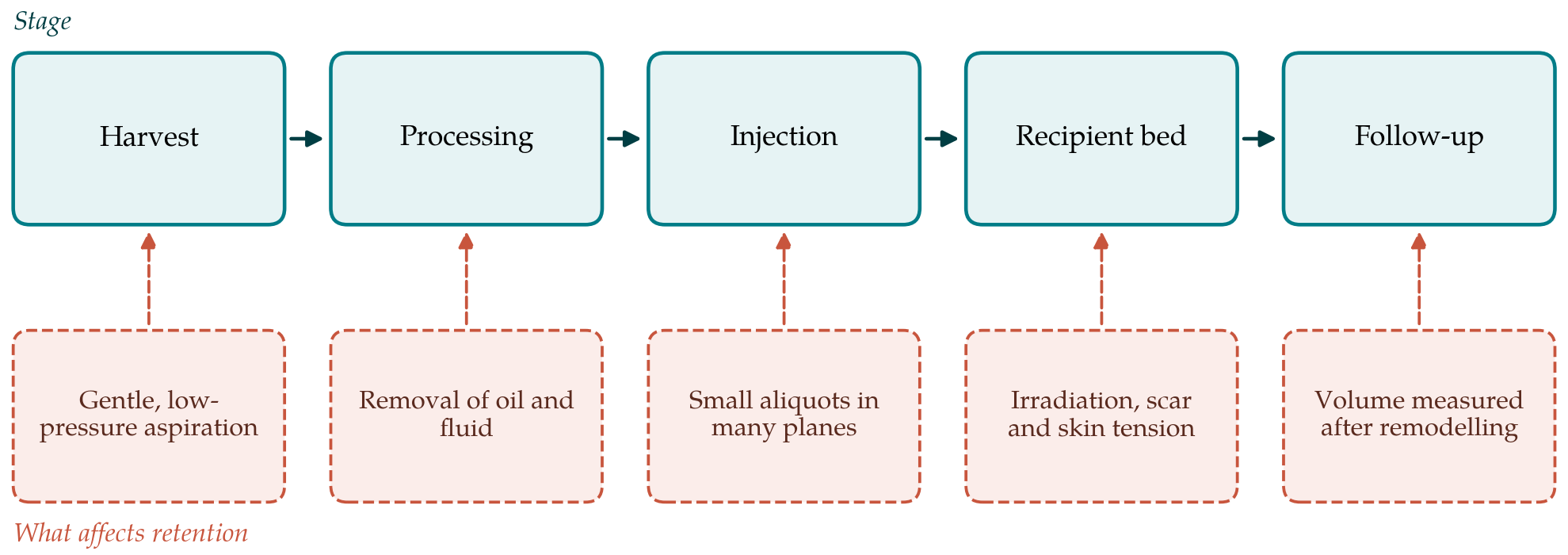
The recipient site may matter as much as the graft. Khouri et al. (2014) set out the argument that survival is limited by the surface available for contact between graft and host and by the rise in tissue pressure when too much is injected into a tight space. A mastectomy flap that is thin, adherent and irradiated offers little room, which is the rationale for treating in several modest sessions.
4.3. Clinical measurement of retention
Clinical retention has been estimated with three-dimensional surface imaging and with magnetic resonance imaging. Choi et al. (2013) used surface imaging in reconstructed breasts and found that retained volume fell progressively over the first months and varied with the amount injected. Published estimates range widely between studies, which reflects differences in the time of measurement, the method, the recipient site and the patients. No single retention figure can honestly be quoted to a patient. The consistent findings are that a substantial part of the injected volume is lost, that most of the loss occurs early, and that repeat sessions are commonly needed. How far previous radiotherapy reduces retention is not settled. Irradiated tissue is stiffer and less vascular, which should work against the graft, yet it is also the tissue in which clinical improvement after grafting has been most often described.
4.4. Cell enrichment
Because stromal cells drive regeneration, adding more of them is an obvious strategy. Yoshimura et al. (2008) introduced cell-assisted lipotransfer, in which part of the aspirate is processed to concentrate its stromal vascular fraction and recombined with the graft. A randomised trial in healthy volunteers found markedly better retention of small fat grafts enriched with culture-expanded stromal cells (Kølle et al., 2013). A prospective comparison in the breast using the uncultured stromal fraction found no benefit (Peltoniemi et al., 2013). The two results are not contradictory, since the cell dose differed greatly, but they leave the clinical value of enrichment unsettled. In reconstruction after cancer the question cannot be separated from that of safety.
5. Oncological Safety
Concern about safety has a laboratory basis and a clinical one. Table 1 sets out the main questions with the state of the evidence on each.
| Question | What the literature indicates | Strength of evidence | Representative sources |
|---|---|---|---|
| How much fat persists? | Substantial, variable early loss; repeat sessions often needed | Moderate; methods differ | Choi et al., 2013 |
| Does technique change retention? | Gentle handling and dispersed placement agreed; no superior processing method | Low to moderate | Strong et al., 2015 |
| Does cell enrichment help? | Conflicting results; benefit seen with expanded cells in volunteers | Low; few controlled studies | Kølle et al., 2013 |
| Is locoregional recurrence increased? | No increase found in matched cohorts and meta-analysis | Moderate; observational only | Krastev et al., 2018 |
| Are some subgroups at higher risk? | A signal after intraepithelial neoplasia; not settled | Low | Petit et al., 2013 |
| Does grafting obscure imaging? | Oil cysts and calcification occur; usually recognisable as benign | Moderate | Carvajal and Patiño, 2008 |
5.1. The biological concern
Adipose tissue is an endocrine organ, and its stromal cells secrete growth factors and promote the formation of new vessels. Those properties are the reason fat grafting improves irradiated skin. They are also the properties a residual tumour cell might exploit. Lohsiriwat et al. (2011) reviewed the experimental literature and found that adipose-derived stromal cells and adipocytes can stimulate the growth and invasiveness of breast cancer cells in culture and in animal models. The authors framed the open issue as whether lipofilling might awaken dormant cells. Experimental conditions differ from the clinical one in cell dose, in the use of active tumour and in the absence of adjuvant treatment, so these studies establish plausibility and nothing more.
5.2. Clinical studies of recurrence
The first large clinical report pooled the experience of three European centres and found few complications and a low rate of recurrence, though with short follow-up and no control group (Petit et al., 2011). The same group then matched patients who had lipofilling with controls who had not. Recurrence did not differ overall, but it was more frequent after lipofilling in the subgroup treated for intraepithelial neoplasia (Petit et al., 2012), a finding repeated in a dedicated matched cohort (Petit et al., 2013). That signal has been neither clearly confirmed nor dismissed by later work.
Subsequent studies were larger. Kronowitz et al. (2016) compared reconstructed breasts with and without lipofilling at a single cancer centre and found no increase in locoregional recurrence. The meta-analysis by Krastev et al. (2018) combined the matched and unmatched studies then available and reached the same conclusion, and a matched cohort with five years of follow-up again showed no difference in relapse (Krastev et al., 2019). The agreement of these studies is reassuring. Their limits are those of all observational research on the question: patients offered lipofilling have already survived disease-free long enough to reach secondary reconstruction, which biases comparison in favour of the procedure unless matching accounts for it, and recurrence is rare enough that modest increases in risk could escape detection.
5.3. Imaging after fat grafting
The fear that prompted the 1987 statement has been largely answered. Fat necrosis, oil cysts and calcifications do appear after grafting, as Carvajal and Patiño (2008) documented on mammography, but similar changes follow any breast operation and experienced radiologists can generally distinguish them from malignancy. The practical cost is an occasional additional scan or biopsy to resolve an indeterminate finding. After mastectomy, where little or no breast tissue remains to be screened, the problem is smaller than in the augmented or conserved breast. Patients should nonetheless be told to expect that lumps may form and may need investigation.
6. Gaps and Future Work
Several things would strengthen the evidence. Retention should be reported with a standard method at a standard interval after the last session, late enough for remodelling to be complete, so that studies can be compared. Trials of processing methods and of cell enrichment should be conducted in reconstructed, irradiated breasts and not inferred from healthy volunteers. On safety, prospective registries with long follow-up, recording tumour biology, the interval from cancer treatment and the volume grafted, are needed to examine the subgroups in which doubt persists: patients treated for intraepithelial disease, those with breast conservation, and those receiving enriched grafts. Patient-reported outcomes deserve the same attention as volume, since the purpose of the procedure is a breast the patient is content with.
7. Conclusion
Fat grafting after mastectomy rests on a coherent biological account of graft survival and on a clinical literature that is broadly favourable but methodologically modest. Part of every graft is lost, the amount is hard to predict, and no processing method or additive has been shown reliably to prevent it. The cohort studies available do not show that lipofilling of the reconstructed breast increases recurrence, and that is the most that observational data can say. Patients can reasonably be offered the procedure with an honest account of both points: several sessions may be needed, and long-term safety is supported by current evidence but not proven beyond question.
Conflict of interest
To be declared by the author(s) before publication.
Funding
To be declared by the author(s) before publication.