1. Introduction
Repair of the nose with forehead skin is among the oldest operations in plastic surgery. It reached European practice through the report of Carpue (1816), and the midline design was refined in the twentieth century by Kazanjian (1946), who closed the donor site directly. The modern operation differs from these in two respects. The flap is centred on one supratrochlear pedicle and not on the midline, which lets the base be narrow and the flap reach further, and the reconstruction is conceived as the rebuilding of three layers and not as the patching of a hole.
Much of that conception comes from Burget and Menick. Their account of nasal subunits (Burget and Menick, 1985) and of lining and support (Burget and Menick, 1989) turned a flap operation into a discipline with rules. Menick (2002) later added an intermediate operation between transfer and division, arguing that it allowed safer thinning and more deliberate shaping. The principles are now standard textbook material (Baker, 2014).
What is harder to find is a single ordered plan that a unit could adopt, teach from and audit against. This article proposes one for full-thickness defects of the lower two thirds of the nose. It describes no patients and reports no outcomes.
2. Background
The protocol rests on two bodies of knowledge: the vascular anatomy that makes a narrow pedicle safe, and the principles that govern how the three layers of the nose are replaced.
2.1. Vascular anatomy of the flap
The supratrochlear artery leaves the orbit at the medial end of the brow, roughly two centimetres from the midline, and climbs vertically toward the hairline. McCarthy et al. (1985) showed that the flap is supplied through a rich anastomotic network around the medial canthus and can survive on a base extended below the brow. Shumrick and Smith (1992) traced the artery from a deep position over the corrugator, through the frontalis, to a subcutaneous course in the upper forehead, and Reece et al. (2008) confirmed this pattern with dynamic imaging. Two design rules follow from these studies. The pedicle can be made narrow provided it is centred on the vessel, and the distal flap, where the artery lies just under the skin, can be thinned with more safety than the proximal part.
2.2. Principles of reconstruction
The nose is seen as a set of convex and concave surfaces bounded by shadows. Burget and Menick proposed that when most of a convex subunit such as the tip or ala is lost, replacing the whole subunit places scars in the natural borders and lets the flap contract into a rounded form (Burget and Menick, 1985). Rohrich et al. (2004) argued from a large series that the rule should be applied flexibly and that healthy tissue need not always be discarded. Both positions accept that contour, not scar position alone, determines whether a nose looks normal.
Lining and support matter as much as cover. Lining may come from hinged septal mucoperichondrial flaps, from skin turned in from the defect margin, from a skin graft, or from the folded distal end of the forehead flap itself (Burget and Menick, 1989; Menick, 2010). Cartilage must be placed along the nostril rim even where none exists in the normal nose, because an unsupported flap contracts upward and inward as it heals. Alternatives to the forehead for smaller defects, such as the bilobed flap (Zitelli, 1989), are outside the present scope, and the choice among methods by size and site is reviewed by Park (2000) and Thornton et al. (2008).
3. The Proposed Protocol
The protocol has six phases. Table 1 lists them with the central decision and the record each should leave, and Figure 1 shows their order in time. Intervals between operations follow those published by Menick (2002), who allowed about a month between each stage, and are to be lengthened whenever healing is in doubt.
| Phase | Main decision | Record kept |
|---|---|---|
| 1. Analysis | Is the wound clear of tumour, clean and ready; which layers and subunits are missing | Defect diagram, photographs, risk factors |
| 2. Planning | Source of lining; source of cartilage; side of pedicle; subunit adjustment | Written plan and templates |
| 3. Transfer (stage 1) | Whether to thin the distal flap now or leave it full thickness | Flap dimensions, lining used, donor closure |
| 4. Intermediate (stage 2) | How far to thin; which support grafts to add or revise | Grafts placed, flap perfusion on re-inset |
| 5. Division (stage 3) | Whether the inset is healed enough to divide the pedicle | Date of division, state of brow inset |
| 6. Late review | Whether revision of contour, airway or scars is wanted | Standard photographs, airway and patient-reported assessment |
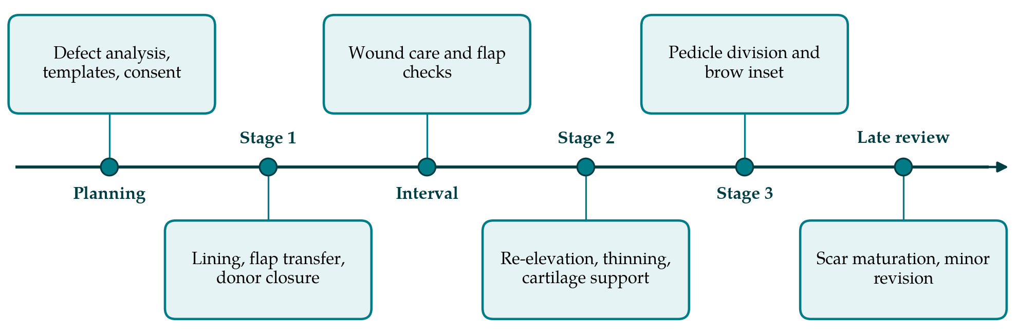
3.1. Phase 1: Analysis of the defect and the patient
Reconstruction should not begin until the wound is ready. After tumour excision this means confirmed histological clearance, and after trauma or infection a clean, stable bed. The defect is then described layer by layer and subunit by subunit on a standard diagram: which skin is missing, whether the alar cartilages, upper lateral cartilages or septum are deficient, and how much lining has gone. The forehead is inspected for scars that cross the intended pedicle and for the height of the hairline. Factors that the literature links with flap complications, of which smoking is the most consistently reported (Little et al., 2009), are recorded and discussed. Consent covers the number of operations, the weeks spent with a visible pedicle and the forehead scar.
3.2. Phase 2: Planning the three layers
A written plan names the source of each layer. For lining, the choice depends on the size of the lining defect and on whether the septum is intact. For support, septal cartilage is preferred for straight struts, conchal cartilage for curved alar grafts, and rib where large amounts are needed. For cover, the surgeon decides whether to enlarge the defect to a full subunit, weighing the two positions described above.
An exact template is essential. Where the opposite side of the nose is intact, a pattern of the normal ala or hemi-tip is made in foil or suture-pack material and reversed; where it is not, the pattern is made on a model or on the reconstructed framework. The template is three-dimensional when formed and is flattened before transfer to the forehead, so that the flap is neither too small, which distorts the nostril, nor too large, which leaves a bulky result. The pedicle side is chosen with a Doppler probe, and the reach is checked with a length of gauze swung from the pivot point at the medial brow.
3.3. Phase 3: First stage, flap transfer
Lining is established first, then primary cartilage grafts where the lining is vascular enough to carry them. The flap is outlined from the flattened template with its axis vertical over the Doppler signal and its pedicle a little over one centimetre wide. In the three-stage method the flap is raised with all its layers, including frontalis, and is not thinned at this operation (Menick, 2009). The traditional two-stage alternative thins the distal flap at transfer and divides the pedicle about three weeks later (Boyd et al., 2000). The protocol asks the surgeon to choose between them in advance and to record the reason: the three-stage sequence for defects needing folded lining, extensive cartilage work or transfer in a smoker, and the two-stage sequence for limited defects in favourable tissue. The forehead is closed directly as far as it will come, and any gap near the hairline is left to heal by secondary intention.
The weeks between operations are part of the method. The raw undersurface of the pedicle is dressed or covered with a thin skin graft to limit oozing, the nasal airway is kept clear of crusts, and the colour of the flap is inspected at each dressing. Spectacles must not rest on the pedicle. Abstinence from tobacco and nicotine is asked for throughout the staged sequence, and the request is repeated at every visit, since the flap is most at risk at the operations still to come.
3.4. Phase 4: Intermediate operation
About a month after transfer the flap is lifted again from the nose, leaving its pedicle intact. Because both the pedicle and the healed distal inset now nourish it, the skin can be elevated with a thin, even layer of fat, and the underlying frontalis and excess subcutaneous tissue removed under direct vision. The exposed bed is then sculpted, delayed cartilage grafts are fixed to shape the tip and to brace the alar rim, and a folded lining is separated from the cover so that cartilage can be placed between them. The thinned skin is returned with quilting sutures to define the alar groove. Perfusion on re-inset is noted before the patient leaves theatre.
3.5. Phase 5: Pedicle division
After a further interval of about a month the pedicle is divided. The proximal stump is trimmed and returned to the medial brow as a small inverted V so that the brows keep their spacing, and the upper margin of the nasal inset is thinned and closed along the subunit border. Division is deferred if any part of the inset has been slow to heal. Where there is doubt, the pedicle can be occluded temporarily and the flap observed before it is cut.
3.6. Phase 6: Late review and revision
The result is not judged until scars have matured over several months. Review then uses standard photographic views, an assessment of nasal breathing and a patient-reported measure, and compares them with the plan made in Phase 2. Minor revisions, such as defining the alar crease, thinning a nostril margin or opening a narrowed airway, are common in published series and should be presented to the patient from the outset as a possible fourth step and not as a failure.
4. Fitting the Protocol to Practice
The protocol is meant for a unit that performs these reconstructions occasionally, where the risk lies in omitted steps more than in technical limits. Its written plan and templates can be prepared by a trainee and checked by the senior surgeon before the first operation, which makes the planning itself a teaching exercise. The records listed in Table 1 fit on a single sheet filed with the operation notes. Units that work with dermatological surgeons can attach the sheet to the referral, so that clearance, defect description and photographs arrive in one form.
Some defects fall outside it. Total and subtotal nasal loss with absent septal support commonly needs free tissue transfer for lining before any forehead flap is raised. A forehead already scarred across both pedicles, or a low hairline that would bring hair to the nasal tip, calls for modified designs. Children and irradiated fields raise further questions. For these the protocol supplies a checklist for analysis but not an operative sequence.
5. Limitations and What Remains to Be Shown
This article proposes a protocol and does not test it. Each element comes from published anatomy or from the technique of experienced authors, but the literature on which it draws consists largely of personal series, and the assembled sequence has not been evaluated as a whole. Whether a unit that adopts it obtains better contour, fewer complications or fewer revisions than before is unknown.
Three comparisons would be informative. One is an audit of complications, namely partial flap loss, infection of cartilage grafts and nostril stenosis, before and after adoption. A second is blinded rating of standard photographs together with a validated patient-reported measure. The third concerns the choice built into Phase 3: the three-stage and two-stage sequences have never been compared in a controlled study, and the additional operation carries its own cost to the patient. The intervals quoted here are those of the original descriptions and are not thresholds established by trial.
6. Conclusion
Staged forehead flap reconstruction succeeds when lining, support and cover are each planned before the first incision and when thinning is postponed until the flap can tolerate it. The six-phase protocol proposed here arranges established anatomy and technique into an order that can be taught and audited, with a stated decision and record at every phase. It remains a proposal until units that adopt it report what happens to their results.
Conflict of interest
To be declared by the author(s) before publication.
Funding
To be declared by the author(s) before publication.