1. Introduction
Lipid nanoparticles are now the most widely used non-viral carrier for nucleic acid drugs. The first approved example, patisiran, delivers a small interfering RNA to hepatocytes to treat hereditary transthyretin amyloidosis (Adams et al., 2018), and the same particle architecture carried the messenger RNA of two large-scale coronavirus vaccines (Polack et al., 2020; Baden et al., 2021). A modern lipid nanoparticle contains four components: an ionisable lipid, a helper phospholipid, cholesterol and a lipid anchored poly(ethylene glycol) chain, mixed with the nucleic acid by rapid microfluidic combination of an ethanol and an aqueous stream (Cullis and Hope, 2017).
The ionisable lipid is the component that distinguishes these particles from older cationic liposomes. It is designed to have an apparent acid dissociation constant near 6.5, so that it carries little charge at the pH of blood, which lengthens circulation and lowers toxicity, yet becomes protonated in the acidifying endosome, where it disrupts the membrane and releases the cargo into the cytoplasm (Jayaraman et al., 2012; Hassett et al., 2019). This single idea, developed over years of lipid screening, underlies every approved product.
The clinical uses to date share a feature: the target cell is either the hepatocyte, reached after intravenous dosing, or an antigen-presenting cell, reached after intramuscular injection. Both are consistent with where lipid nanoparticles naturally go. Treating diseases of the lung, the nervous system, the heart or solid tumours would require the particles to act elsewhere, and that has turned out to be the central unsolved problem of the platform. This review examines why the liver dominates and what is being tried to move beyond it. It presents no new experimental data.
2. Scope and Approach
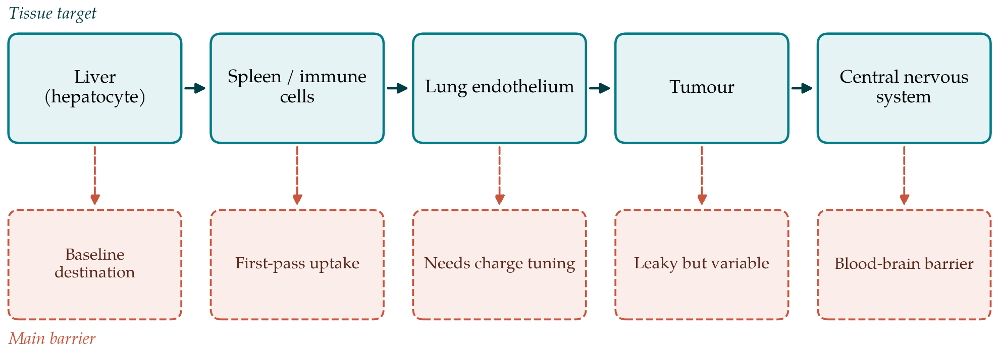
This is a narrative review drawing on the primary reports that established the platform, on widely cited reviews of lipid nanoparticle design and of nanoparticle biodistribution, and on representative studies of extrahepatic targeting. It does not attempt a systematic survey of the rapidly growing preclinical literature, and it does not pool quantitative results. The aim is to organise the known barriers by tissue and by mechanism so that the requirements for an extrahepatic product can be stated clearly. Figure 1 pairs each principal target tissue with the barrier that most limits delivery to it.
3. Why the Liver Is the Default Destination
Two mechanisms send intravenously injected lipid nanoparticles to the liver. The first is the protein corona. On contact with plasma the particle adsorbs a layer of proteins whose identity, more than the engineered surface, governs what happens next (Walkey and Chan, 2012). For lipid nanoparticles that contain certain ionisable lipids, apolipoprotein E binds to the corona and is recognised by the low-density lipoprotein receptor on hepatocytes, which provides an efficient uptake route (Akinc et al., 2010). The dependence on an endogenous targeting ligand means the liver is reached without any deliberate targeting. The identity and proportion of the ionisable lipid change which plasma proteins are recruited and therefore how strongly this hepatocyte route operates, which is one reason small changes in composition can shift biodistribution so markedly.
The second mechanism is the mononuclear phagocyte system. Macrophages of the liver and spleen are specialised to clear particulate matter from blood, and nanoparticles in the relevant size range are captured by them regardless of surface chemistry (Blanco et al., 2015). The poly(ethylene glycol) coat slows this capture but does not prevent it, and it brings its own complication: anti-PEG antibodies exist in the population and can accelerate clearance on repeated dosing (Verhoef and Anchordoquy, 2013). The practical consequence is that a large share of an injected dose is removed by liver and spleen before it can reach a chosen tissue, and raising the dose to compensate raises toxicity.
4. The Rate-Limiting Step: Endosomal Escape
Reaching a cell is not the same as delivering to its cytoplasm. After endocytosis a lipid nanoparticle is enclosed in an endosome, and unless the cargo crosses that membrane it is routed to the lysosome and degraded. Careful measurements indicate that only a small percentage of internalised small interfering RNA escapes the endosome, so that most of what a cell takes up is wasted (Gilleron et al., 2013; Sahay et al., 2013). The protonation of the ionisable lipid in the acidic endosome is believed to drive escape by forming cone-shaped ion pairs with endogenous anionic lipids that destabilise the bilayer (Hafez et al., 2001).
Endosomal escape is poorly understood and hard to measure, and it is the step that most limits potency. Its inefficiency has a bearing on extrahepatic ambitions: a tissue that is harder to reach also tends to receive fewer particles, so the escape penalty compounds the delivery penalty. Improvements in ionisable lipid structure have raised potency by orders of magnitude over the generations of lipids (Hassett et al., 2019), but escape remains the step where most of the dose is lost, in the liver as elsewhere.
The difficulty of escape is compounded by endocytic recycling. Sahay et al. (2013) observed that much of the small interfering RNA taken into a cell is exported again within the recycling endosome before it can act, so the effective dose is reduced both by failure to escape and by active expulsion of what was internalised. Together these findings explain why gains in potency have come disproportionately from improving the intracellular fate of the cargo rather than from increasing the amount that enters cells, and why a tissue reached with a smaller injected fraction is doubly disadvantaged.
5. Strategies for Redirecting Lipid Nanoparticles
Approaches to extrahepatic delivery fall into a few groups, summarised in Table 1. They differ in how much they rely on the particle finding its own way versus being actively steered, and in whether they act on biodistribution or on cell entry.
| Strategy | Mechanism | Reported reach | Main limitation |
|---|---|---|---|
| Selective organ targeting by charge | Add a charged lipid to shift corona and destination Cheng et al., 2020 | Spleen, lung in animals | Added lipid may raise toxicity |
| Antibody or ligand conjugation | Attach a targeting molecule to the surface Kedmi et al., 2018 | Immune cells, endothelium | Corona can mask the ligand |
| Receptor-directed small molecules | Use a ligand such as GalNAc for a specific receptor | Hepatocyte (established) | Few validated extrahepatic ligands |
| Altered PEG and lipid ratio | Tune coating to change circulation and uptake | Shifts liver-spleen split | Narrow and often transient effect |
| Local administration | Deliver into lung, muscle, eye or nerve directly | Site of injection | Many tissues not accessible |
The charge-based approach deserves particular mention because it reframed the problem. Rather than attaching a targeting ligand, it adds a supplementary charged lipid whose proportion predictably moves the particle to the spleen or the lung, apparently by changing which plasma proteins adsorb (Cheng et al., 2020). This demonstrated that biodistribution can be programmed by composition alone, and it has become a reference point for the field. Active targeting with antibodies has succeeded in directing particles to specific leukocyte populations (Kedmi et al., 2018), but the corona that forms in blood can bury the targeting molecule, which is why passive composition effects have in some cases outperformed deliberate ligands.
6. Barriers in Specific Tissues
Each extrahepatic target presents its own obstacles beyond the general problems of corona, clearance and escape.
6.1. Spleen and immune cells
The spleen is in one sense the easiest extrahepatic target, because its macrophages already capture a share of any injected dose. The goal is usually to reach a defined population, such as T cells or antigen-presenting cells, for vaccination or immunotherapy. Charge-based targeting enriches the spleen efficiently (Cheng et al., 2020), and antibody conjugation can address particular subsets (Kedmi et al., 2018). The difficulty is selectivity among the many cell types the organ contains.
6.2. Lung
The lung can be reached systemically, by shifting particle charge so that the capillary endothelium captures the dose, or locally by inhalation. Inhaled delivery faces the mucus layer, clearance by airway macrophages and the shear of nebulisation, which can damage the particle. Formulating a lipid nanoparticle that survives aerosolisation and still escapes the endosome in airway epithelium is an active and unresolved problem, of obvious relevance to inherited lung disease.
6.3. Tumours
Solid tumours have long been approached through the enhanced permeability and retention effect, the idea that leaky tumour vessels let nanoparticles accumulate. The reliability of that effect in human tumours, as opposed to rodent models, has been seriously questioned (Wilhelm et al., 2016), and the heterogeneity of tumour vasculature means delivery is uneven. For nucleic acid cargoes the further requirement of cytoplasmic escape in the malignant cell, rather than uptake by tumour-associated macrophages, raises the bar again. Where the therapeutic intent is to express an antigen or an immunomodulator inside tumour-associated immune cells rather than the malignant cells themselves, the natural tropism of the particle for phagocytes becomes an asset rather than an obstacle, and some of the most advanced oncology applications of the platform exploit exactly that.
6.4. Central nervous system
The blood-brain barrier excludes most nanoparticles from the brain after intravenous dosing, and no lipid nanoparticle has a validated route across it. Research directions include direct administration into the cerebrospinal fluid and the use of receptor-mediated transcytosis systems developed for other carriers (Pardridge, 2012). This remains the hardest of the targets considered here.
7. Manufacturing, Stability and Immunogenicity
An extrahepatic product must also be manufacturable and stable. Microfluidic mixing scales, but any added targeting chemistry adds synthetic and analytical steps and new release specifications. The approved messenger RNA vaccines require storage at low temperature, and improving the thermal stability of lipid nanoparticles is a goal in its own right. Immunogenicity is a two-sided concern: for a vaccine the innate stimulation provided by the particle is useful, whereas for a chronically dosed therapeutic the same stimulation, together with anti-PEG antibodies, limits repeat administration (Verhoef and Anchordoquy, 2013). A targeting strategy that works in a single-dose animal experiment may fail on repeated dosing in patients.
8. Gaps and Future Work
Several gaps recur across the tissues. The protein corona is still characterised incompletely, yet it governs destination more than the engineered surface, so methods to predict and control it would help every target. Endosomal escape needs quantitative assays that report the cytoplasmic fraction directly rather than a downstream effect. Preclinical models, especially of tumour vasculature and of the human corona, translate poorly, and this weakens the evidence for passive targeting in particular. Finally, the field would benefit from reporting biodistribution and the delivered, escaped dose in common units, so that competing strategies can be compared rather than each described in its own terms.
9. Conclusion
Lipid nanoparticles reach the liver and the immune system reliably because the chemistry that makes them work also sends them there. Moving the platform to the lung, the tumour and the brain means overriding the protein corona, surviving the phagocyte system and still accomplishing endosomal escape in the intended cell. Charge-based organ selection has shown that composition alone can redirect the dose, which is the most encouraging recent development, but durable, dose-repeatable extrahepatic delivery in humans has not been demonstrated. The barriers are now well enough defined that progress is likely to come from measuring the corona, the escaped fraction and the true biodistribution rather than from screening lipids alone.
None of this diminishes what the platform has already achieved. A delivery system that moves a fragile nucleic acid intact through blood into a cell, and does so at a scale of billions of doses, is a genuine advance, and the liver and lymphoid tissues it reaches are the targets of serious diseases in their own right. The argument of this review is narrower: that the next set of indications depends on biology the particle does not yet control, and that honest reporting of where the dose goes and how little of it escapes is the prerequisite for addressing that biology rather than working around it.
Conflict of interest
To be declared by the author(s) before publication.
Funding
To be declared by the author(s) before publication.