1. Introduction
Blood glucose falls in every newborn during the first hours after the cord is clamped and then rises as feeding is established and counter-regulatory pathways mature. Operational thresholds for intervention during this transition have been debated for decades, and current guidance for late-preterm and term infants concentrates on screening those with recognised risk factors and supporting feeding (Adamkin, 2011). For the great majority this is sufficient. The low glucose resolves within two or three days and no cause beyond the circumstances of birth is ever found.
Congenital hyperinsulinism is the important exception. Inappropriate insulin secretion from pancreatic β-cells drives glucose into tissues and at the same time suppresses lipolysis and ketogenesis, so the brain is deprived of both its principal fuel and the alternatives. Neurodevelopmental impairment is frequent in published cohorts, including those with transient forms of the disease (Menni et al., 2001; Avatapalle et al., 2013). Affected infants are often born at term, are well grown or large, and present on the postnatal ward and not in intensive care. The first clinician to see them may meet the condition only a few times in a career.
This article proposes a stepwise diagnostic pathway for that clinician. It is a synthesis of published guidance, set out in the order in which decisions arise at the cot side. It does not describe a patient and reports no experience of using the pathway. The procedure is proposed and has not been validated here.
2. Background
Two bodies of knowledge inform the pathway: the physiology that distinguishes transitional from persistent hypoglycaemia, and the genetics that determine what is done once hyperinsulinism is confirmed.
2.1. Transitional and persistent hypoglycaemia
Stanley et al. (2015) argued that the normal fall in glucose after birth is itself a mild, self-limiting form of hyperinsulinism, in which the glucose threshold for insulin release is set lower than in later life. This explains why ketones are low in healthy newborns during the first day and why a single low glucose in that window says little about underlying disease. The Pediatric Endocrine Society recommendations build on the same reasoning: evaluation for a persistent disorder is best deferred until after the first 48 hours, unless hypoglycaemia is severe, and the diagnosis rests on metabolic markers measured at the time of hypoglycaemia (Thornton et al., 2015). A prolonged but ultimately self-resolving form associated with perinatal stress, growth restriction or birth asphyxia lies between the two and may last for weeks (Hoe et al., 2006).
2.2. Genetic forms and why they matter
The commonest and most severe forms result from loss-of-function variants in ABCC8 and KCNJ11, which encode the two subunits of the ATP-sensitive potassium channel of the β-cell; the first such variants were identified in the sulfonylurea receptor gene by Thomas et al. (1995). Activating variants in GLUD1 cause the hyperinsulinism and hyperammonaemia syndrome (Stanley et al., 1998), and several rarer dominant forms are known. A large series showed that the genotype predicts the response to diazoxide and the pancreatic histology (Snider et al., 2013). Recessive channel variants on both alleles cause diffuse disease. A single paternally inherited recessive variant, combined with somatic loss of the maternal allele in a region of the pancreas, causes a focal lesion that can be cured by limited resection (Arnoux et al., 2011). The diagnostic pathway therefore has two aims: to establish hyperinsulinism quickly, and to identify the infant with focal disease.
3. The Proposed Pathway
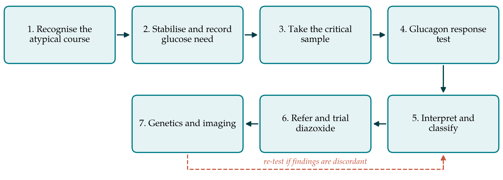
The pathway has seven steps. Table 1 lists each with its purpose and the record it should leave in the notes, and Figure 1 shows the sequence. The steps follow the recommendations of the Pediatric Endocrine Society (Thornton et al., 2015), the British framework for term infants (British Association of Perinatal Medicine, 2017) and the international guideline on hyperinsulinism (De Leon et al., 2024). Where these documents give numerical thresholds, the local protocol in force should be used.
| Step | Purpose | Record in the notes |
|---|---|---|
| 1. Recognise the atypical course | Separate transitional low glucose from a possible persistent disorder | Risk factors, age in hours, laboratory glucose values |
| 2. Stabilise and record glucose need | Protect the brain; quantify the glucose infusion rate | Infusion rate, route of access, feeds given |
| 3. Critical sample | Capture fuels and hormones while glucose is low | Time, glucose at sampling, tests sent |
| 4. Glucagon response | Show that liver glycogen is inappropriately preserved | Glucose before and at intervals after glucagon |
| 5. Interpret and classify | Decide whether hyperinsulinism is established, and its likely type | Written interpretation; differential diagnoses excluded |
| 6. Refer and trial diazoxide | Engage a specialist centre; test responsiveness | Advice received, response assessment, adverse effects |
| 7. Genetics and imaging | Find the genotype; identify focal disease | Samples from infant and parents; imaging plan |
3.1. Step 1: Recognise the atypical course
The pathway begins when hypoglycaemia departs from the transitional pattern. The features that should prompt it are a low glucose that is severe, symptomatic, recurrent despite adequate feeding, or still present after 48 hours; a need for intravenous glucose; and hypoglycaemia in an infant with no conventional risk factor. Macrosomia without maternal diabetes, a family history of hypoglycaemia, and physical signs of Beckwith-Wiedemann syndrome or of hypopituitarism add weight. Bedside glucose meters are imprecise at low concentrations, so values that will drive decisions should be confirmed on a laboratory or blood gas analyser (British Association of Perinatal Medicine, 2017).
3.2. Step 2: Stabilise and record the glucose requirement
Treatment is never delayed for the sake of diagnosis. Glucose is restored and maintained according to the unit protocol. The pathway asks for one additional act: calculation and documentation of the glucose infusion rate needed to keep the infant normoglycaemic, expressed in milligrams per kilogram per minute and including enteral intake where relevant. A healthy newborn liver releases roughly 4 to 6 mg/kg/min. A requirement well above that range points strongly to excess insulin action, and the British framework uses a figure above 8 mg/kg/min as a trigger for investigation (British Association of Perinatal Medicine, 2017). High requirements often exceed what a peripheral vein tolerates, and the need for central access should be anticipated.
3.3. Step 3: Take the critical sample
The diagnosis depends on blood taken while the plasma glucose is below the threshold set in the guideline in use, which for the Pediatric Endocrine Society is 2.8 mmol/L (50 mg/dL) (Thornton et al., 2015). This may occur spontaneously or, in a stable infant under close observation, during a supervised reduction in glucose delivery planned with specialist advice. The sample includes laboratory glucose, insulin, C-peptide, β-hydroxybutyrate, free fatty acids, cortisol, growth hormone, lactate, ammonia and an acylcarnitine profile, with urine for organic acids. Tubes and request forms should be assembled in advance and kept at the cot side, because the opportunity is brief and the glucose must then be corrected at once.
3.4. Step 4: Measure the glycaemic response to glucagon
Immediately after the critical sample, glucagon is given by the route and in the amount specified by the local protocol, and glucose is measured at intervals over the following 30 to 40 minutes. In a fasted infant without hyperinsulinism the liver has little glycogen left to mobilise. A rise of more than 1.7 mmol/L (30 mg/dL) shows that glycogen has been inappropriately retained and is strong evidence of insulin excess (Thornton et al., 2015). The test doubles as treatment of the hypoglycaemic episode.
3.5. Step 5: Interpret and classify
Hyperinsulinism is established when, during hypoglycaemia, ketones and free fatty acids are suppressed, the glucagon response is exaggerated and the glucose requirement is high. Insulin itself is an unreliable marker. It is often only just detectable, and assays vary; Ferrara et al. (2016) showed that suppressed β-hydroxybutyrate and free fatty acids and the glucagon response identify the condition more dependably than the insulin concentration. Raised ammonia suggests the GLUD1 form. Low cortisol or growth hormone in a single critical sample has poor specificity for true deficiency and should lead to formal testing, not to a diagnosis (Kelly et al., 2008). Ketotic hypoglycaemia, raised lactate or an abnormal acylcarnitine profile redirect the work-up towards other metabolic and endocrine causes. The interpretation is written down with the differential diagnoses considered.
3.6. Step 6: Refer and assess the response to diazoxide
Once hyperinsulinism is established the infant should be discussed with a centre experienced in the condition, and this is true whatever the apparent severity. Diazoxide, which holds the potassium channel open, is the first-line drug, and a trial lasting several days is the conventional test of responsiveness (De Leon et al., 2024). Prescribing follows specialist advice and the product information. The pathway requires that responsiveness be judged by a stated criterion, namely the ability to maintain normal glucose on ordinary feeds through an age-appropriate fast, and that adverse effects be looked for. Fluid retention is expected, and pulmonary hypertension has been reported in neonates (Herrera et al., 2018). Most infants with channel variants that abolish function do not respond.
3.7. Step 7: Genetic testing and imaging
Blood from the infant and both parents is sent for analysis of ABCC8 and KCNJ11 as soon as diazoxide unresponsiveness is suspected, and without waiting for the trial to end. Rapid sequencing of these genes changes management, because a single paternal recessive variant predicts focal disease with high probability (Banerjee et al., 2011). Such infants are referred for positron emission tomography with 18F-fluoro-L-DOPA, which localises focal lesions accurately enough to guide surgery (Hardy et al., 2007). Infants with diffuse disease continue medical treatment under specialist care.
4. Fitting the Pathway to a General Neonatal Service
The pathway is written for units that see hyperinsulinism rarely. Three practical arrangements make it usable. A pre-packed critical sample kit, with labelled tubes, a request form and a one-page instruction sheet agreed with the laboratory, removes the commonest reason for a missed sample. Laboratories differ in which assays they run on site, and the turnaround for β-hydroxybutyrate and free fatty acids should be known in advance; a bedside ketone meter gives an immediate indication while formal results are awaited. A named route of referral, including out-of-hours contact details for the regional endocrine or hyperinsulinism service, should be written into the hypoglycaemia guideline so that step 6 is not left to improvisation.
Parents need a clear account of why blood is being taken at the moment their infant's glucose is low, and why the infant may need to stay in hospital when feeding seems to be going well. Breastfeeding should be protected throughout. A safety fast before discharge, as recommended for any neonate who has needed prolonged treatment for hypoglycaemia (Thornton et al., 2015), closes the pathway for infants in whom hyperinsulinism has been excluded or has resolved.
5. Limitations and What Remains to Be Shown
The pathway has not been tested. It arranges existing recommendations in a sequence, and its claim to usefulness is that the sequence is easier to follow under pressure than the source documents. Whether it shortens the time to diagnosis, increases the proportion of infants in whom a critical sample is obtained at the first opportunity, or reduces the time to referral could be examined by audit before and after its introduction. Whether it improves neurodevelopmental outcome is a harder question, since the condition is rare and the relation between early glycaemia and later development is itself uncertain (McKinlay et al., 2015).
The thresholds quoted are those of consensus documents and are not agreed universally. The pathway may over-investigate infants with stress-related hyperinsulinism that would have resolved, and the cost and distress of doing so have not been weighed. It assumes access to assays, genetic testing and specialised imaging that many health systems cannot provide, and it does not address preterm or growth-restricted infants, in whom the differential diagnosis is wider.
6. Conclusion
Congenital hyperinsulinism is rare, treatable and easily missed among the many term infants with transient low glucose. The diagnosis can be made on a general neonatal unit if the clinician recognises the atypical course, documents the glucose requirement, and obtains the critical sample and glucagon response before correcting the hypoglycaemia. Early referral and prompt genetic testing then determine whether the infant has a focal lesion amenable to cure. The seven steps proposed here set that reasoning out in order. They await evaluation in practice and are offered as a starting point for local guidelines.
Conflict of interest
To be declared by the author(s) before publication.
Funding
To be declared by the author(s) before publication.