1. Introduction
Every student of embryology learns that the vertebrate body is built from three germ layers. The ectoderm gives the epidermis and the nervous system, the mesoderm gives muscle, bone, blood and kidney, and the endoderm gives the lining of the gut and its associated organs (Gilbert and Barresi, 2016). The scheme dates from the early nineteenth century, when Pander and then von Baer described the layers of the chick embryo, and it has outlived the theories of its founders because it works. It predicts which tissues arise together, and it supplies the first branching points in almost every protocol for differentiating pluripotent stem cells.
The scheme carries two meanings that are seldom distinguished. One is topological: the layers are sheets of cells with definite positions once gastrulation has rearranged the embryo. The other is genealogical: the layers are the first restriction of developmental potential, so that after gastrulation a cell and all its descendants belong to one layer and form only that layer's tissues. The second meaning is the stronger claim, and it is the one that lineage tracing can test.
This Perspective argues that the genealogical reading has been weakened by the results of recent single-cell lineage tracing to the point where it should no longer be the default, and that the boundary between germ layers is better understood as a boundary of position and signalling history than of descent.
2. What Lineage Recording Now Makes Visible
Classical fate maps were made by labelling a cell or a patch of cells with dye and observing where the label ended up. In the zebrafish they showed that cells of the blastula are not yet restricted to a tissue and that a predictable fate map emerges only at the onset of gastrulation (Kimmel et al., 1990). Such maps follow few cells and say little about what the descendants become at the molecular level.
The newer methods remove both limits. In one family, genome editing with CRISPR-Cas9 writes cumulative, heritable mutations into a synthetic or endogenous target sequence, so that each cell carries a barcode recording its ancestry (McKenna et al., 2016). When the barcode is expressed as RNA it can be read in the same single-cell sequencing run as the transcriptome, giving the lineage and the state of each cell together (Raj et al., 2018). Related designs have been applied to the mouse embryo (Chan et al., 2019), and the field is reviewed by Wagner and Klein (2020).
3. Three Observations That Strain the Classical Reading
The evidence comes from several systems and predates the newest methods in part. Table 1 collects the main observations, and three of them are discussed here.
| Observation | System | Why it strains the classical reading | Source |
|---|---|---|---|
| Founder cells give mixed tissue types | C. elegans | Early lineage branches do not correspond to germ layers | Sulston et al., 1983 |
| Common precursors of neural tube and somites persist | Mouse | Ectoderm and mesoderm separate after gastrulation in the trunk | Tzouanacou et al., 2009 |
| Visceral endoderm cells join the gut tube | Mouse | An embryonic tissue has part extra-embryonic ancestry | Kwon et al., 2008 |
| Gut cells of two origins converge in expression | Mouse | State does not reveal lineage | Chan et al., 2019 |
| Cranial crest forms skeleton and connective tissue | Vertebrates | An ectodermal population yields mesoderm-like derivatives | Le Douarin and Kalcheim, 1999 |
| Clones span distinct transcriptional states | Zebrafish | State tree and lineage tree differ | Wagner et al., 2018 |
3.1. Progenitors that straddle ectoderm and mesoderm
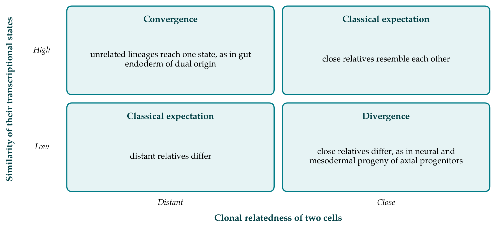
The spinal cord is the standard example of an ectodermal derivative and the somites of a mesodermal one. Retrospective clonal analysis in the mouse by Tzouanacou et al. (2009) showed that single cells contribute descendants to both, and continue to do so as the body axis extends, well after the germ layers are supposed to have separated. The cells responsible, called neuromesodermal progenitors, reside near the posterior end of the embryo and express both the neural factor SOX2 and the mesodermal factor Brachyury (Henrique et al., 2015). In the trunk, then, the split between neural ectoderm and paraxial mesoderm is a late and local decision. The endoderm, by contrast, did segregate early in the same clonal analysis, which shows that the three layers do not behave alike.
3.2. Tissues with two ancestries
The gut endoderm of the mouse was long held to derive wholly from the epiblast, with the visceral endoderm that surrounds the early embryo destined for the yolk sac. Live imaging by Kwon et al. (2008) showed otherwise: epiblast-derived cells intercalate into the visceral endoderm layer, and descendants of the visceral endoderm remain in the embryonic gut tube. Single-cell profiling later found that cells of the two origins become very similar in gene expression as the gut forms (Nowotschin et al., 2019), and molecular recording confirmed that cells of extra-embryonic ancestry sit among embryonic ones with nearly the same transcriptome (Chan et al., 2019). Here a single tissue, with a single state, has two lineage histories. A definition of endoderm by descent would have to exclude some of the cells that line the gut.
3.3. The neural crest
The neural crest arises at the border of the neural plate, within the ectoderm, and migrates throughout the body. In the head it forms cartilage, bone, dentine and connective tissue, cell types that elsewhere come from mesoderm (Le Douarin and Kalcheim, 1999). The anomaly is old enough that Hall (2000) proposed calling the crest a fourth germ layer. More recent work in the mouse indicates that cranial crest cells transiently re-express factors associated with pluripotency before they form mesenchyme (Zalc et al., 2021). On that account the crest does not so much violate a germ layer restriction as lift it.
Figure 1 places these cases in a common frame. If lineage and state were two views of one hierarchy, cells would fall only on one diagonal: close relatives would be alike and distant relatives different. The observations above occupy the other two corners.
4. The Case for Keeping the Layers
Several objections can be made to the argument so far, and they have force. The first is that the exceptions are few. Most cells do descend from the layer the textbooks assign them to, and a rule that holds for the great majority of cases is worth keeping. The second is that some of the exceptions are matters of definition. Neuromesodermal progenitors can be regarded as cells that have not yet completed gastrulation, since the posterior growth zone continues the movements of the primitive streak; on that view the layers still separate at gastrulation, and gastrulation simply lasts longer than was thought.
The third objection is evolutionary. The germ layers are recognisable across animals and the genes that specify them are widely conserved, which suggests ancient and real units and not a convenience of description. Even here, however, the layers are not cleanly separate, since endoderm and mesoderm arise in many species from a common mesendodermal population (Rodaway and Patient, 2001). The fourth is technical. Lineage barcodes are recovered from only a fraction of cells, editing can saturate or erase earlier marks, and a clone that appears to span two layers may reflect an ancestor labelled before the layers formed. Claims of late bipotency need the timing of the mark to be known.
These points limit the conclusion without overturning it. The nematode shows that the issue is not one of rare exceptions in complicated embryos: in the invariant lineage of Caenorhabditis elegans, most of the early founder cells produce mixtures of tissue types, and only the gut derives from a single founder (Sulston et al., 1983). And an appeal to prolonged gastrulation concedes the main point, which is that the moment of restriction varies by region and by layer.
5. A Proposal: Three Descriptions in Place of One
The difficulty arises from asking one term to carry three kinds of information. The proposal made here is to separate them. Germ layer would denote position and context during gastrulation: which sheet a cell lies in and therefore which signals it has received. Lineage would denote ancestry, measured by clonal analysis or molecular recording and stated with the time at which the common ancestor lived. State would denote the molecular profile of the cell at the time of observation. In most cells the three agree, which is why the classical scheme has served well. Where they disagree, as in the cases above, the disagreement is itself the finding and should not be hidden by assigning the cell to a layer. The boundary between layers is then real without being a wall: it marks a difference in the environment that cells meet at a formative period, which biases, without fixing, what their descendants do.
6. Implications
Three practical consequences follow. Cell atlases built from single-cell transcriptomes often arrange cell types in a hierarchy whose first division is by germ layer. That arrangement is a classification of states, and it should not be presented as a pedigree unless lineage has been measured. Protocols for differentiating pluripotent stem cells usually pass through a germ layer intermediate, yet axial tissues may be reached more faithfully through a bipotent progenitor, as the work on neuromesodermal progenitors suggests. And in teaching, the layers can be introduced as the outcome of gastrulation movements, with the classical lists of derivatives given as the usual result and the known exceptions as part of the account.
7. Conclusion
The germ layers remain a sound description of the gastrulating embryo. What the recent lineage studies call into question is the inference from layer to ancestry and from ancestry to cell type. Progenitors that feed two layers, tissues with two origins and a crest that forms skeleton all show that position, descent and molecular state can come apart. Recording them as separate properties loses nothing of the classical scheme where it holds, and it makes room for the cases, now measurable one cell at a time, in which it does not.
Conflict of interest
To be declared by the author(s) before publication.
Funding
To be declared by the author(s) before publication.