1. Introduction
Bisphosphonates are the most widely prescribed treatment for osteoporosis, and their capacity to reduce vertebral and hip fractures is supported by large randomised trials. From 2005 onward, clinicians began to report an unusual pattern of femoral fracture in patients who had taken these drugs for several years. Odvina et al. (2005) described low-energy fractures with delayed healing and biopsy evidence of severely suppressed bone turnover. Reports from Singapore drew attention to subtrochanteric insufficiency fractures with a characteristic radiographic appearance in women on alendronate (Goh et al., 2007), and series from New York described a simple transverse fracture with thickened cortices in the femoral shaft (Lenart et al., 2008; Neviaser et al., 2008).
A task force of the American Society for Bone and Mineral Research (ASBMR) gave the entity a case definition in 2010 and revised it in the light of further evidence (Shane et al., 2010; Shane et al., 2014). The fractures are rare in absolute terms, and for most patients with osteoporosis the benefit of treatment outweighs the risk by a wide margin (Black et al., 2020). For the orthopaedic surgeon, though, the problem is not prescribing policy. It is that the patient with an evolving atypical fracture often presents weeks or months before the bone breaks, and the opportunity to prevent a complete fracture is lost if the presentation is not recognised.
This article proposes a pathway for recognising and managing atypical femoral fracture in an orthopaedic department. It is built from published guidance and surgical series. It is a proposed procedure and has not been applied to patients or validated here; no case is reported. Its purpose is to turn scattered recommendations into a sequence that a department can adopt, adapt and audit.
2. Background
Three bodies of evidence shape the pathway: what an atypical fracture is, how strongly it is tied to treatment, and how it behaves once it has formed.
2.1. The case definition
Under the revised ASBMR definition the fracture must lie along the femoral diaphysis, from just distal to the lesser trochanter to just proximal to the supracondylar flare, and at least four of five major features must be present. These are: association with minimal or no trauma; a fracture line that originates at the lateral cortex and is substantially transverse, though it may become oblique as it crosses medially; a complete fracture extending through both cortices, possibly with a medial spike, or an incomplete fracture involving only the lateral cortex; no or minimal comminution; and localised periosteal or endosteal thickening of the lateral cortex at the fracture site, often described as beaking or flaring (Shane et al., 2014). Minor features, none of them required, include generalised cortical thickening of the shafts, prodromal pain, bilateral involvement and delayed healing. Femoral neck, intertrochanteric and periprosthetic fractures and pathological fractures through tumour are excluded.
2.2. Association with antiresorptive treatment
The link with bisphosphonates is supported by consistent observational evidence, although causation in the individual patient cannot be proved. A Swedish national study with radiographic adjudication found a strong relative association between bisphosphonate use and atypical fracture with a small absolute risk (Schilcher et al., 2011). A secondary analysis of the major randomised trials found too few subtrochanteric and diaphyseal fractures to confirm or exclude an effect (Black et al., 2010). Data from a large health maintenance organisation in California showed that incidence rises with duration of use (Dell et al., 2012), and later work indicated that risk falls quickly after the drug is stopped (Schilcher et al., 2014). The proposed mechanism is that prolonged suppression of remodelling allows microdamage to accumulate at the lateral cortex, where tensile stress is highest, but this remains a hypothesis (Shane et al., 2014). Atypical fractures also occur in people who have never taken an antiresorptive drug.
2.3. Natural history and the opposite femur
The lesion behaves as a stress fracture. It begins as a focal thickening of the lateral cortex, may develop a transverse radiolucent line, and can progress to a complete fracture after trivial load. Pain in the thigh or groin precedes complete fracture in a large proportion of reported patients (Kwek et al., 2008). Cortical stress lesions with a visible lucent line and accompanying pain appear more likely to progress than painless thickening alone (Koh et al., 2010). The opposite femur is affected in a substantial minority of patients, sometimes without symptoms, which is the reason every published recommendation calls for it to be imaged (Shane et al., 2010).
3. The Proposed Pathway
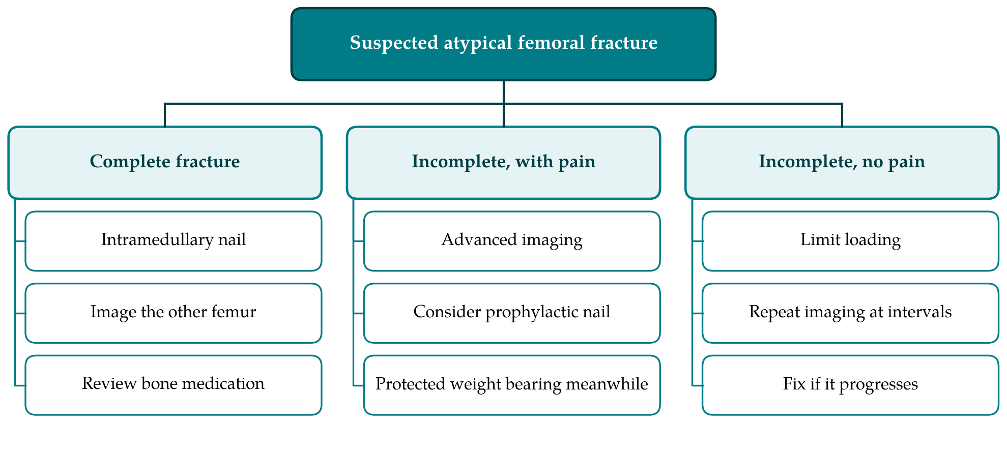
The pathway has six steps. Table 1 lists each with its purpose and the published basis for it, and Figure 1 shows the branching that follows from the classification of the lesion. The steps are set out in order below. Where the supporting evidence is weak, the text says so.
| Step | Purpose | Published basis |
|---|---|---|
| 1. Recognise the prodrome | Identify thigh or groin pain in a patient on antiresorptive treatment | Kwek et al., 2008 |
| 2. Image both femora | Full-length views to find cortical thickening and contralateral lesions | Shane et al., 2010 |
| 3. Apply the case definition | Separate atypical from ordinary osteoporotic and pathological fractures | Shane et al., 2014 |
| 4. Stratify the lesion | Complete; incomplete with pain; incomplete without pain | Koh et al., 2010 |
| 5. Choose surgical or conservative care | Stabilise complete fractures; prevent progression of incomplete ones | Banffy et al., 2011; Egol et al., 2014 |
| 6. Coordinate medical review | Reassess antiresorptive treatment, calcium and vitamin D with the prescriber | Adler et al., 2016 |
3.1. Step 1: Recognise the prodrome
Any patient who has taken a bisphosphonate or another potent antiresorptive agent for several years and reports a dull or aching pain in the thigh or groin should be regarded as having a possible stress lesion of the femur until imaging shows otherwise. The pain is typically worse on weight bearing and may have been present for weeks. In practice it is commonly ascribed to lumbar spine disease, hip arthritis or trochanteric bursitis. The pathway therefore begins with a prompt in the history: duration and type of osteoporosis treatment should be asked about in every older patient with unexplained thigh pain.
3.2. Step 2: Image both femora
Anteroposterior and lateral radiographs should include the whole length of both femora, from hip to knee. A radiograph of the pelvis or hip alone will miss a diaphyseal lesion. The reader looks for a focal bump on the lateral cortex, with or without a transverse lucent line. If radiographs are normal or equivocal and suspicion remains, magnetic resonance imaging or a radionuclide bone scan can show a stress reaction before cortical change is visible, and the long femur images produced by some dual-energy X-ray absorptiometry scanners have been used for the same purpose (Shane et al., 2014).
3.3. Step 3: Apply the case definition
The radiographs are assessed against the major features listed above. The step matters because ordinary osteoporotic fractures of the subtrochanteric region and shaft are more common than atypical ones and are spiral or comminuted in pattern, and because metastatic disease must not be mistaken for either. Recording which features are present, in the notes and in the operative record, also makes later audit and reporting to pharmacovigilance schemes possible.
3.4. Step 4: Stratify the lesion
Lesions are placed in one of three groups. A complete fracture needs fixation. An incomplete fracture with pain, and particularly one with a visible lucent line, carries a meaningful risk of completion (Koh et al., 2010). An incomplete fracture without pain, seen as cortical thickening alone, is the least likely to progress. Both femora are stratified separately, since a patient may have a complete fracture on one side and an incomplete lesion on the other.
3.5. Step 5: Choose surgical or conservative care
For complete fractures the task force recommends intramedullary nailing, which protects the whole femur and allows healing by endochondral repair, a process less dependent on the remodelling that antiresorptive drugs suppress than the direct healing sought with rigid plate fixation (Shane et al., 2010). Surgeons should expect technical difficulty from the bowed, brittle femur and a narrow canal, and should counsel patients that union is usually achieved but is slower than after an ordinary femoral shaft fracture (Egol et al., 2014).
For incomplete fractures with pain, the task force advises that prophylactic nailing be considered; where the pain is minimal, a trial of conservative treatment with limited weight bearing is reasonable, with fixation if symptoms and imaging have not improved after two to three months (Shane et al., 2010). A comparative series supports this caution, since most incomplete fractures managed without surgery in that report progressed to complete fracture, and hospital stay was shorter after prophylactic fixation than after fixation of a completed fracture (Banffy et al., 2011). For incomplete fractures without pain, the pathway follows the task force in advising reduced loading and repeat imaging until the lesion has resolved. The choice in each case belongs to the surgeon and the patient, and the evidence behind it consists of case series, not trials.
3.6. Step 6: Coordinate medical review
An orthopaedic pathway should not end at the wound. The task force recommends that potent antiresorptive treatment be stopped once an atypical fracture is diagnosed, that calcium and vitamin D status be assessed and corrected, and that the further management of osteoporosis be reviewed (Shane et al., 2014). These decisions lie with the physician responsible for the patient's bone health, and the pathway's requirement is only that the referral is made and documented before discharge. The use of teriparatide to promote healing has been described in case reports and small series; the task force regarded the evidence as limited and inconclusive, and this article makes no recommendation on it. General guidance on reviewing the need for continued bisphosphonate treatment after several years, including the concept of a drug holiday for patients at lower risk, is given by a separate ASBMR task force (Adler et al., 2016) and draws on the extension of the alendronate trials (Black et al., 2006).
4. Fitting the Pathway to a Department
The pathway asks for little that a department does not already have. Its first two steps depend on the people who see the patient first: emergency clinicians, general practitioners and fracture clinic staff. A short prompt on the request form for hip and femur radiographs, asking about antiresorptive treatment, and a standing arrangement with radiology that full-length views of both femora follow any report of focal lateral cortical thickening, would address the two commonest points of failure. Steps three to five can be captured on a single proforma that records the features of the case definition, the stratification of each femur and the agreed plan. Step six is most reliably delivered where a fracture liaison service or an orthogeriatric team already reviews patients with fragility fractures.
5. Limitations and What Remains to Be Shown
The pathway is proposed and untested. It combines recommendations that are themselves based largely on observational studies, case series and expert consensus, and following it cannot be assumed to improve outcomes until that has been measured. A department adopting it could audit several things: the proportion of patients with an atypical fracture who had imaging of the opposite femur; the interval between first presentation with prodromal pain and diagnosis; the proportion of incomplete lesions that progressed to complete fracture under conservative care; time to union after fixation; and whether medical review took place. Comparison with the period before adoption would give a first indication of its value.
Several matters remain uncertain in the literature and are not resolved by a pathway. The threshold at which a painless incomplete lesion warrants fixation is not defined. The benefit of anabolic treatment for healing is unproven. The best management of osteoporosis after an atypical fracture, in a patient who remains at high risk of ordinary fragility fractures, is a matter of specialist judgement. Rarity makes randomised trials difficult, so registries with radiographic adjudication are likely to remain the main source of better evidence (Schilcher et al., 2011).
6. Conclusion
Atypical femoral fracture is rare, but it announces itself in advance more often than most fractures do, and both the warning and the lesion in the opposite femur are frequently overlooked. The six-step pathway proposed here arranges existing guidance into a sequence that begins with a question about treatment history and ends with a documented medical review. It is offered as a framework for local adaptation and audit. Whether it shortens the time to diagnosis or reduces the number of incomplete lesions that go on to break is a question for departments that choose to adopt and evaluate it.
Conflict of interest
To be declared by the author(s) before publication.
Funding
To be declared by the author(s) before publication.