1. Introduction
Refractive surgery for myopia flattens the central cornea by removing stromal tissue, and the cornea that remains has to carry the intraocular pressure for the rest of the patient's life. When it cannot, the result is progressive steepening and thinning, described after laser in situ keratomileusis (LASIK) by Seiler et al. (1998) and now known as postoperative ectasia. The risk rises with the amount of tissue removed, which is why high myopia has always been the difficult end of the treatment range (Randleman, Woodward et al., 2008).
Small incision lenticule extraction (SMILE) was introduced as a flapless alternative. A femtosecond laser cuts the two surfaces of a refractive lenticule inside the stroma, and the surgeon removes it through an arcuate incision a few millimetres long (Sekundo et al., 2011). The tissue above the lenticule, called the cap, is never lifted. Early accounts of the technique presented this as a biomechanical advantage, and the claim has been repeated in most descriptions of the procedure since (Reinstein et al., 2014).
Whether the advantage is real, and how large it is in eyes that need a thick lenticule, is the subject of this review. The question has practical weight. If SMILE preserves strength, surgeons may reasonably offer it at corrections where they would hesitate to cut a flap. If it does not, the same residual-bed reasoning applies to both operations and candidacy should be judged alike.
2. Scope and Approach
This is a narrative review of the published literature. It draws on the papers that introduced SMILE, the theoretical and computational models of postoperative corneal strength, clinical comparisons that used commercial air-puff devices, laboratory studies of excised tissue, earlier reviews and one meta-analysis, and published accounts of ectasia after the procedure. No formal search protocol was followed and no pooled estimate is offered. Where studies are described, the description is of their design and the direction of their findings, and readers who need the figures should go to the sources.
High myopia has no single definition in this literature. Many refractive surgery papers use a spherical equivalent of 6 dioptres or more, and some analyse the higher corrections as a subgroup of a broader series. The review therefore treats high myopia as the upper part of the treated range and pays attention to what each study says about the relation between lenticule thickness and biomechanical change.
3. The Structural Argument
The case for SMILE rests on the way stiffness is distributed through the depth of the cornea. Collagen lamellae in the anterior third are narrow, interwoven and inserted into Bowman's layer, whereas those of the posterior stroma lie in flatter, less connected sheets. Measurements on donor corneas show that cohesive tensile strength falls with depth, with the anterior part of the stroma considerably stronger than the posterior part (Randleman, Dawson et al., 2008). The general account of how lamellar cuts redistribute stress, and how the healing response fails to restore the original architecture, is given by Dupps and Wilson (2006).
A LASIK flap severs the anterior lamellae around almost the whole circumference. The flap heals at its edge but its lamellae do not regain tension, so the load is carried by the residual bed alone. In SMILE the cap is cut only along the small incision and at the lenticule edge, and the tissue removed comes from deeper, weaker stroma. Figure 1 sets the three main procedures side by side. Surface ablation cuts no lamellae at depth but removes the strongest tissue; LASIK both cuts and removes anterior and mid-stromal tissue; SMILE removes mid-stromal tissue and leaves the anterior lamellae mostly continuous.
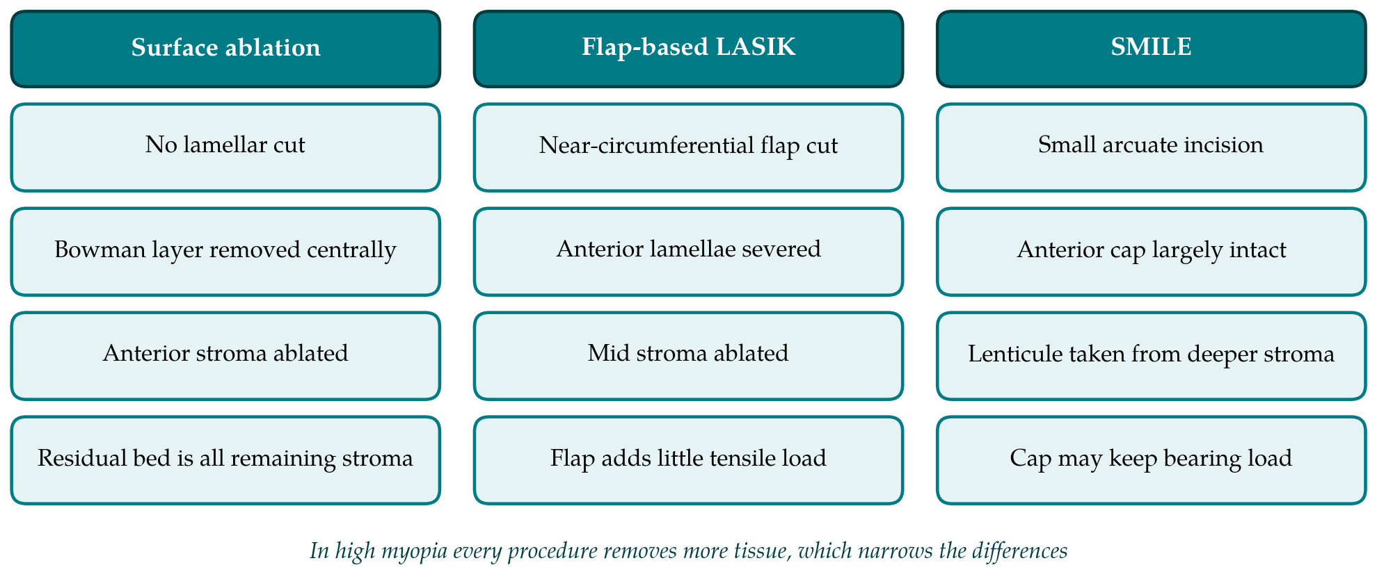
Reinstein et al. (2013) turned this reasoning into a mathematical model. Using the published depth profile of tensile strength, they calculated the total strength remaining after each procedure for a range of corrections and corneal thicknesses, and found that the modelled cornea kept more of its strength after SMILE than after LASIK or surface ablation for the same correction. The finite-element analysis of Sinha Roy et al. (2014) reached a compatible conclusion from patient-specific geometry: stresses in the residual bed were lower after the simulated flapless procedure, and the cap continued to bear load in a way that a flap did not.
Both models are careful pieces of work, and both depend on assumptions that deserve stating. The depth profile of strength came from a modest number of donor corneas and from cohesive strength, which is not the same property as in-plane tensile stiffness. The models treat the cap as mechanically continuous with the periphery, although the lenticule side cut and the interface left after extraction may reduce the coupling. They also assume that the cap depth and lenticule position are those planned. A model can show what follows from its premises. It cannot show that a living cornea behaves accordingly, and for that the clinical and laboratory evidence has to be examined.
4. Clinical Measurement With Air-Puff Devices
Two commercial instruments supply nearly all the clinical data. Both deform the cornea with a pulse of air and record the response, and neither measures a material property such as elastic modulus. Their outputs depend on corneal thickness, curvature and intraocular pressure as well as on tissue stiffness, which is the central difficulty in interpreting change after an operation that alters the first two.
4.1. Ocular Response Analyzer
The Ocular Response Analyzer records the pressures at which the cornea applanates on the way in and on the way out, and reports their difference as corneal hysteresis together with a derived corneal resistance factor (Luce, 2005). Hysteresis reflects viscous damping more than stiffness. Studies with this device agree that both indices fall after SMILE, as they do after LASIK. A prospective fellow-eye comparison by Agca et al. (2014) found no meaningful difference between the two procedures, and Vestergaard et al. (2014) reported a similar picture when comparing the flap-based and small-incision forms of lenticule extraction. Wu et al. (2014) and Wang et al. (2014) described a smaller reduction after SMILE, with the difference in the latter study emerging mainly at higher corrections.
4.2. Dynamic Scheimpflug tonometry
The Corvis ST films the deformation with a high-speed Scheimpflug camera and derives timing, velocity and amplitude parameters from the image sequence. Early comparisons using the first-generation parameters found little to separate SMILE from flap-based surgery (Pedersen et al., 2014), while Osman et al. (2016) reported smaller changes after SMILE. The original parameters were strongly dependent on pressure and thickness, a limitation documented in healthy eyes by Vinciguerra et al. (2016). Later software added stiffness parameters intended to be less sensitive to those influences (Roberts et al., 2017). These were derived and tested in untreated eyes, and none is a validated measure of weakening after surgery.
Table 1 summarises the lines of evidence and what each can and cannot support. The pooled analysis by Guo et al. (2019) is consistent with the individual studies: SMILE appeared to preserve the measured indices better than flap-based LASIK, with no clear advantage over surface ablation, and with heterogeneity between studies that the authors acknowledged. The narrative review of Damgaard et al. (2018) came to a more cautious reading, noting that randomised and fellow-eye designs tended to show smaller differences than non-randomised comparisons.
| Line of evidence | What it shows | Main limitation | Sources |
|---|---|---|---|
| Depth-dependent strength in donor tissue | Anterior stroma is stronger than posterior stroma | Few corneas; cohesive, not in-plane, strength | Randleman, Dawson et al., 2008 |
| Mathematical and finite-element models | More strength retained after SMILE for equal correction | Assume an intact, fully coupled cap | Reinstein et al., 2013; Sinha Roy et al., 2014 |
| Ocular Response Analyzer studies | Indices fall after both procedures; mixed differences | Hysteresis tracks thickness and damping | Agca et al., 2014; Wu et al., 2014 |
| Dynamic Scheimpflug studies | Small or no difference in several comparisons | Parameters depend on pressure and geometry | Pedersen et al., 2014; Osman et al., 2016 |
| Laboratory tests on excised tissue | Flapless cut preserves more stiffness than a flap cut | Animal or donor tissue, no healing | Spiru et al., 2017; Kanellopoulos, 2018 |
| Reports of ectasia after SMILE | Ectasia can follow SMILE | No denominator; many eyes had abnormal topography | Moshirfar et al., 2017 |
5. Laboratory Evidence
Bench studies avoid the confounding of air-puff measurement by loading the tissue directly. Spiru et al. (2017) cut porcine corneas with either the flap-based or the small-incision form of lenticule extraction, using the same refractive correction, and tested them by two-dimensional extensometry. The corneas treated through a small incision resisted deformation better, which supports the idea that an uncut cap contributes to strength. Kanellopoulos (2018) compared SMILE with LASIK in human donor corneas and reported that the difference between them was smaller than theory predicted. The two results are not contradictory. Porcine corneas lack a true Bowman's layer and differ in thickness and lamellar organisation, and excised tissue swells and cannot heal, so neither experiment reproduces a living human eye months after surgery.
6. Why High Myopia Is a Special Case
Three features of highly myopic eyes complicate the picture. The first is simple arithmetic. Lenticule thickness rises with the correction and with the optical zone, and a minimum lenticule edge thickness is added to allow clean dissection, so for a given correction SMILE may remove slightly more central tissue than an excimer ablation. The stromal bed beneath the lenticule can be thinner than the bed left under a thin LASIK flap. The argument for SMILE is that the cap compensates, and at high corrections that argument carries more of the weight.
The second feature is that the amount of tissue altered predicts ectasia independently of residual bed thickness. Santhiago et al. (2014) showed this for LASIK with the percent tissue altered metric, which adds flap thickness to ablation depth. There is no agreed equivalent for SMILE, because it is not settled how much of the cap should be counted as altered. A surgeon who counts none of it will accept corrections that a surgeon who counts all of it would decline, and the published models do not resolve the disagreement.
The third feature is the measurement problem at its worst. Highly myopic eyes lose the most thickness and change curvature the most, so the geometric contribution to any air-puff parameter is largest exactly where the biomechanical question is most pressing. Studies that report a greater fall in indices at higher corrections are describing something expected from the reduction in thickness alone, and separating that from a change in tissue properties needs either statistical adjustment, which rests on relations derived in unoperated eyes, or a method that measures stiffness directly.
7. Ectasia After SMILE
The clinical outcome that biomechanical preservation is supposed to prevent has been reported after SMILE. Moshirfar et al. (2017) collected the published cases and observed that a large share of the affected eyes had preoperative topography that was abnormal or suspicious in retrospect. That pattern is familiar from the LASIK literature, where unrecognised early keratoconus is the leading risk factor (Randleman, Woodward et al., 2008). Case reports cannot give an incidence, since the number of procedures from which they arose is unknown and reporting is voluntary. What they establish is narrower and still useful: a flapless procedure does not make a susceptible cornea safe, and screening standards should not be relaxed on biomechanical grounds.
8. Gaps and Future Work
The literature leaves several questions open. Most clinical comparisons are small and follow patients for months, whereas ectasia may appear years later. Few are randomised, and fellow-eye designs, which control for individual variation, remain the minority. High myopia is usually a subgroup. Reporting of cap thickness, lenticule thickness, optical zone and residual bed is inconsistent, which makes it hard to compare eyes that had the same amount of tissue altered.
Better instruments may help more than more studies with the present ones. Brillouin microscopy maps a local elastic property through the depth of the cornea without contact (Scarcelli and Yun, 2008), and optical coherence elastography and related methods aim to do the same from induced displacement (Kling and Hafezi, 2017). Either could show directly whether the cap carries load after SMILE. Until such measurements are available in treated eyes with long follow-up, the comparison between procedures at high corrections will continue to rest on models and on indices that mix geometry with material behaviour.
9. Conclusion
The structural reasoning behind SMILE is sound, and the mathematical and finite-element models built on it predict a stronger cornea than after a flap-based procedure of equal correction. Clinical measurement has confirmed the prediction only in part. All procedures reduce the measured indices, the reduction grows with the tissue removed, and the advantage of SMILE over femtosecond LASIK ranges from modest to undetectable depending on the study design and the device. In high myopia the theoretical benefit should be greatest, yet this is where thick lenticules, thin residual beds and geometry-dependent instruments make the evidence hardest to read. A reasonable position for the present is that SMILE may spare some corneal strength, that the size of the effect in highly myopic eyes is not established, and that candidacy should rest on the same careful screening as for any lamellar procedure.
Conflict of interest
To be declared by the author(s) before publication.
Funding
To be declared by the author(s) before publication.