1. Introduction
Amelogenesis imperfecta comprises a clinically and genetically diverse set of conditions in which the enamel of all or nearly all teeth, in both dentitions, forms abnormally in the absence of a wider syndrome (Crawford et al., 2007). Reported prevalence differs widely between populations. Mutations in a growing list of genes have been identified, among them AMELX, ENAM, MMP20, KLK4 and FAM83H, and the mode of inheritance may be autosomal dominant, autosomal recessive or X-linked (Smith et al., 2017).
For the affected young person the consequences are practical. Teeth are yellow or brown, rough, sensitive to temperature and touch, and may chip or wear quickly. Anterior open bite and delayed eruption are more frequent than in the general population. The appearance of the teeth carries a social cost: adults with the condition report more social avoidance and distress than unaffected relatives (Coffield et al., 2005). Treatment therefore begins early and often ends in restoration of the whole dentition.
Clinical guidance is thin. A review of restorative treatment in amelogenesis imperfecta found the literature dominated by case reports, with few comparative studies (Strauch and Hahnel, 2018). Digital tools have meanwhile changed how extensive rehabilitations are planned and made. This article proposes a stepwise protocol that applies those tools to the young patient with amelogenesis imperfecta. It describes a method and the reasoning behind it. It does not report the treatment of any individual, and the protocol has not been validated here.
2. Background
Three bodies of work inform the design: the classification of enamel defects and what it implies for bonding, the evidence on restoration survival in this condition, and the capabilities and limits of digital prosthodontic methods.
2.1. Enamel phenotype and adhesion
The classification in widest use distinguishes hypoplastic, hypomaturation and hypocalcified forms, with a fourth group combining hypomaturation and hypoplasia with taurodontism (Witkop, 1988). The distinction matters at the chairside. In hypoplastic forms the enamel is thin but well mineralised, and it etches and bonds much as normal enamel does. In hypomaturation and hypocalcified forms the enamel retains excess protein and is poorly mineralised, etching patterns are irregular, and bonds are less dependable (Seow and Amaratunge, 1998). Pretreatment with sodium hypochlorite to remove surface protein has improved bond strength to hypocalcified enamel in laboratory testing (Saroğlu et al., 2006), though clinical confirmation is limited.
2.2. Survival of restorations
Direct composite restorations in young people with amelogenesis imperfecta fail sooner than in unaffected peers, and failure is more frequent in the hypomineralised forms (Pousette Lundgren and Dahllöf, 2014). A randomised trial of ceramic crowns placed in adolescents and young adults with the condition reported good short-term survival and reduced sensitivity, with few pulpal complications (Pousette Lundgren et al., 2015). These findings support indirect restoration earlier than was once customary. They do not show which material or preparation design is best, and follow-up remains short in relation to the lifetime of the patient.
2.3. Digital methods
Intraoral scanners capture tooth and soft-tissue surfaces without impression material, which is an advantage when teeth are sensitive and gag reflexes strong (Mangano et al., 2017). Design software allows the proposed tooth form to be related to facial photographs, as in the digital smile design approach (Coachman and Calamita, 2012), and the design can be printed or milled at each stage. A systematic review of complete digital workflows in fixed prosthodontics found the clinical evidence encouraging but sparse (Joda et al., 2017). The protocol below treats digital methods as a means of keeping one design consistent from planning to delivery. It makes no assumption that they are more accurate than conventional ones.
3. The Proposed Protocol
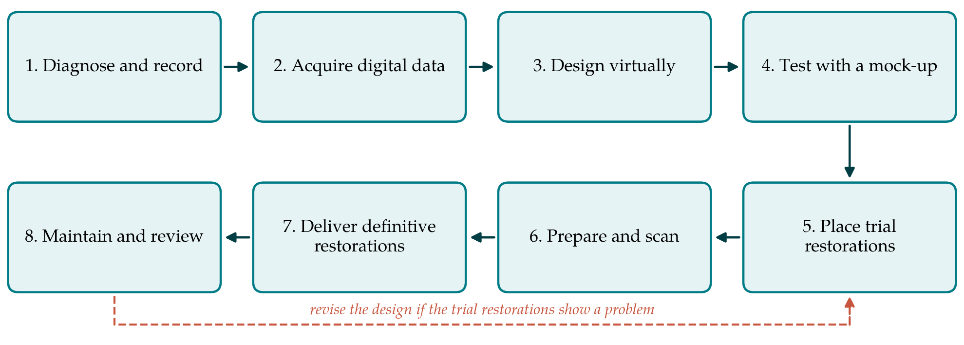
The protocol has eight steps. Figure 1 shows the sequence, and Table 1 lists the digital tool and the record associated with each step. The steps are then described in order.
| Step | Principal tool | Record retained |
|---|---|---|
| 1. Diagnose and record | Clinical and radiographic examination | Phenotype, family history, baseline indices |
| 2. Acquire digital data | Intraoral scanner; standard photographs | Arch scans, bite registration, image set |
| 3. Design virtually | Computer-aided design software | Approved design file and vertical dimension |
| 4. Test with a mock-up | Printed model and silicone index | Photographs and patient comments |
| 5. Place trial restorations | Milled polymer restorations | Review notes over the test period |
| 6. Prepare and scan | Mock-up-guided preparation; scanner | Preparation scans and shade record |
| 7. Deliver definitive restorations | Milled ceramic; adhesive cementation | Bonding protocol and occlusal record |
| 8. Maintain and review | Repeat scans superimposed on baseline | Wear, margin and periodontal findings |
3.1. Step 1: Diagnose and record
Treatment starts with the diagnosis. The clinician records the enamel phenotype, the family history and any associated findings such as taurodontism, open bite or unerupted teeth, and considers referral for genetic assessment. Radiographs show pulp size and enamel thickness. Baseline measures of sensitivity, plaque, gingival health and oral health-related quality of life are taken so that later change can be judged against them. Caries and gingival inflammation are brought under control before any restorative planning.
3.2. Step 2: Acquire digital data
Both arches and the intercuspal relationship are scanned, and a standard series of facial and intraoral photographs is taken. Where the vertical dimension is to be changed, a registration in centric relation at the proposed dimension is scanned as well. Rough, pitted enamel and moisture can disturb image stitching, so the scan is checked for gaps before the patient leaves. The files become the reference against which every later stage is compared.
3.3. Step 3: Design virtually
The new tooth forms are designed on the scans with reference to the photographs. Because enamel is thin or worn, the design is largely additive: material is added to restore the original volume of the teeth, and the occlusal vertical dimension is commonly increased to create room for it. A moderate increase is generally well tolerated when it is tested first in provisional form (Abduo and Lyons, 2012). The principle follows the adhesive rehabilitation sequence described for eroded dentitions by Vailati and Belser (2008), in which restorative space is gained by opening the bite and not by cutting teeth.
3.4. Step 4: Test with a mock-up
The design is printed and transferred to the mouth with a silicone index and a temporary resin. The mock-up lets patient, family and clinician see the proposed appearance, hear the effect on speech and check the lip line before anything irreversible is done. Changes requested at this visit are made in the design file, not by adjusting the resin alone, so that the file remains the single source for later steps.
3.5. Step 5: Place trial restorations
Trial restorations are milled from polymer blocks to the approved design and bonded with little or no preparation. They serve as a test of the new occlusion and vertical dimension over a period of months, protect sensitive teeth, and give an adolescent an acceptable appearance while growth and eruption continue. For a patient who is still growing, this phase may be deliberately prolonged, with the definitive stage deferred until gingival margins are stable. Problems that emerge, such as fractures, discomfort or phonetic difficulty, are corrected in the design, as the return loop in Figure 1 indicates.
3.6. Step 6: Prepare and scan
Definitive preparation is carried out through the trial restorations or a fresh mock-up, so that reduction is measured from the intended final surface and not from the existing tooth (Magne and Belser, 2004). This keeps removal of tooth tissue to the minimum that the material requires, an important consideration when pulps are large; conventional full-coverage preparations remove far more of the crown than partial-coverage designs do (Edelhoff and Sorensen, 2002). The preparations are scanned, and the scan is aligned to the approved design so that the definitive restorations reproduce it.
3.7. Step 7: Deliver definitive restorations
The definitive restorations are milled, tried in and cemented adhesively under isolation. Choice of material depends on available thickness, enamel quality and occlusal load; glass-ceramics, zirconia and resin-based blocks each have a place, and the evidence in this condition does not single out one. The bonding procedure is matched to the substrate. Where sound enamel remains, etch-and-rinse adhesion is the reference approach (Van Meerbeek et al., 2003). Where enamel is hypomineralised, margins are placed on better tissue if possible and deproteinisation is considered, in the knowledge that its clinical benefit is not established.
3.8. Step 8: Maintain and review
A protective splint is provided where parafunction is suspected, and recall intervals are set by periodontal and caries risk. At review the arches are rescanned and the scans superimposed on the delivery scan, which gives an objective record of wear, chipping and gingival change. The same baseline measures taken at Step 1 are repeated.
4. Fitting the Protocol to the Young Patient
Three features of this age group shaped the sequence. The first is biological: pulps are wide and clinical crowns may be short, so the protocol relies on additive design, adhesive retention and mock-up-guided reduction. The second is developmental. Gingival levels and jaw relationships are still changing in adolescence, which is why the trial phase can be extended and why orthodontic or surgical opinion on an open bite is sought before the definitive stage. The third is the time horizon. Any restoration placed in the late teens will be replaced more than once, and each step is chosen to leave as much tooth as possible for those later cycles. Stored design files make remaking a fractured unit simpler.
Cost and access are real constraints. The protocol assumes a scanner, design software and a laboratory or clinic with milling and printing equipment. Where these are unavailable, the same sequence can be followed with conventional impressions and a waxed design; what the digital route adds is continuity of one file across stages and an objective record at review.
5. Limitations and What Remains to Be Shown
This protocol is a proposal built from published principles, and it has not been tested here. The evidence beneath several steps is modest. Bonding to hypomineralised enamel rests mainly on laboratory studies. The comparative survival of ceramic, zirconia and resin-based restorations in amelogenesis imperfecta is unknown beyond the short term. Scanner accuracy on rough, hypoplastic enamel has received little direct study. The claim that a digital sequence reduces chair time or remakes in this group is plausible and unproven.
A clinical evaluation would need to follow a defined cohort and record restoration survival and complications, pulpal outcomes, periodontal health, sensitivity and patient-reported quality of life at fixed intervals, with phenotype documented for each participant. Given the rarity of the condition, multicentre collaboration and consistent reporting are likely to be necessary. Where individual cases are published, the CARE guideline offers a structure that would make them easier to compare (Gagnier et al., 2014).
6. Conclusion
Young people with amelogenesis imperfecta need extensive restoration at an age when conservative treatment matters most. The eight-step protocol proposed here uses digital tools to carry a single additive design from planning, through a reversible trial phase, to definitive adhesive restorations and objective review. It draws together established ideas and does not introduce a new technique. Whether it yields better or more durable results than conventional sequences is a question for prospective clinical study, and the outcomes such a study should measure have been indicated above.
Conflict of interest
To be declared by the author(s) before publication.
Funding
To be declared by the author(s) before publication.