1. Introduction
Indwelling urinary catheters are among the most widely used medical devices, and bacteriuria follows catheterisation with near inevitability if the catheter stays in place long enough. Most of that bacteriuria is asymptomatic and requires no treatment, a point on which clinical guidance is explicit (Hooton et al., 2010; Gould et al., 2010). One complication, however, is mechanical rather than inflammatory, and it is caused predominantly by a single organism. Proteus mirabilis colonises catheters and generates a crystalline deposit that obstructs the lumen, leading to bypassing, retention, and in some patients to pyelonephritis or septicaemia when the obstructed system is disturbed (Stickler, 2008).
The organism is also increasingly reported with resistance to several antimicrobial classes. Proteus is intrinsically resistant to nitrofurantoin, tetracyclines and colistin, so the acquisition of extended-spectrum beta-lactamases or carbapenemases narrows the usable options sharply. This note considers what the combination of crystalline biofilm formation and multidrug resistance means in practice, and where the laboratory methods in routine use fall short of describing it. It reports no experiments and presents no new data.
2. Why Crystalline Biofilm Is a Distinct Problem
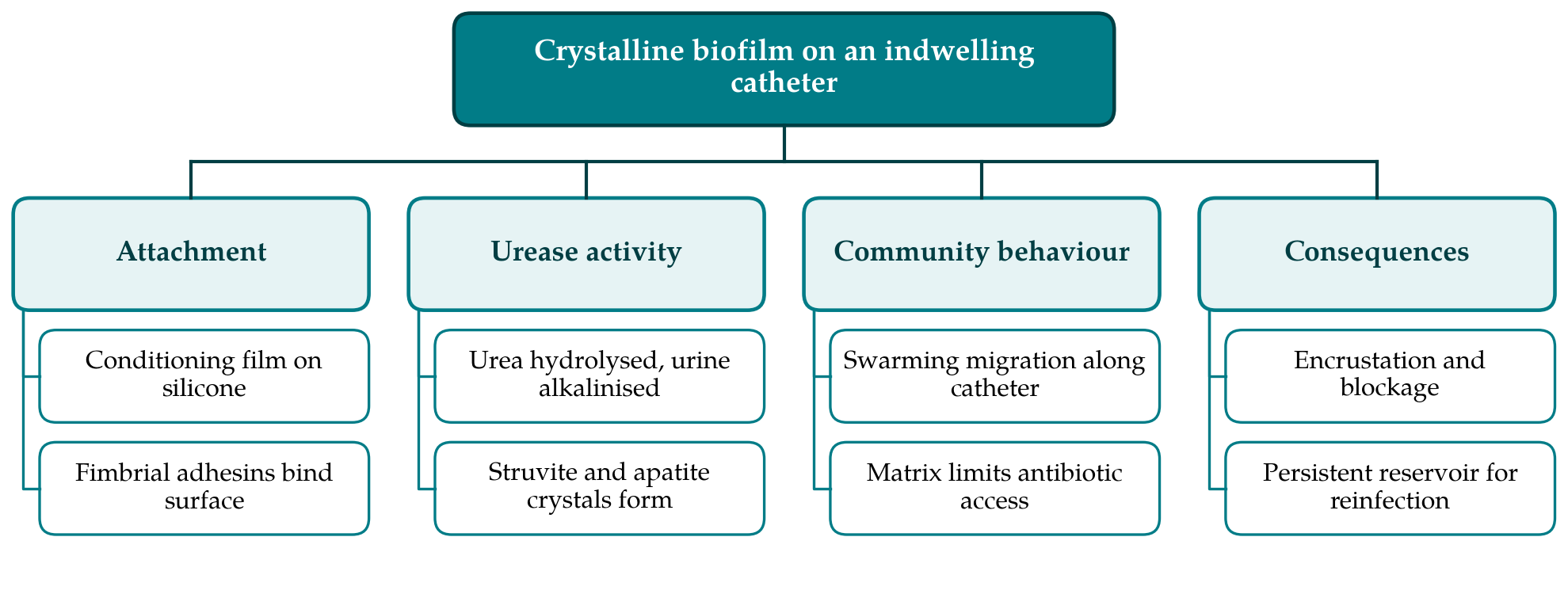
Biofilms on medical devices are a general phenomenon, and the reasons they tolerate antimicrobial agents, including slow growth, restricted penetration and persister subpopulations, apply here as elsewhere (Costerton et al., 1999; Hoiby et al., 2010). What distinguishes Proteus is urease. The enzyme hydrolyses urea to ammonia far faster than the ureases of most other urinary isolates, the pH of the urine rises, and at a sufficiently alkaline pH magnesium ammonium phosphate and calcium phosphate come out of solution. Crystals become incorporated into the extracellular matrix and the deposit hardens. The association between urease-positive bacteriuria and catheter obstruction was established in clinical observations some decades ago (Mobley and Warren, 1987) and the physical and chemical basis has been set out in detail since (Stickler and Feneley, 2010).
Two further features of the organism contribute. Differentiation into elongated, hyperflagellated swarmer cells allows migration over catheter surfaces, so colonisation is not confined to the point of entry, and a range of fimbrial adhesins mediate attachment to silicone and to the conditioning film of host proteins that coats it within minutes of insertion. The pathogenesis has been reviewed comprehensively (Jacobsen et al., 2008; Armbruster and Mobley, 2012; Armbruster et al., 2018). Figure 1 summarises the sequence from attachment to blockage.
3. What Resistance Adds, and What the Routine Report Omits
Antimicrobial susceptibility is determined on planktonic cells growing in a standardised broth at a controlled pH. A catheter lumen colonised by Proteus differs from that environment in several ways simultaneously: the pH is alkaline, mineral phases are present, cells are embedded in matrix, and growth rates are heterogeneous. Each of these can alter the activity of an antimicrobial agent, and aminoglycoside activity in particular is pH dependent. A report of susceptibility therefore predicts the response of the organism in urine better than it predicts the fate of an established crystalline deposit, and clearing bacteriuria without removing or replacing the catheter is correspondingly unlikely to resolve the obstruction.
Classification of isolates compounds the difficulty of comparing studies. Where papers apply the interim standard definitions of multidrug resistance proposed by an international working group (Magiorakos et al., 2012) the results can be set side by side; where local criteria are used they cannot. Table 1 lists the mismatches between what is commonly measured and what determines the clinical course, with the practical consequence of each.
| What is measured | What determines the outcome | Practical consequence |
|---|---|---|
| Planktonic susceptibility in broth | Activity against matrix-embedded cells at alkaline pH | Susceptible result need not predict clearance |
| Crystal violet biomass on polystyrene | Mineral deposition on silicone in flowing urine | Assay ranks strains on the wrong property |
| Single endpoint reading | Time to blockage under continuous flow | Rate of encrustation is not captured |
| Bacteriuria at a threshold count | Whether the device carries a mature biofilm | Treating the urine leaves the reservoir |
| Local resistance categories | Comparable, defined resistance phenotypes | Studies cannot be pooled Magiorakos et al., 2012 |
A second consequence concerns how specimens are taken. Urine drawn from a blocked or long-standing catheter reflects the organisms shed from the device rather than those in the bladder, and a culture taken through an encrusted lumen may report the biofilm community instead of the infecting flora. Guidance accordingly advises replacing the catheter before sampling where a catheter-associated infection is suspected and treatment is intended (Hooton et al., 2010). Where that step is omitted, both the identification and the susceptibility result may describe a population that antibiotic therapy was never going to reach.
4. Assay Conditions Worth Standardising
The microtitre plate assay with crystal violet staining is the usual first approach to quantifying biofilm, and the standardised description of its conditions and pitfalls remains a useful reference (Stepanovic et al., 2007). For Proteus on catheters it answers a narrower question than is often assumed, because it measures attached biomass on polystyrene in static broth, and neither the substrate nor the chemistry resembles a catheter carrying urine. Three changes would make results more transferable. Growing the organism in artificial urine rather than nutrient broth preserves the urea on which the whole mechanism depends. Using segments of the catheter material under study, silicone or latex with its particular coating, reproduces the surface the organism meets. And reporting pH and deposited mineral alongside biomass records the property that actually blocks the lumen.
Reporting conventions matter as much as conditions. Studies that state the medium, the surface, the incubation time, the washing procedure and the resistance phenotype by an agreed definition can be compared with one another; those that report an optical density and a strain label cannot. Flow models that measure time to blockage are more demanding and are confined to relatively few laboratories, but they remain the closest laboratory approximation to the clinical problem and give a result that clinicians can interpret directly.
5. Conclusion
Crystalline biofilm formation and multidrug resistance are separate properties of Proteus mirabilis that compound one another at the bedside: the first makes the device the problem, the second narrows what can be done about any accompanying infection. The clinical implications are already clear from existing guidance, which emphasises avoiding unnecessary catheterisation, removing catheters promptly and not treating asymptomatic bacteriuria. The laboratory implication is that the standard measures describe the organism incompletely. Assays conducted in artificial urine on the relevant catheter material, reporting mineral deposition as well as biomass and using agreed resistance definitions, would give a more faithful account and would allow results from different groups to be read together.
Conflict of interest
To be declared by the author(s) before publication.
Funding
To be declared by the author(s) before publication.