1. Introduction
Abdominal magnetic resonance imaging (MRI) asks for something difficult. Sequences that last many seconds must be acquired with the breath held, repeatedly, by people who are often elderly, breathless or in pain. When the breath-hold fails, the image is degraded by ghosting and blurring, and the examination may have to be repeated or reported with reservations. Motion artefact is a long-standing problem of the technique as a whole, and a standard review of the field describes it as one with many partial solutions and no complete one (Zaitsev et al., 2015).
Reconstruction based on deep learning is the most recent addition to the list. Within a few years it has moved from research papers to products offered by the major manufacturers, on the strength of evidence that trained networks can reconstruct diagnostic images from data too sparse or too noisy for conventional methods (Hammernik et al., 2018; Knoll et al., 2020). Departments acquiring the software face a practical question that the technical literature does not answer for them: how to change abdominal protocols so that the new reconstruction reduces motion artefact, and how to be satisfied that nothing of diagnostic value is lost in the exchange.
This article proposes a procedure for doing so. It is a method paper: it describes stages, reasons and records, and reports no measurements, examinations or reader results. The procedure has not been validated, and the final sections say what validation would require.
2. Background
The design of the procedure depends on three things: how motion produces artefact, what the established countermeasures do, and what deep learning reconstruction does and does not change.
2.1. How motion corrupts the image
MRI data are collected in spatial-frequency space over time, and the reconstruction assumes that the object is the same throughout. When it is not, the inconsistencies between lines of data appear as artefact, whose form depends on the motion and on the order of sampling. Periodic motion during a Cartesian acquisition produces discrete ghosts displaced along the phase-encoding direction; irregular motion produces diffuse blurring and loss of edge sharpness (Zaitsev et al., 2015). Table 1 lists the sources that matter in the abdomen with their usual remedies. One deserves particular note. Transient breathlessness shortly after injection of gadoxetate disodium can spoil the arterial phase of a liver examination, the one phase that cannot be repeated (Davenport et al., 2013).
| Source of motion | Typical artefact | Conventional remedy | What deep learning reconstruction can add |
|---|---|---|---|
| Respiration, failed breath-hold | Ghosting along the phase-encoding direction; blurring | Shorter breath-hold, triggering, navigators | Shorter acquisition through higher acceleration |
| Respiration, free breathing | Streaks with radial sampling | Golden-angle radial with motion sorting (Feng et al., 2016) | Removal of undersampling streaks; faster reconstruction |
| Cardiac pulsation | Ghosts across the left hepatic lobe | Change of phase direction, saturation bands | Little direct effect |
| Peristalsis | Blurring of bowel wall and nearby organs | Antiperistaltic agent, single-shot sequences | Denoising of fast single-shot images |
| Breathlessness after contrast | Degraded arterial phase | Several short arterial phases, radial acquisition | Support for very short or continuous acquisitions |
| Bulk body movement | Global blurring, misregistration | Coaching, repeat scan, PROPELLER (Pipe, 1999) | Research-stage correction networks (Küstner et al., 2019) |
2.2. Conventional countermeasures
Existing remedies work in one of three ways. Some stop the motion or wait for it: breath-holding, respiratory triggering and navigator gating. Some make the acquisition less sensitive to it: radial and PROPELLER trajectories sample the centre of k-space repeatedly, so that motion is averaged into mild blurring or streaks in place of coherent ghosts (Pipe, 1999). The rest make the acquisition faster. Parallel imaging with SENSE or GRAPPA uses the spatial sensitivity of receiver coils to replace some phase-encoding steps (Pruessmann et al., 1999; Griswold et al., 2002), and compressed sensing exploits image sparsity to go further (Lustig et al., 2007). Combined with golden-angle radial sampling, these gave rise to free-breathing dynamic imaging and to reconstructions that sort data by respiratory state (Feng et al., 2014; Feng et al., 2016). Each has a cost: noise amplification, residual aliasing, long computation or a longer examination.
2.3. What deep learning reconstruction does
Most clinical deep learning reconstructions belong to one family. An iterative reconstruction is unrolled into a fixed number of steps, each alternating a data-consistency operation, which keeps the estimate faithful to the measured samples, with a learned regulariser in the form of a convolutional network. The variational network, the deep cascade and model-based deep learning are the reference designs (Hammernik et al., 2018; Schlemper et al., 2018; Aggarwal et al., 2019). Trained on pairs of fully sampled and undersampled data, such networks suppress noise and aliasing at acceleration factors where parallel imaging fails. Their effect on motion is indirect. They do not model motion; they make it possible to acquire the same image in less time, and a shorter breath-hold is one that more people can manage.
Direct correction is a separate line of work. Image-to-image networks have been trained to map motion-corrupted images to clean ones, and hybrid schemes use a network to guide a physical model of the motion (Küstner et al., 2019; Haskell et al., 2019). These are promising and largely unavailable in clinical products. Both kinds share a known weakness. A learned prior can produce images that look sharp and plausible while departing from the data, and small structures or perturbations unlike anything in the training set can be removed or altered (Antun et al., 2020). The best-known public training set consists of knee and brain examinations (Zbontar et al., 2018), so abdominal performance cannot be assumed from published benchmarks.
3. The Proposed Procedure
The procedure has six stages, grouped in Figure 1 under acquisition, reconstruction and evaluation. It assumes a department with a scanner on which a deep learning reconstruction, supplied by the manufacturer or developed in research, is available for abdominal sequences, and a physicist or senior radiographer able to modify protocols.
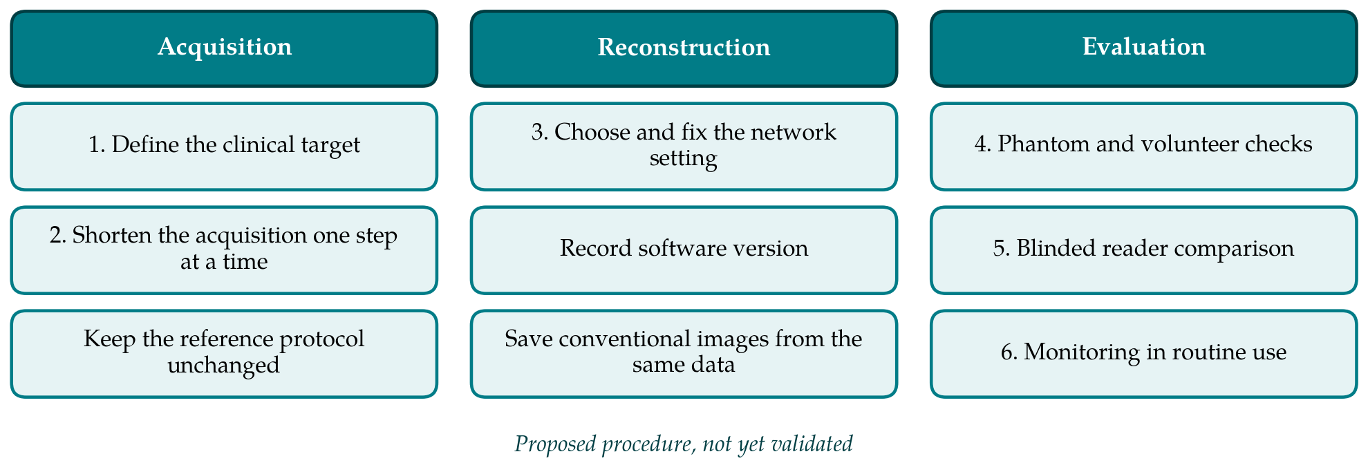
3.1. Stage 1: Define the clinical target
The first decision is which motion problem is to be solved, for which patients and on which sequences. The answer should come from the experience the department has of failed examinations. Typical candidates are the breath-held T1-weighted gradient-echo sequence used for dynamic liver imaging, T2-weighted single-shot and fast spin-echo sequences, and diffusion-weighted imaging. A target is stated in operational terms, for example that the dynamic sequence should be acquired in a breath-hold short enough for people who cannot hold for the present duration. A sequence with no identified motion problem is left alone.
3.2. Stage 2: Redesign the acquisition
Deep learning reconstruction reduces motion artefact only if the acquisition is changed to take advantage of it. The options are a higher parallel-imaging or compressed-sensing factor, fewer signal averages, or partial Fourier sampling, each of which shortens the scan and lowers the signal-to-noise ratio that the network is then asked to restore. Changes are made one at a time from the existing protocol, which is kept unaltered as the reference. Contrast-determining parameters, that is repetition time, echo time, flip angle and fat suppression, are left as they are, so that any change in lesion conspicuity can be attributed to the reconstruction and not to altered weighting. Where a radial free-breathing sequence is available, it is treated as a separate candidate and not mixed with the breath-hold variant (Feng et al., 2014).
3.3. Stage 3: Choose and configure the reconstruction
Commercial implementations expose few controls, usually a strength or denoising level and sometimes an edge-enhancement option. The setting is chosen on test data before any comparison and then fixed, because adjusting it case by case makes the result irreproducible. The software version, the setting and the sequences to which it is applied are written down; a version update is treated as a new reconstruction requiring at least the technical checks of the next stage. Throughout the evaluation the conventionally reconstructed images from the same raw data are also saved wherever the system allows. They cost nothing in scan time and provide the only direct check on whether a feature seen, or not seen, on the deep learning images is present in the data.
3.4. Stage 4: Technical verification
Before clinical use, the candidate protocol is run on a structured phantom to confirm geometric accuracy, the absence of new structured artefact and the behaviour of fine detail and low-contrast inserts at the chosen setting. Global similarity indices such as the structural similarity index are convenient for comparing reconstructions of the same data (Wang et al., 2004), but they summarise the whole image and are insensitive to the loss of a small lesion, so they are recorded without being treated as evidence of diagnostic adequacy. A small number of volunteer examinations, under the arrangements the department has for volunteer scanning, then establishes that the shortened sequence runs as intended and that the breath-hold has in fact become shorter.
3.5. Stage 5: Blinded reader comparison
The central test is a paired comparison in clinical examinations. Each person undergoes the reference sequence and the candidate sequence in the same session, in an order that is varied to avoid a systematic effect of fatigue or contrast timing. For post-contrast sequences, where both cannot occupy the same phase, the comparison is restricted to phases that can be duplicated, or to consecutive examinations of the same person. At least two radiologists, unaware of the method, score each series on predefined scales for motion artefact, sharpness, noise, artificial texture and overall diagnostic quality, and separately record the focal lesions they detect. Lesion detection against a reference reading matters more than preference scores, because readers tend to prefer smooth images. The sample size, the scales and the analysis are fixed in a written plan, and the work is reported in line with the CLAIM checklist (Mongan et al., 2020).
3.6. Stage 6: Monitoring in routine use
If the comparison supports adoption, the candidate protocol replaces the reference for the stated indication, and monitoring begins. Two simple measures are proposed. One is the proportion of examinations in which a sequence is repeated or flagged by the reporting radiologist as limited by motion, tracked before and after the change. The other is a log of discrepancies in which a finding on follow-up or on another modality was not visible on the deep learning images, each reviewed against the conventional reconstruction if it was kept. Monitoring is renewed after any software update or change of coil.
4. Fitting the Procedure to a Department
The procedure can be scaled. A department with limited research capacity may confine itself to one sequence, perform the technical checks, and run the reader comparison on a modest consecutive series as a service evaluation under local governance. A larger centre can add free-breathing radial acquisitions, quantitative sequences and formal lesion-detection analysis. In both, the essential discipline is the same: one change at a time, a retained reference, fixed settings and readers who do not know which image they are scoring. Radiographers need to be told what has changed and why, since the benefit depends on the shorter breath-hold being coached and used. Old instructions given for a new sequence gain nothing.
5. Limitations and What Remains to Be Shown
The procedure is a proposal and has not been tested. Whether its stages are sufficient, or more than a busy department will carry out, can be learned only by using it. Its reader comparison measures image quality and lesion detection within a single session and cannot show effects on diagnosis or clinical outcome. It also inherits the difficulty of any evaluation of learned reconstruction: the absence of a ground truth. In clinical examinations there is no motion-free, fully sampled image against which to judge, and agreement between two reconstructions of the same motion-affected data proves little.
Three further matters are open. The first is failure behaviour. When motion is severe, a network trained on motion-free data is operating outside its training distribution, and whether it then degrades visibly or produces a clean-looking but wrong image is the question that most needs study (Antun et al., 2020). The second is generalisation across field strengths, coils, contrast agents and body habitus, which product documentation seldom describes in detail. The third is the place of direct motion-correction networks. If these reach clinical products, the procedure would need an additional stage to test them on data with known, deliberately induced motion (Haskell et al., 2019). None of this argues against adoption. It argues for adoption with the reference images kept and the results watched.
6. Conclusion
Deep learning reconstruction reduces motion artefact in abdominal MRI mainly by making short acquisitions usable, and it does so only when protocols are redesigned to shorten the time for which stillness is required. The six-stage procedure proposed here gives a department an orderly way to make that change: define the problem, alter the acquisition one step at a time, fix the reconstruction settings, verify technically, compare under blinded reading with lesion detection as the main measure, and monitor afterwards. It treats the learned reconstruction as a component to be checked against the data, not as a guarantee of quality. The procedure has yet to be applied, and reports of its use, including instances where the new images proved worse, would be the most useful next contribution.
Conflict of interest
To be declared by the author(s) before publication.
Funding
To be declared by the author(s) before publication.