1. Introduction
A radiograph taken in an emergency department at night is usually interpreted first by the clinician who requested it, often a doctor in training or a nurse practitioner, and acted on at once. The report of the radiologist arrives later, sometimes after the person examined has gone home. The radiographer who acquired the image has looked at it carefully, because judging its technical adequacy requires that, and has frequently formed a view about whether it is normal. In many departments that view goes unrecorded.
Preliminary image evaluation (PIE), also called preliminary clinical evaluation or radiographer commenting, is the practice of recording it. The radiographer writes a short statement, at the time of imaging, saying whether an abnormality is suspected and what and where it is. The comment is advisory. It does not replace the definitive report, and the referrer remains responsible for the immediate decision. The professional body for radiographers in the United Kingdom has for more than a decade stated that this should be a normal part of practice (Society and College of Radiographers, 2013).
This article asks whether the practice should now be adopted more widely, and argues that it should, subject to conditions that are set out below.
2. From Red Dot to Written Comment
The idea began with a simple experiment in a London hospital, in which radiographers placed a red sticker on films they thought abnormal, and the casualty officers were found to benefit (Berman et al., 1985). Red dot schemes spread through British departments over the next two decades and became close to universal, while schemes requiring a written comment remained uncommon (Snaith and Hardy, 2008). The red dot has obvious weaknesses. It says that something is wrong and not what or where. Its absence is ambiguous, since it may mean a normal image, an uncertain radiographer or one who does not take part.
A written comment answers each of these. It names the finding, it can state positively that no abnormality was seen, and it can be stored with the image. It also asks more of the radiographer. When the two were compared directly, radiographers who flagged abnormal images accurately were less accurate when asked to describe the abnormality in words (Hardy and Culpan, 2007). The step from red dot to PIE is therefore a real change in the task, and it is the reason that wider adoption is a question of training and governance and not only of policy.
3. The Case for Wider Adoption
The first argument is the size of the problem that PIE addresses. Analyses of diagnostic error in emergency departments have found that missed fractures form the largest single group, and that most arise because an abnormality visible on the radiograph was not recognised by the clinician reading it (Guly, 2001). The injuries overlooked are often subtle ones in the foot, knee, elbow, hand and wrist (Wei et al., 2006), and the contributing factors, such as inexperience, fatigue, time pressure and the absence of a second reader, are those of out-of-hours work (Pinto et al., 2016). A comment from the radiographer supplies a second reader at exactly the moment one is missing.
The second argument is that radiographers can do it. A systematic review of red dot and triage schemes in clinical practice found the accuracy of radiographers in distinguishing normal from abnormal emergency radiographs to be high (Brealey et al., 2006). In a direct comparison on appendicular skeletal radiographs, radiographers interpreted more accurately than emergency nurse practitioners, and both groups improved with a short course (Piper and Paterson, 2009). At the far end of the spectrum, radiographers with postgraduate training who issue definitive reports perform at a level comparable to radiologists for defined examinations (Brealey et al., 2005; Woznitza et al., 2018). Focused teaching has been reported to raise the accuracy of comments by newly qualified staff (Stevens and Thompson, 2018).
The third argument is capacity. A national review of radiology reporting in England documented delays in reporting (Care Quality Commission, 2018), and the subsequent independent review of diagnostic services called for a major expansion of the imaging workforce and fuller use of the skills of each professional group (Richards, 2020). PIE creates no new posts and no new reports. It uses attention that has already been paid to the image. In Australia the capabilities expected of every registered practitioner include identifying urgent or unexpected findings and conveying them to those responsible for care (Medical Radiation Practice Board of Australia, 2020), and the standards of proficiency for radiographers in the United Kingdom expect registrants to be able to recognise abnormal appearances on the images they produce (Health and Care Professions Council, 2023).
4. The Counter-Arguments
The objections deserve a serious hearing, and Table 1 summarises them with the responses developed in this section.
| Objection | Substance of the concern | Response |
|---|---|---|
| Insufficient accuracy | Written comments are less accurate than red dots (Hardy and Culpan, 2007) | Training before participation; begin with the appendicular skeleton |
| False reassurance | A normal comment may stop the referrer looking carefully | State advisory status; referrer still reviews the image |
| Legal exposure | Radiographers fear liability for a wrong comment | Employer-approved scheme with defined scope and protocol |
| Workload | Comments take time in a busy department | Short structured format; measure the time cost |
| Uneven participation | Voluntary schemes are used by few staff (Lancaster and Hardy, 2012) | Make the comment an expected part of the examination |
| Professional boundaries | Interpretation is regarded as the role of the radiologist | Agreed team-working framework (RCR and SCoR, 2012) |
The strongest objection is accuracy. A wrong comment can mislead in either direction, and a false negative is the more dangerous, because a junior clinician told that a film shows no abnormality may look less hard. The published accuracy figures come largely from motivated volunteers and from test image banks enriched with abnormal cases, and may flatter routine practice. This is a reason to define the scope narrowly at first, to the appendicular skeleton where the evidence is best, to require training before participation, and to make the advisory status of the comment explicit. It is not a reason to prefer the present arrangement, in which the same junior clinician reads the same film with no second opinion at all.
Work on commenting schemes in practice indicates that lack of time, lack of confidence, uncertainty about legal standing and limited feedback keep participation low where it is voluntary (Lancaster and Hardy, 2012). The answer to the legal concern is institutional: a radiographer commenting within an employer-approved scheme, after the required training and within its stated scope, is acting within the role, as professional guidance makes clear (Society and College of Radiographers, 2013). A scheme that depends on who is on shift is not a safety system.
The remaining objection is about professional territory. Radiologists have at times regarded image interpretation by radiographers as an encroachment, and the worry that a preliminary comment will come to be treated as a report is not unreasonable where reports are delayed. The joint position of the two United Kingdom professional bodies is that interpretation is properly a team activity with defined roles (RCR and SCoR, 2012). PIE fits that model if the report of the radiologist remains the definitive record and is used to audit the comments.
5. What Wider Adoption Would Require
If the case is accepted, the conditions for doing it well can be stated fairly precisely. Figure 1 sets out six that support a reliable scheme. Training comes first. A scheme should specify a course, an assessment against a test bank and periodic refreshment. The comment itself should follow a standard form, stating the examination, whether an abnormality is suspected, its nature and site, and that the comment is preliminary.
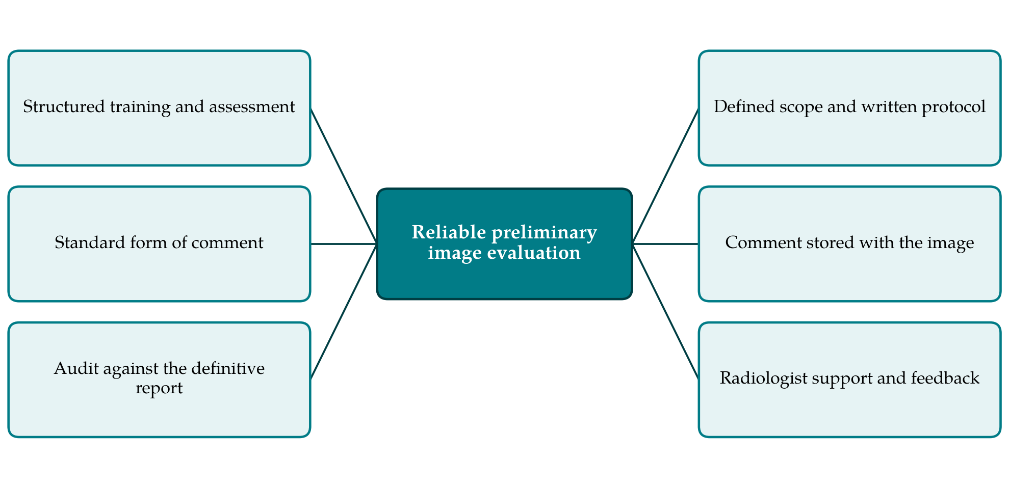
Audit closes the loop. Every comment can be compared with the definitive report once that is issued, and agreement rates fed back to individuals and to the department. Governance means a written protocol approved by the employer, naming the examinations in scope, the training required and the route for raising an urgent finding directly with the referrer. The comment must be recorded in the imaging or record system where the referrer will see it with the image. Finally, radiologists need to be partners in the scheme, contributing to teaching and receiving the audit results.
6. Implications for Education, Regulation and Research
For educators, wider adoption implies that preliminary evaluation is treated as a core graduate outcome, assessed as rigorously as positioning or radiation protection. For regulators and professional bodies outside the United Kingdom and Australia, it implies deciding whether the scope of radiographic practice includes communicating a suspected abnormality. In many jurisdictions it currently does not. The argument made here is for adoption where the legal and educational conditions exist or can be created.
For researchers, the main gap is evidence of effect. The literature shows that radiographers can comment accurately under study conditions. It says far less about whether PIE in routine use reduces missed injuries, recalls or harm, or what it costs. Comparisons of departments before and after introduction, with missed-fracture and recall rates as outcomes, would answer it. How PIE should relate to automated detection software, which is entering emergency radiography with the same aim, is a further question that has yet to be studied.
7. Conclusion
The opinion of the radiographer about the image exists whether or not anyone asks for it. Preliminary image evaluation is a means of putting that opinion where it can prevent an error, at the time the error would be made. The evidence that trained radiographers can provide it is sound for the appendicular skeleton and reasonable beyond, the need is evident from what is known about missed injuries and reporting delay, and the objections point to the conditions for success more than to grounds for refusal. Wider adoption is justified where training, a standard comment, audit and governance are in place, and should be accompanied by measurement of its effect on care. Without those conditions the practice would be a gesture; with them it is an inexpensive addition to the safety of emergency imaging.
Conflict of interest
To be declared by the author(s) before publication.
Funding
To be declared by the author(s) before publication.