1. Introduction
Integral membrane proteins fold and function inside a solvent that is thin, anisotropic and chemically mixed. The lipids around them differ in chain length, saturation, headgroup charge and intrinsic curvature, and cells regulate that mixture closely. That lipid composition affects the activity of channels, transporters and receptors has been documented for decades (Lee, 2004), and physical theory offers several explanations in terms of bilayer thickness, elasticity and lateral pressure (Phillips et al., 2009). What has been harder to obtain is a direct measurement of how a given lipid environment changes the stability of a membrane protein, because the classical tools of protein folding, chemical and thermal denaturation, work poorly on proteins that aggregate once removed from the membrane (Bowie, 2005).
Single-molecule force spectroscopy (SMFS) avoids that difficulty by using mechanical force as the denaturant. In the atomic force microscope version of the experiment, the tip picks up one terminus of a protein sitting in a supported membrane and the cantilever is retracted, so that the polypeptide is extracted segment by segment while the force is recorded. The first application to bacteriorhodopsin showed that individual molecules unfold along reproducible pathways with force peaks that could be assigned to particular transmembrane helices (Oesterhelt et al., 2000). Because the protein starts in a bilayer, the bilayer is part of what is being measured.
This article reviews what SMFS has established about the dependence of membrane protein unfolding on lipid composition, and where the evidence remains thin. It is written for readers who know either membrane biophysics or force spectroscopy but not necessarily both.
2. Scope and Approach
The article is a narrative review of the published literature. It draws on the founding experimental papers, on the theoretical work used to interpret force spectra, on the established reviews of the field (Kedrov et al., 2007; Engel and Gaub, 2008; Bippes and Müller, 2011) and on the membrane biophysics literature that predicts how lipids should matter. No formal search protocol was applied, and the selection reflects a judgement about which studies bear most directly on the question. The emphasis is on α-helical proteins, for which most of the relevant work has been done, and on the atomic force microscope, with magnetic tweezers considered where they offer a contrasting geometry.
3. How Force Spectroscopy Reports on a Membrane Protein
An account of lipid effects has to begin with what the instrument measures, since each derived quantity carries its own assumptions about the membrane.
3.1. The unfolding experiment
Membranes containing the protein, either native patches or reconstituted proteoliposomes, are adsorbed onto freshly cleaved mica in buffer. The tip is pressed onto the surface so that a terminal segment attaches to it, usually by non-specific adsorption, and the cantilever is then withdrawn at constant speed. Each time a stable structural segment gives way, the force drops and a further length of polypeptide is released, which produces the sawtooth pattern first described for the tandem domains of titin (Rief et al., 1997). The rising flank of each peak is fitted with the worm-like chain model of polymer elasticity (Marko and Siggia, 1995), and the fitted contour length gives the number of amino acids stretched between tip and membrane at that moment.
In bacteriorhodopsin the main peaks correspond to the extraction of transmembrane helices in pairs, with smaller peaks indicating that single helices and loops sometimes unfold separately (Oesterhelt et al., 2000). The same logic has since been applied to other rhodopsins, transporters, channels and receptors, and the positions of the peaks are generally characteristic of the protein (Kedrov et al., 2007). Two features of the trace are therefore available for comparison between lipid conditions: where the peaks fall, which reports on the boundaries of the stable segments, and how high and how frequent they are, which reports on the strength of the interactions that hold each segment in place.
3.2. From force peaks to energy landscapes
An unfolding force is not a property of the protein alone. It depends on how quickly the load is applied, because thermal fluctuations have more time to carry the system over the barrier when the pulling is slow. Bell (1978) proposed that an applied force lowers an activation barrier in proportion to the distance between the bound state and the transition state, and Evans and Ritchie (1997) showed that the most probable rupture force should then rise linearly with the logarithm of the loading rate. Repeating the experiment at several pulling speeds, known as dynamic force spectroscopy, yields two parameters for each structural segment: the distance to the transition state and the unfolding rate extrapolated to zero force. Later treatments replaced the sharp-barrier assumption with explicit landscape shapes and allow the barrier height to be estimated as well (Dudko et al., 2006).
Equilibrium free energies can also be recovered from repeated non-equilibrium pulls through the work relation of Jarzynski (1997), and a practical reconstruction procedure for pulling experiments was set out by Hummer and Szabo (2001). These analyses matter for the lipid question because a lipid might change a peak height by altering the barrier, by moving the transition state, or by changing the stiffness of the surrounding material, and only measurements across loading rates can separate those possibilities. All of them rest on a calibrated force, and the usual thermal-noise calibration of the cantilever spring constant (Hutter and Bechhoefer, 1993) carries an uncertainty that is not negligible beside the differences expected between lipid conditions.
4. How the Bilayer Can Alter Mechanical Stability
Membrane biophysics supplies several mechanisms by which lipids act on embedded proteins. They are summarised in Table 1 together with the signature each would be expected to leave in a force spectrum, and Figure 1 sets out the same relations as a map from bilayer variable to readout.
| Bilayer property | Proposed mechanism | Expected signature | Representative sources |
|---|---|---|---|
| Hydrophobic thickness | Mismatch with the transmembrane length strains protein and lipid | Changed force to extract helices; shifted segment boundaries | Mouritsen and Bloom, 1984; Killian, 1998 |
| Lateral pressure profile | Pressure redistributed with depth favours some conformations | Stability of particular segments altered | Cantor, 1997 |
| Bilayer elasticity | Energetic cost of deforming the membrane near the protein | Altered unfolding rates across segments | Andersen and Koeppe, 2007 |
| Cholesterol content | Ordering of acyl chains and direct binding to the protein | Higher forces and slower unfolding of most segments | Zocher et al., 2012 |
| Headgroup charge | Electrostatic interaction with charged loops and termini | Changed anchoring of interfacial segments | Lee, 2004 |
| Specific bound lipids | Lipids held at defined sites on the protein surface | Local stabilisation of individual peaks | Lee, 2004 |
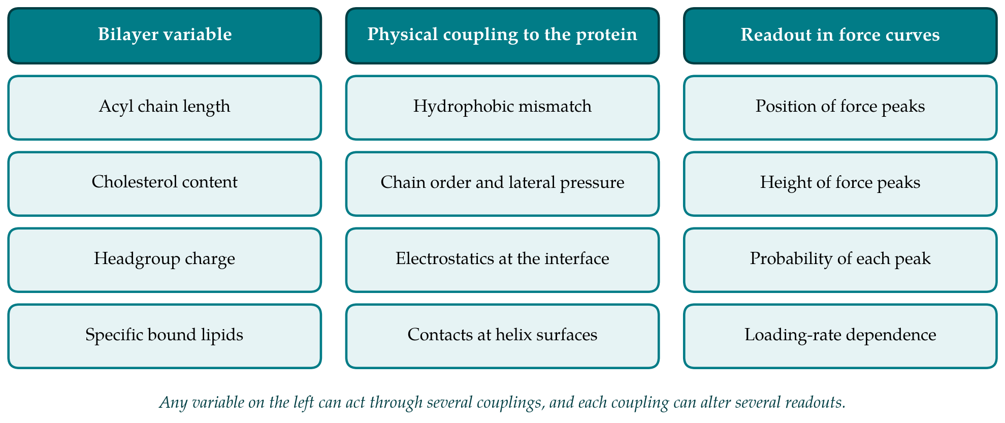
4.1. Thickness, pressure and elasticity
The oldest of these ideas is hydrophobic matching. In the mattress model of Mouritsen and Bloom (1984), a protein whose hydrophobic span differs from that of the surrounding bilayer forces either the lipids or the protein to deform, at an energetic cost, and Killian (1998) reviewed the experimental consequences for helix tilt, aggregation and activity. For an extraction experiment the prediction is straightforward in direction if not in size: a helix that sits comfortably in its bilayer should be harder to pull out than one that is already strained, and the residues that anchor a helix at the interface may differ between thick and thin membranes.
A second mechanism works through the lateral pressure profile. The interfacial tension at the headgroup region is balanced by repulsive pressure among the chains, and Cantor (1997) pointed out that changing chain unsaturation or adding small amphiphiles redistributes this pressure with depth, which shifts the equilibrium between protein conformations that differ in cross-sectional shape. A third treats the bilayer as an elastic sheet that must bend and compress to meet the protein, so that bilayer stiffness enters the energetics of any conformational change that alters the protein's boundary (Andersen and Koeppe, 2007; Phillips et al., 2009). These continuum effects are non-specific. They should act on every protein in the membrane, though not equally on every segment.
4.2. Cholesterol and specific lipid binding
Cholesterol illustrates why general and specific effects are hard to separate. It orders neighbouring acyl chains, thickens fluid bilayers and increases their bending rigidity, all of which are continuum effects, and it also occupies defined grooves on the surface of several proteins. The same duality applies to the annular lipids that exchange rapidly at the protein surface and to the smaller number of lipids bound tightly enough to be retained through purification (Lee, 2004). A change in an unfolding force after cholesterol is added to a membrane therefore does not, taken alone, say which mechanism is responsible. An answer needs comparison with other sterols or with lipids that reproduce the bulk effect without the binding.
5. What the Experiments Show
The experimental record is considered here in three groups: proteins unfolded from their native membranes, proteins reconstituted into bilayers of chosen composition, and proteins unfolded within the plane of a membrane mimetic.
5.1. Native membranes
Much of what is known comes from membranes that nature supplies in nearly pure form, above all the purple membrane of Halobacterium salinarum, in which bacteriorhodopsin is packed into a two-dimensional lattice with a small complement of archaeal lipids. Work on such samples established that unfolding pathways are reproducible, that they respond to temperature, pH, mutation, ligand binding and oligomeric state, and that each structural segment can be characterised by its own kinetic parameters (Kedrov et al., 2007; Engel and Gaub, 2008). These findings show that SMFS is sensitive enough to detect the kind of change a lipid might cause. They do not isolate the lipid contribution, since in a native patch the lipid composition is fixed and bound up with the crystalline packing of the protein.
5.2. Reconstituted bilayers
Controlling composition requires reconstitution. The clearest example is the study of the human β2-adrenergic receptor by Zocher et al. (2012), who unfolded the receptor from phospholipid liposomes prepared with and without the cholesterol analogue cholesteryl hemisuccinate and applied dynamic force spectroscopy to each structural segment. They reported that the sterol increased the kinetic, energetic and mechanical stability of nearly all segments of the receptor, a result consistent with structural and functional evidence that cholesterol stabilises this class of proteins. The study is valuable because it compares like with like: one protein, one instrument, one analysis, and a single compositional variable.
It also shows the limits of the approach. Reconstitution changes more than the lipid. The density of protein in the membrane, its orientation, its oligomeric state and the residual detergent may all differ between preparations, and each can influence how readily a terminus is picked up and how the molecule unfolds. A cholesterol analogue with a charged headgroup is not cholesterol. And the comparison of two compositions does not establish a dose-response relation or distinguish binding from bulk ordering. Published comparisons of this controlled kind remain few relative to the number of proteins that have been unfolded.
5.3. Unfolding within the membrane plane
Extraction by the atomic force microscope pulls the polypeptide out of the bilayer along the membrane normal. In terms of the two-stage model of Popot and Engelman (1990), it reverses helix association and helix insertion together, so the measured work includes the cost of transferring hydrophobic segments from lipid to water. Magnetic tweezers allow a different geometry. Min et al. (2015) attached DNA handles to both termini of the rhomboid protease GlpG held in bicelles and applied force laterally, which let the helices separate while remaining in the membrane mimetic. Unfolding occurred at forces low enough for refolding to be observed, and an energy landscape for the second stage of folding could be mapped. The lipid dependence of that second stage is the more relevant quantity for protein stability in a cell, and the review of Neuman and Nagy (2008) sets out how the force range and resolution of the two instruments differ.
6. Methodological Constraints on Comparing Lipid Conditions
Several features of the experiment make a comparison between two membranes harder than a comparison between two mutants in the same membrane. The first is the support. A bilayer adsorbed on mica interacts with the substrate through a thin water layer, and that interaction depends on headgroup charge and ionic strength. Changing the lipid may therefore change how firmly the membrane is held, how mobile its lower leaflet is, and whether the bilayer is in a fluid or a gel state at the temperature of measurement, quite apart from any effect on the protein. The asymmetry between leaflets that characterises cell membranes is in any case lost in most reconstituted samples.
The second is force calibration. Spring constants determined by the thermal method differ from one cantilever to the next and from one calibration to the next (Hutter and Bechhoefer, 1993). When lipid conditions are measured on different days with different cantilevers, a systematic offset in force can be mistaken for a lipid effect, or can conceal one. Designs that interleave conditions, reuse a cantilever across them, or include an internal mechanical reference reduce this risk, and reports that omit such details are difficult to weigh.
The third is resolution. Yu et al. (2017) used modified ultrashort cantilevers to unfold bacteriorhodopsin with far better time resolution and force precision than had been available, and resolved many more intermediates than earlier work, some separated by only a few amino acids. The implication for lipid studies is that a conventional cantilever averages over closely spaced states, so that a shift in the population of those states could appear as a change in the height or width of one broad peak. Conclusions about which segment a lipid stabilises are bounded by the resolution at which the segments were defined.
7. Gaps and Directions for Further Work
Taken as a whole, the literature supports three statements. Force spectroscopy can detect changes in the stability of individual segments of a membrane protein. Theory predicts that lipid composition should produce such changes through several distinct mechanisms. And where composition has been varied under controlled conditions, a change of the expected kind has been reported. What is missing is breadth: series in which chain length, sterol content or headgroup charge is varied in steps for a single protein, with loading-rate dependence measured at each step.
Defined membrane mimetics should help. Nanodiscs enclose a small patch of bilayer of chosen composition within a protein belt and hold a controlled number of protein molecules (Bayburt and Sligar, 2010), which removes the uncertainty about protein density and oligomeric state that accompanies liposome reconstitution, though the belt introduces a boundary of its own. Site-specific attachment of the terminus to the tip would replace non-specific adsorption and make the pulling geometry reproducible. Reporting practices also need attention: lipid composition after reconstitution, measurement temperature relative to the phase transition, cantilever calibration and the number of independent preparations are not consistently stated, and without them results from different laboratories cannot be combined. Finally, the two pulling geometries answer different questions, and applying both to the same protein in the same lipid would show how much of an apparent lipid effect lies in helix extraction and how much in helix packing.
8. Conclusion
Mechanical unfolding of single membrane proteins offers a direct, segment-resolved view of stability in a lipid environment, and the interpretive framework that links force peaks to barriers and rates is mature. The proposition that lipid composition alters that stability is well founded in theory and supported by a small number of careful experiments, most clearly for cholesterol and a G protein-coupled receptor. It has not yet been tested systematically across lipid variables or across proteins. The instruments and membrane mimetics needed for such tests exist, and the main requirement now is experimental design that isolates the lipid from the many other things that change when a membrane is rebuilt.
Conflict of interest
To be declared by the author(s) before publication.
Funding
To be declared by the author(s) before publication.