1. Introduction
Plants cannot leave a hot place, so their cells must absorb the consequences of heat where they stand. The best studied part of the response is transcriptional: heat stress transcription factors drive the synthesis of chaperones that hold unfolded proteins in a recoverable state (Ohama et al., 2017). A second part acts on messenger RNA that already exists. Within minutes of a temperature shift, bulk translation initiation slows, polysomes release their transcripts, and microscopically visible granules containing RNA and RNA-binding proteins appear in the cytoplasm (Chantarachot and Bailey-Serres, 2018).
These granules have no membrane. For most of their history they were described simply as aggregates or ribonucleoprotein particles. That changed after the demonstration that P granules in the germline of Caenorhabditis elegans behave as liquid droplets that fuse, drip and dissolve (Brangwynne et al., 2009). The idea that cells organise part of their chemistry by liquid-liquid phase separation spread quickly through cell biology (Banani et al., 2017), and plant biologists have adopted it with enthusiasm (Emenecker et al., 2020).
Enthusiasm is not evidence, and the vocabulary of phase separation is now applied to plant heat responses more freely than the experiments always justify. This review asks a narrow question: what has actually been shown about the condensation of RNA-binding proteins in heat-stressed plant cells, and how strong is the case that condensation contributes to survival?
2. Scope and Approach
The article is a narrative review of the published literature. It draws on primary studies of plant stress granules and their protein components, on the foundational work in yeast and mammalian cells from which the concepts were borrowed, and on methodological critiques of the phase separation field. No formal search protocol was followed and no claim of completeness is made. Papers were chosen because they introduced a concept, supplied a piece of evidence that later work relied on, or stated a limitation clearly. Proteins that condense in response to warm, non-stressful temperatures are mentioned only where they clarify the argument, and nuclear condensates involved in transcription and RNA processing are left to other reviews (Emenecker et al., 2020).
3. From Heat Shock Granules to Stress Granules
The first cytoplasmic granules reported in heat-stressed plant cells were seen by electron microscopy in tomato cell cultures and leaves (Nover et al., 1983). They were dense, abundant and built largely from small heat shock proteins. Later biochemical work suggested that these heat shock granules were associated with untranslated housekeeping transcripts, which led to the attractive proposal that they stored messenger RNA through the stress period (Nover et al., 1989).
That proposal did not survive closer inspection. Using fluorescent markers in tomato and Arabidopsis cells, Weber et al. (2008) showed that three structures coexist after heat: processing bodies marked by decapping factors, stress granules containing translation initiation factors and the RNA-binding proteins RBP47 and UBP1, and heat shock granules made of small heat shock proteins. Only the first two contained polyadenylated RNA. The heat shock granule of the earlier literature was therefore a chaperone assembly, and the RNA belonged to a different particle. The same study found that stress granule formation was blocked when polysomes were stabilised with cycloheximide, as it is in animal cells, which tied the granules to the pool of transcripts leaving translation.
The proteins that mark plant stress granules had been characterised earlier for other reasons. RBP45, RBP47 and UBP1 are oligouridylate-binding proteins with three RNA recognition motifs, related to the mammalian stress granule protein TIA-1 (Lorkovic et al., 2000). Their relocation to cytoplasmic foci is not specific to heat. Under low oxygen, UBP1C gathers a large share of cellular transcripts into granules and releases them on reoxygenation (Sorenson and Bailey-Serres, 2014). Heat, then, is one of several stresses that feed the same machinery, and findings from one stress have often been generalised to the others without direct test.
4. The Physical Picture
Liquid-liquid phase separation is the demixing of a solution into a dense phase and a dilute phase once the concentration of some component passes a saturation threshold. For proteins the driving force is multivalency: many weak contacts per molecule, supplied by repeated folded domains or by intrinsically disordered regions of low sequence complexity (Banani et al., 2017). Work on the mammalian protein FUS showed that such a region is sufficient to form droplets in vitro and that the droplets can harden over time into less dynamic material (Patel et al., 2015). A later analysis of the FUS family identified tyrosine and arginine residues as the principal sticky positions and the residues between them as spacers that set material properties (Wang et al., 2018).
Temperature enters this picture directly, because the saturation concentration of a protein depends on it. A protein whose solubility falls sharply over a few degrees can in principle act as its own thermometer. The clearest demonstration comes from budding yeast, where the poly(A)-binding protein Pab1 demixes on heat shock through its folded RNA-binding domains, with the disordered region tuning the transition temperature, and where mutations that shift the transition alter growth during stress (Riback et al., 2017). That study matters for plant work because it supplied a standard: a quantitative link between the temperature at which a purified protein condenses and the fitness of the organism carrying it.
Stress granules in living cells are more complicated than droplets of one protein. Mammalian and yeast granules have a stable core surrounded by a more dynamic shell, their assembly and disassembly are modulated by ATP-driven machines, and RNA contributes to assembly as a scaffold in its own right (Protter and Parker, 2016). Any account of plant granules that mentions only the protein is therefore incomplete from the start.
5. Evidence From Plants
Several kinds of evidence are offered for phase separation of RNA-binding proteins in heat-stressed plants. They differ a good deal in what they can establish, and Table 1 summarises them before each is discussed.
| Kind of evidence | What it shows | Main limitation | Representative sources |
|---|---|---|---|
| Fluorescent foci in heated cells | The protein relocates to discrete bodies | Foci arise by several mechanisms; expression is often elevated | Weber et al., 2008 |
| Recovery after photobleaching | Molecules exchange with the surroundings | Exchange also occurs at binding sites without demixing | McSwiggen et al., 2019 |
| Droplets of purified protein | The sequence can demix under chosen conditions | Concentration, salt and crowding may not match the cell | Zhu et al., 2022 |
| Granule isolation and proteomics | Which proteins and metabolites co-purify | Says nothing about material state or order of assembly | Kosmacz et al., 2019 |
| Mutant thermotolerance | The protein is needed for survival | Does not separate condensation from other functions | Gutierrez-Beltran et al., 2015 |
| Sequence variants that shift condensation | Condensation itself is linked to phenotype | Rarely attempted; variants may alter RNA binding too | Riback et al., 2017 |
5.1. Composition of the granules
Affinity isolation of granules from heat-treated Arabidopsis seedlings expressing tagged RBP47b recovered the expected RNA-binding proteins and translation factors together with chaperones, signalling proteins and a set of small molecules (Kosmacz et al., 2019). The inventory resembles that of yeast and mammalian granules closely enough to suggest a conserved core. It also contains many proteins with no obvious connection to RNA, which is a reminder that a list of co-purifying components does not distinguish scaffolds from clients or from passengers trapped during isolation.
5.2. Scaffolds with disordered regions
Several plant proteins have been proposed as granule scaffolds. Tudor staphylococcal nuclease localises to both stress granules and processing bodies in heated Arabidopsis cells, and plants lacking both of its genes tolerate stress poorly (Gutierrez-Beltran et al., 2015). Later work attributed its recruiting function to an intrinsically disordered region and connected the protein to activation of the energy-sensing kinase SnRK1 (Gutierrez-Beltran et al., 2021). The glycine-rich RNA-binding proteins RBGD2 and RBGD4 form droplets in vitro through tyrosine residues in their low-complexity domains, enter stress granules on heating, and are required for normal heat resistance (Zhu et al., 2022). Members of the ALBA family have likewise been reported to condense into cytoplasmic granules at high temperature, where they bind transcripts of heat stress transcription factors and shield them from exonucleolytic decay (Tong et al., 2022).
The study of the RBGD proteins comes closest to the standard set in yeast, because it altered the residues thought to drive demixing and followed the consequence in the plant. Even there the inference has a weak point. Aromatic residues in a low-complexity domain may contribute to RNA binding and to protein partnerships as well as to condensation, so a variant that fails to form droplets may fail in other ways at the same time.
5.3. Fate of the sequestered transcripts
What happens to messenger RNA during heat is known in outline. A large fraction of the Arabidopsis transcriptome is degraded soon after the temperature rises, by a pathway that needs the exonuclease XRN4 and the RNA-binding protein LARP1 (Merret et al., 2013). Transcripts encoding ribosomal proteins behave differently: they are held in granules and returned to translation during recovery, and that release depends on the disaggregase HSP101 (Merret et al., 2017). The second observation is among the more persuasive arguments for a storage function, since it links a defined class of transcripts, a defined structure and a defined chaperone. Figure 1 places these events in sequence and sets beside each the evidence that a complete account would require.
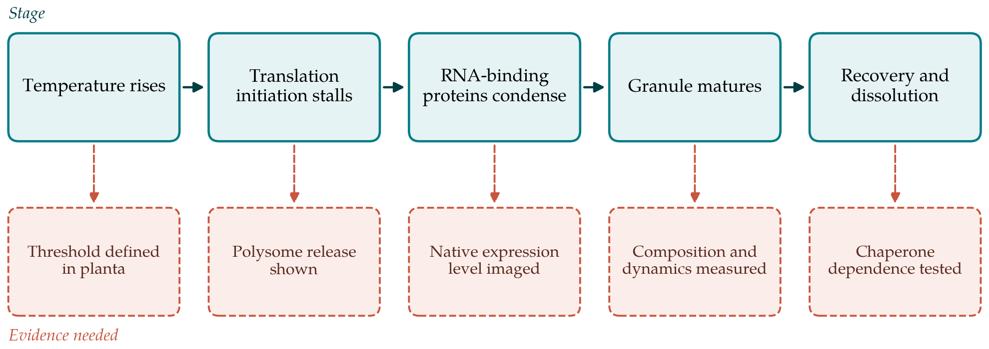
The temperature dependence of assembly has received less attention than its molecular parts. Live imaging in Arabidopsis indicates that granules appear once an absolute temperature is exceeded and not in proportion to the size of the shift (Hamada et al., 2018). A threshold of this kind is what a phase transition predicts, although a threshold in an upstream signalling step would produce the same behaviour.
6. How Strong Is the Case?
Methodological reviews of the wider field have warned that the usual trio of round foci, fluorescence recovery and sensitivity to 1,6-hexanediol does not demonstrate liquid-liquid phase separation (Alberti et al., 2019; McSwiggen et al., 2019). Foci can form by binding to a pre-existing scaffold. Recovery after photobleaching reports exchange, which occurs at any site with a finite residence time. Hexanediol disturbs membranes and enzymes as well as weak hydrophobic contacts. Droplets in a test tube show that a sequence is capable of demixing at the concentration and in the buffer chosen by the experimenter, which is often far from cellular conditions.
Plant studies are exposed to each of these objections and to some of their own. Transient expression from strong promoters in leaf epidermis or protoplasts is common and raises protein concentration by an unknown factor, which matters greatly for a process defined by a saturation threshold. Heat is usually applied as an abrupt shift to a temperature the plant would seldom meet in the field without a gradual approach. Granules are imaged in a few accessible cell types, and little is known about meristems, pollen or developing seed, where heat does the most agronomic damage.
None of this means that the phase separation interpretation is wrong. The physical argument is sound, the sequence features are present, and the yeast precedent shows that adaptive, heat-triggered demixing exists in nature. The fair summary is that for plants the mechanism is plausible and partly supported, while the functional claim, that the plant survives because the protein condenses, rests on a small number of experiments.
7. Gaps and Directions for Future Work
The most useful single advance would be wider use of separation-of-function variants: changes to a scaffold protein that move its transition temperature while leaving RNA binding intact, expressed from the native locus and scored for thermotolerance. A second need is quantitative imaging at endogenous expression levels, so that saturation concentrations inside cells can be compared with those measured in vitro. A third is attention to recovery. Dissolution after stress, its dependence on HSP101 and related chaperones, and the possibility that granules which persist too long become harmful have been studied far less than assembly.
Work in crops lags well behind Arabidopsis. Whether natural variation in the low-complexity regions of granule proteins contributes to differences in heat tolerance between cultivars is an open and tractable question. The parallel case of ELF3, in which the length of a polyglutamine tract within a prion-like domain tunes a response to warm temperature (Jung et al., 2020), suggests that such variation can carry adaptive weight, though ELF3 is a transcriptional regulator and not a stress granule component.
8. Conclusion
Four decades of work have established that heat-stressed plant cells sort part of their messenger RNA into membraneless granules built around RNA-binding proteins, that these granules are distinct from chaperone aggregates, and that several of their components are needed for full thermotolerance. The interpretation of the granules as phase-separated condensates fits what is known and has generated productive experiments. It has not yet been tested in plants with the rigour applied in yeast, and reports in this area would serve readers better by stating which of the available kinds of evidence they provide and which they do not.
Conflict of interest
To be declared by the author(s) before publication.
Funding
To be declared by the author(s) before publication.