1. Introduction
The claim that most gut bacteria cannot be cultured has not survived contact with serious attempts to culture them. Large-scale efforts using many media and long incubations recovered hundreds of species from human stool, including taxa previously known only from sequence (Lagier et al., 2016), and work built around spore selection and targeted media showed that a substantial majority of the abundant species in a stool sample can be brought into culture (Browne et al., 2016). The resulting collections now underpin a large part of functional gut microbiology (Forster et al., 2019; Zou et al., 2019).
What these efforts also demonstrated is that success depends on technique rather than luck. The organisms concerned are mostly obligate anaerobes of the Bacillota and Bacteroidota, many of them sensitive to oxygen at concentrations well below atmospheric, several dependent on fermentation products or on host-derived compounds that defined media lack. A stool sample handled in air for an hour, streaked on blood agar and placed in a jar will yield facultative organisms and a thin selection of the robust anaerobes, and will be reported as having low diversity.
This article describes a workflow for a laboratory with an anaerobic chamber or a well-run jar system that wishes to recover fastidious anaerobes from stool reliably. It is a procedure assembled from the published literature and from established anaerobic practice, presented step by step with the reason for each choice. It is proposed rather than validated: no isolation campaign is reported here, and the final section states what would need to be shown before the workflow could be recommended without qualification.
2. Background to the Technique
Modern anaerobic bacteriology descends from the roll tube method developed by Hungate for rumen organisms, in which medium is reduced, dispensed under oxygen-free gas and solidified as a film on the tube wall, so that the culture never meets air (Hungate, 1969). Later refinements made the approach usable in ordinary laboratories: the serum bottle modification replaced specialised glassware with crimp-sealed bottles (Miller and Wolin, 1974), and commentaries of the period set out the practical principles that still govern the work, chief among them that the reducing agent and the redox indicator matter more than the brand of gas mixture (Bryant, 1972).
Two ideas from the recent literature shape the workflow below. The first is that no single medium represents the community. Culturomics arrived at that conclusion by brute force, applying many media and incubation conditions to the same sample and identifying every colony that grew (Lagier et al., 2012; Lagier et al., 2018). The second is that the laboratory should expect to supply what the colon supplies. Strategies for recovering previously uncultivated bacteria converge on providing the missing factor, whether that is a specific substrate such as mucin, a reducing environment, or a diffusible compound produced by a neighbouring organism (Vartoukian et al., 2010).
Two organisms illustrate the range of difficulty. Faecalibacterium prausnitzii, among the most abundant species in the healthy colon, is markedly oxygen sensitive, was long considered difficult, and was reclassified only after its growth requirements were characterised in detail (Duncan et al., 2002); it is of continuing interest because of its association with intestinal health (Sokol et al., 2008), and it tolerates low oxygen tension better in the presence of an extracellular electron shuttle such as riboflavin, which is a hint about why it grows at the mucosal interface (Khan et al., 2012). Akkermansia muciniphila, by contrast, was isolated by giving it what it eats: a medium with mucin as the sole carbon and nitrogen source (Derrien et al., 2004). Both cases support the same conclusion. The organism is not uncultivable; the medium was wrong.
3. The Proposed Workflow
The workflow has six stages, shown in Figure 1 and summarised with their critical control points in Table 1. The sequence assumes a fresh stool sample, an anaerobic chamber with a nitrogen, carbon dioxide and hydrogen atmosphere, and access to a mass spectrometer or to Sanger sequencing. Where a chamber is unavailable, jars with palladium catalyst and a gas-generating sachet can substitute for incubation but not for colony picking, which is where most of the loss occurs in jar-based laboratories.
| Stage | Critical control point | Common failure | Record |
|---|---|---|---|
| Collection and transport | Anaerobic container, same-day processing | Oxygen exposure before plating | Time voided, time plated |
| Dilution | Pre-reduced buffer equilibrated in the chamber | Dilution in aerobic saline | Diluent lot, series used |
| Plating | Panel of media, not a single agar Lagier et al., 2016 | One rich medium only | Media, batch, redox indicator |
| Incubation | Long and short reads of the same plates | Plates discarded at 48 hours | Days to visible colony |
| Picking | Picking performed inside the chamber | Plates opened on the bench | Colony morphology, plate of origin |
| Purification | At least two restreaks before identification | Mixed cultures carried forward | Purity checks and Gram stain |
| Identification | Mass spectrometry with sequencing fallback Seng et al., 2009 | Database lacks the genus | Spectrum, score, gene sequence |
| Archiving | Duplicate cryovials with glycerol | Single vial, viability untested | Freezer position, revival check |
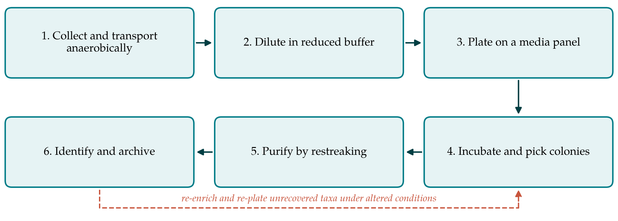
Stage one is collection and transport. Stool should be taken into an anaerobic transport container or into a tube pre-filled with reduced, pre-gassed medium, and processed the same day. The first hour after voiding is where the most oxygen-sensitive organisms are lost, and no later step recovers them. If the sample must be held, it should be held at ambient temperature under anaerobic conditions rather than refrigerated in air; if a portion is to be frozen for later culture, it should be mixed with glycerol to a final concentration of about ten per cent and frozen promptly, accepting that freezing selects against some non-sporulating taxa.
Stage two is dilution. A weighed portion of stool is homogenised in pre-reduced anaerobically sterilised buffer that has been equilibrated in the chamber overnight, and a decimal dilution series is prepared inside the chamber. Diluting in aerobic saline on the bench is the most frequent avoidable error in this work, and it is invisible in the result: the plates grow, and the laboratory concludes that the sample was of low diversity.
Stage three is plating on a panel. The panel should combine a rich non-selective base such as brain heart infusion or Wilkins-Chalgren supplemented with yeast extract, haemin and vitamin K, a blood-based medium, a medium with a complex host-derived substrate such as mucin, a bile-containing selective medium for Bacteroidota, and at least one defined medium with a single fermentable substrate. Short-chain fatty acids and rumen fluid or its substitutes remain useful additions for organisms that depend on cross-feeding. Plates should be poured in advance, reduced in the chamber for at least a day, and inspected for an oxidised indicator before use.
Stage four is incubation and reading. Plates are read early, at two to three days, and then held for two to three weeks, since slow-growing taxa are routinely discarded by laboratories working to a forty-eight hour cycle. Reading the same plates twice is deliberate: fast organisms are picked before they overgrow the plate, and slow ones are given time to appear.
Stage five is picking and purification. Colonies are picked inside the chamber, chosen for morphological variety rather than for abundance, since abundance on a plate reflects what the medium favours. Each isolate is restreaked at least twice on the medium that produced it, and purity is confirmed by colony morphology and a Gram stain before any identification is attempted. Isolates that fail to grow on restreak are candidates for a co-culture attempt alongside an isolate from the same plate, which is the simplest practical response to a dependence on a diffusible factor.
Stage six is identification and archiving. Matrix-assisted laser desorption ionisation time-of-flight mass spectrometry identifies most isolates within minutes and is the sensible first pass (Seng et al., 2009). Its weakness is its reference library, which is built from clinical organisms and is thin for strictly anaerobic commensals, so isolates without a confident match go to 16S rRNA gene sequencing, interpreted against the thresholds conventionally used to delimit species and genera (Yarza et al., 2014). Every confirmed isolate is frozen in duplicate in glycerol at minus eighty degrees Celsius or below, and one vial of each is revived once to confirm viability before the isolation campaign is considered complete.
4. Fitting the Workflow to a Laboratory
Three adjustments make the procedure usable under common constraints. Where no chamber is available, an anaerobic jar system with a catalyst and an indicator strip will support incubation, and a nitrogen- flushed glove bag can serve for picking; the bag is awkward but it keeps the plates out of air, which is the point. Where throughput rather than depth is wanted, the panel can be reduced to a rich medium, a mucin medium and a bile-selective medium, accepting a narrower recovery. Where the aim is a personal collection for gnotobiotic work, the arrayed-plate approach used to build such collections is a better fit than colony-by-colony isolation, because it preserves the relative composition of the donor community (Goodman et al., 2011).
Sequencing has a place in the workflow, but as an instrument rather than an alternative. A 16S profile of the starting sample, compared with the identifications obtained, shows which abundant taxa the panel is missing, and the missing taxa then direct the next round of media design. Used this way the comparison turns an isolation campaign into an iterative process rather than a single pass, which is the return loop shown in Figure 1.
5. Limitations and What Remains to Be Shown
The workflow has not been validated here, and three claims would have to be tested before it could be put forward as better than existing practice. The first is coverage: what proportion of the genera present in a stool sample by sequencing is recovered, measured against a defined benchmark rather than against an impression. The second is reproducibility: whether two operators following the written procedure on portions of the same sample recover comparable sets of organisms. The third is cost in effort: how many colonies must be picked and identified per novel isolate obtained, since that number determines whether a laboratory can sustain the work.
Two limits are intrinsic rather than remediable by validation. Stool represents the luminal community of the distal colon and under-represents mucosa-associated and small-intestinal organisms, so no stool workflow recovers the full gut community. And any medium selects. A panel widens the selection without removing it, and an organism that depends on a partner it never meets on an agar plate will not be recovered by any of the steps described here.
6. Conclusion
Recovering fastidious anaerobes from stool is a matter of disciplined technique rather than specialised insight. The decisive points are few: keep the sample out of air from the moment it is produced, dilute and pick inside a reduced atmosphere, use several media rather than one, read plates late as well as early, and confirm identity and viability before the isolate is counted. The workflow set out here gathers those points into a sequence that a laboratory can follow and record. Its coverage and reproducibility are open questions, and both can be settled with a sample, a benchmark and two operators.
Conflict of interest
To be declared by the author(s) before publication.
Funding
To be declared by the author(s) before publication.