1. Introduction
Periodontitis is a chronic inflammatory disease in which a dysbiotic subgingival biofilm provokes a host response that destroys the periodontal ligament and alveolar bone (Hajishengallis, 2015). Its association with diabetes mellitus has been recognised for decades. Löe (1993) called periodontal disease the sixth complication of diabetes, and later work has shown that the relationship runs in both directions: hyperglycaemia raises the risk and severity of periodontitis, and severe periodontitis is associated with poorer glycaemic control and with diabetic complications (Preshaw et al., 2012; Lalla and Papapanou, 2011).
The degree of glycaemic control matters more than the diagnosis of diabetes as such. People whose diabetes is well controlled have periodontal risk close to that of people without diabetes, whereas risk rises as glycated haemoglobin (HbA1c) rises (Mealey and Oates, 2006). The 2018 classification of periodontitis builds this into the grading of a case: in a person with diabetes, an HbA1c of 7.0% or above places the case in the grade with the highest expected rate of progression (Tonetti et al., 2018).
Diagnosis still rests on probing depth, clinical attachment level, bleeding on probing and radiographs (Papapanou et al., 2018). These are records of past destruction. They need a trained examiner and a dental chair, and they say little about whether tissue is breaking down at present. Saliva can be collected without discomfort by non-dental staff, and it contains host and microbial products that reach the mouth through gingival crevicular fluid (Kaufman and Lamster, 2002). A salivary test that identified active periodontal inflammation would be attractive in diabetes clinics, where patients are seen regularly and a dentist is seldom present.
This article reviews the published evidence on salivary biomarkers of periodontal inflammation in adults with poorly controlled type 2 diabetes. It asks which markers have been studied, how consistent the findings are, and what stands in the way of clinical use.
2. Approach to the Literature
This is a narrative review. It draws on consensus reports, earlier reviews and frequently cited primary studies on salivary and crevicular fluid markers in periodontitis and in diabetes, read alongside the methodological literature on biomarker evaluation. No formal search protocol was registered and no pooled estimates are offered. Studies of gingival crevicular fluid are included where they clarify mechanisms, since most salivary markers of periodontal origin enter the mouth by that route. The term poorly controlled is used as the primary studies use it, which is itself a source of inconsistency: authors have set the boundary at different HbA1c values, and some rely on fasting glucose alone.
3. Why Hyperglycaemia Should Alter the Salivary Profile
Several mechanisms link sustained hyperglycaemia to an exaggerated periodontal response. Advanced glycation end products accumulate in periodontal tissues and bind their receptor, RAGE, on endothelial cells and monocytes, which sustains the release of pro-inflammatory cytokines. In a diabetic mouse model, blocking RAGE reduced periodontitis-associated bone loss (Lalla et al., 2000). Neutrophil function is altered, apoptosis of matrix-producing cells increases, and the resolution of inflammation is delayed (Graves and Kayal, 2008). Salvi et al. (1997) described a hyper-responsive monocyte phenotype in people with diabetes, with raised crevicular fluid prostaglandin E2 and interleukin-1β for a given level of plaque.
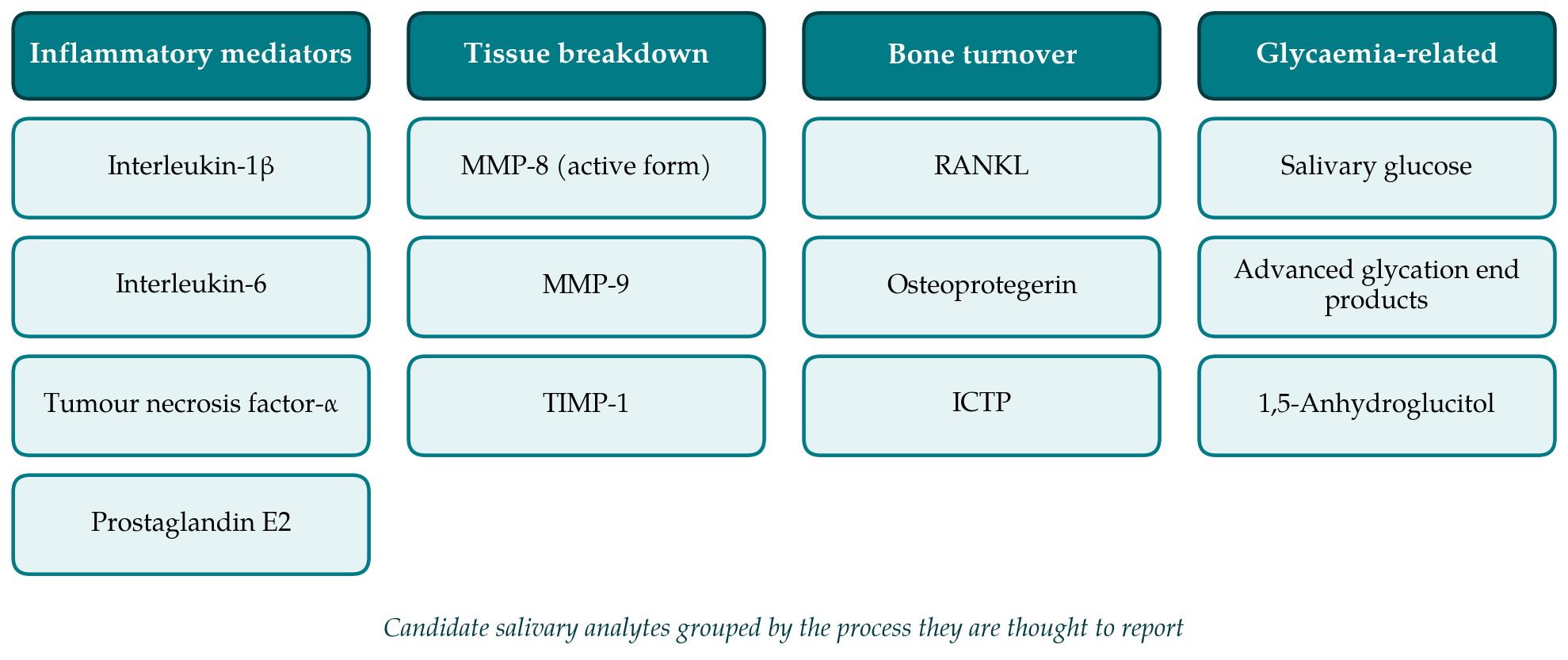
A review of pathogenic mechanisms prepared for a joint European and American workshop concluded that the evidence most strongly supports raised interleukin-1β, tumour necrosis factor-α and interleukin-6, a shifted ratio of RANKL to osteoprotegerin, and oxidative stress as mediators of the diabetic periodontal phenotype (Taylor et al., 2013). If these mediators are raised in the tissues, they should be raised in crevicular fluid and, after dilution, in whole saliva. That expectation is the rationale for the studies reviewed below. Figure 1 groups the candidate analytes by the biological process they are thought to reflect.
Diabetes also changes saliva in ways unrelated to the periodontium. Salivary flow is often reduced, and the composition of the secretion differs from that of people without diabetes. A lower flow concentrates solutes, so that a raised concentration of a marker may reflect less water and not more inflammation. Any study in this population has to deal with that problem explicitly.
4. Candidate Biomarkers
Table 1 summarises the principal groups of salivary markers and the state of the evidence for each. The sections that follow take them in turn.
| Group | Examples | Evidence in periodontitis | Position in diabetes |
|---|---|---|---|
| Cytokines | IL-1β, IL-6, TNF-α | IL-1β consistently raised; others variable (Miller et al., 2006) | Crevicular IL-1β rises with poorer control (Engebretson et al., 2004) |
| Prostanoids | Prostaglandin E 2 | Raised in crevicular fluid; few salivary studies | Raised in crevicular fluid in diabetes (Salvi et al., 1997) |
| Proteinases | MMP-8, MMP-9, TIMP-1 | Active MMP-8 well supported (Sorsa et al., 2016) | Raised with periodontitis; added effect of diabetes unclear |
| Bone turnover | RANKL, osteoprotegerin, ICTP | Associated with progression in combination panels | Altered ratio proposed; salivary data sparse |
| Metabolic | Glucose, AGEs, 1,5-anhydroglucitol | Not markers of periodontitis | Reflect metabolic state, not periodontal status |
| Microbial | Red-complex bacterial DNA | Add to host markers in prediction (Kinney et al., 2011) | Not shown to differ consistently |
4.1. Cytokines and prostanoids
Interleukin-1β is the most reproducible salivary cytokine in periodontitis. In an early cross-sectional comparison, Miller et al. (2006) reported higher salivary interleukin-1β and matrix metalloproteinase-8 in adults with periodontitis than in healthy controls, and later multi-analyte panels have repeatedly placed interleukin-1β among the analytes that best separate health from disease (Giannobile et al., 2009). In type 2 diabetes, Engebretson et al. (2004) found that crevicular fluid interleukin-1β was higher in patients with poorer glycaemic control after allowing for periodontal status, which fits the hyper-inflammatory model.
Salivary interleukin-6 and tumour necrosis factor-α behave less predictably. Both circulate at low concentrations, both are produced by adipose tissue, and both are raised systemically in obesity and insulin resistance, so a salivary rise in a person with type 2 diabetes cannot be attributed to the periodontium with confidence. Studies that measure salivary interleukin-6 in people with periodontitis with and without diabetes illustrate how difficult it is to separate the contributions of the two conditions. Prostaglandin E2 has a sound mechanistic basis but has been studied mainly in crevicular fluid.
4.2. Matrix metalloproteinases
Matrix metalloproteinase-8, the neutrophil collagenase, is the principal enzyme that degrades type I collagen in the inflamed periodontium (Sorsa et al., 2004). It is released in a latent form and activated in the tissues, and assays that detect the active form distinguish periodontitis from gingivitis better than assays for total enzyme. Chairside lateral-flow tests for active matrix metalloproteinase-8 in mouthrinse or saliva have been developed and evaluated in several populations (Sorsa et al., 2016). Among the candidates, this enzyme is the closest to a usable test.
What poor glycaemic control adds is less certain. Neutrophil priming in hyperglycaemia would be expected to raise the enzyme further, and some studies report such a gradient, but others find that periodontal status accounts for the difference and that diabetes contributes little once inflammation is taken into account. Differences in antibody specificity between assays, which do not all recognise the same molecular forms, make direct comparison of reported concentrations unsafe.
4.3. Markers of bone turnover
Alveolar bone loss is the defining lesion of periodontitis, so mediators of osteoclast formation are natural candidates. The balance between RANKL and its decoy receptor osteoprotegerin shifts towards resorption in periodontitis, and hyperglycaemia is thought to push it further (Taylor et al., 2013). The pyridinoline cross-linked carboxyterminal telopeptide of type I collagen (ICTP), a fragment released when bone collagen is degraded, has been examined as a marker of active loss. In a longitudinal study, Kinney et al. (2011) showed that combinations of salivary host markers and periodontal pathogens predicted disease progression better than any single measure. Salivary data on these markers in people with poorly controlled diabetes are few, and the concentrations involved are close to the detection limits of common assays.
4.4. Analytes related to glycaemia
Salivary glucose, advanced glycation end products and 1,5-anhydroglucitol have been proposed as non-invasive indicators of metabolic control. They are relevant here for a different reason. They are not markers of periodontal inflammation, but they could in principle be measured in the same sample to show the metabolic context in which a periodontal marker is being read. Their correlation with blood measures is imperfect and varies between studies, and none is accepted as a substitute for HbA1c. Their inclusion in a panel should be regarded as exploratory.
5. Sources of Inconsistency
The literature is hard to synthesise because studies differ at almost every step. Case definitions of periodontitis have changed over time, and many reports predate the current staging and grading system (Tonetti et al., 2018). Thresholds for poor control vary. Comparison groups are sometimes people without diabetes and sometimes people with well-controlled diabetes, which answer different questions. Most designs are cross-sectional, so they show association at one moment and cannot show that a marker tracks disease activity or responds to treatment.
Collection and handling add further variation. Unstimulated whole saliva collected by passive drooling is the usual reference method, but stimulated saliva, swabs and mouthrinses are also used, and each yields different concentrations (Navazesh, 1993). Time of day, recent eating, tooth brushing and bleeding into the sample all affect results. Proteins degrade quickly in saliva unless samples are chilled and inhibitors added. Few reports state whether concentrations were corrected for flow rate or total protein, a correction that matters particularly when flow is reduced by diabetes or by medication.
Confounding is the third difficulty. Smoking suppresses gingival bleeding and alters neutrophil function. Obesity raises systemic cytokines. The number of remaining teeth determines how much periodontal tissue is available to contribute to saliva, and people with long-standing poorly controlled diabetes tend to have fewer teeth. Statins, metformin and anti-inflammatory drugs may each influence the markers. Studies that match or adjust for only some of these factors can reach opposite conclusions from similar data.
6. From Association to a Usable Test
A biomarker passes through recognised phases before it is fit for clinical use, from exploratory comparison of cases and controls, through assay validation, to prospective evaluation in the intended population and finally to evidence that using the test improves outcomes (Pepe et al., 2001). Most salivary work in diabetes sits in the first phase. The reporting standard for diagnostic accuracy studies asks for a prespecified threshold, a clearly described reference standard, consecutive or random recruitment and a full account of indeterminate results (Bossuyt et al., 2015). Few salivary studies meet these requirements, and thresholds are usually chosen after the data are seen, which inflates apparent accuracy.
The intended use also needs to be stated. A test meant to prompt dental referral from a diabetes clinic needs high sensitivity for moderate and severe periodontitis and can tolerate false positives. A test meant to monitor response to periodontal treatment needs to change reliably within an individual. These are different properties, and a marker may have one without the other. Reviews of salivary diagnostics have stressed that panels combining host and microbial markers are more promising than single analytes, and that validation in independent cohorts is the step most often missing (Giannobile et al., 2009; Dawes and Wong, 2019).
There is a clinical reason to pursue the question. A Cochrane review found that periodontal treatment was followed by a modest short-term reduction in HbA1c, while rating the quality of that evidence as low (Simpson et al., 2015). Consensus guidance now advises that people with diabetes be told of their periodontal risk and referred for periodontal assessment (Chapple and Genco, 2013; Sanz et al., 2018). A validated salivary test could make that advice easier to follow in settings without dental staff. It would complement the periodontal examination and could not replace it.
7. Gaps and Future Work
Four gaps stand out. Longitudinal studies are needed in which saliva is sampled repeatedly from adults with type 2 diabetes across a range of HbA1c values, with periodontal status recorded under the current classification. Collection protocols should be standardised and flow rate reported. Assays for the same analyte should be compared against one another on shared samples so that concentrations from different laboratories can be related. Finally, studies should be designed around a stated clinical use and analysed against a threshold fixed in advance.
Two questions of interpretation also deserve direct study. One is whether a salivary marker adds information beyond what age, smoking, HbA1c and a brief self-report questionnaire already provide. The other is whether marker levels fall after periodontal treatment in people whose glycaemic control does not change, which would show that the marker follows the periodontal lesion and not the metabolic state.
8. Conclusion
Salivary interleukin-1β and active matrix metalloproteinase-8 are consistently associated with periodontal inflammation, and the biology of hyperglycaemia gives good reason to expect a stronger signal when type 2 diabetes is poorly controlled. The published evidence does not yet establish that diabetes changes the diagnostic performance of these markers, nor that any salivary test improves the care of people with diabetes. The obstacles are methodological more than biological: inconsistent definitions, unstandardised collection, uncorrected confounding and a shortage of prospective validation. Until those are addressed, salivary biomarkers remain research tools in this population, and clinical periodontal examination remains the basis of diagnosis.
Conflict of interest
To be declared by the author(s) before publication.
Funding
To be declared by the author(s) before publication.