1. Introduction
Knowing which ewes and does are pregnant, and ideally how many young each carries, lets a keeper feed according to need, rebreed or sell barren animals and prepare for lambing or kidding. Before imaging was available the options were non-return to oestrus, abdominal palpation late in gestation and a series of laboratory and mechanical methods of limited accuracy or convenience (Ishwar, 1995). Real-time B-mode ultrasound replaced them. Early Scottish and Australian work showed that an experienced operator could diagnose pregnancy and count fetuses in large numbers of sheep quickly and with few errors (White et al., 1984; Fowler and Wilkins, 1984), and commercial scanning services grew up in countries with large flocks.
Those services depend on a skilled contractor, a handling crate and flocks mated over a known period. Smallholder and pastoral flocks have none of these. Males often run with females all year, mating dates are unknown, and animals are caught by hand. What has changed is the equipment: hand-held probes that display on a mobile device are now inexpensive enough for a district veterinary office. An instrument is not a method, however. This article proposes a written protocol for using such scanners to diagnose pregnancy in sheep and goats in the field. It is offered as a design for others to test, and no field results are reported.
2. Background
The protocol draws on what is known about the two scanning approaches, the timing of ultrasonic signs of pregnancy, and the sources of error.
2.1. Transrectal and transabdominal scanning
A linear probe of about 5 to 7.5 MHz introduced into the rectum lies close to the uterus and detects the fluid-filled embryonic vesicle, and somewhat later the embryo and its heartbeat, earlier than any external method (Buckrell et al., 1986; Schrick and Inskeep, 1993). Studies in ewes and does place reliable transrectal diagnosis from roughly the fourth week after mating (García et al., 1993; Martinez et al., 1998). Accuracy at this stage is sensitive to preparation. Karen et al. (2004) found that fasting ewes before the examination and lifting the abdomen improved transrectal results, an observation that shows how much the technique asks of the handler. As gestation advances the uterus descends over the pelvic brim and out of reach of a rectal probe.
Transabdominal scanning uses a sector or convex probe of about 3.5 to 5 MHz on the hairless skin of the inguinal region, directed towards the pelvic inlet. It is quick, causes little disturbance and is reliable from about six weeks of gestation, when uterine fluid, placentomes and fetal parts are readily seen (Buckrell, 1988; Haibel, 1990). Fetal counting is most dependable in the middle third of pregnancy; later the fetuses are too large and too far forward to be separated on one image (White et al., 1984). Comparative accounts of the methods are given by Karen et al. (2001) and, for goats, by Medan et al. (2004).
2.2. Where errors arise
False negative results come mainly from scanning too early, from a full rumen or intestinal gas between probe and uterus, and from failing to sweep the whole field. False positives arise when the urinary bladder or a fluid-filled uterus with no fetus, as in hydrometra of the doe, is taken for pregnancy, and from embryonic loss after a correct early diagnosis (Haibel, 1990). Operator experience affected accuracy in the earliest evaluations (Fowler and Wilkins, 1984) and remains the least controlled variable when scanners are handed to new users.
2.3. Differences between sheep and goats
The two species are scanned in the same way and show the same signs, with gestation lasting about five months in each. Two differences bear on a field protocol. Does are prone to hydrometra, or pseudopregnancy, in which the uterus fills with clear fluid in the absence of a conceptus, so that fluid without a fetus or placentomes carries a different meaning in a goat than in a ewe (Haibel, 1990). And woolled sheep need a bare or parted patch of skin for probe contact, whereas most goats and hair sheep do not. Litter size also differs between breeds and affects how much the keeper gains from a fetal count.
3. The Proposed Protocol
The protocol makes three design choices. It uses the transabdominal approach only, because it needs no rectal entry, suits both species and all body sizes, and can be taught quickly. It accepts a later earliest date of diagnosis in exchange. And it allows the operator to answer "uncertain", with a fixed rule for what follows. Table 1 sets out the steps.
| Step | Action | Record |
|---|---|---|
| 1. History | Ask when males had access and when the female last gave birth | Earliest and latest possible mating date |
| 2. Preparation | Withhold feed overnight where practical; restrain standing | Fasted or not; body condition score |
| 3. Probe contact | Apply gel to bare skin of right inguinal region, in front of the udder | Probe type and frequency |
| 4. Landmark | Find the bladder, then angle forward and medially | Bladder seen: yes or no |
| 5. Sweep | Fan slowly through the caudal abdomen; repeat from left side | Sides scanned |
| 6. Classify | Apply the image criteria for the three results | Pregnant, not pregnant or uncertain |
| 7. Follow up | Rescan uncertain and early negative animals after two weeks | Date due and final result |
3.1. Before scanning
The operator first establishes the window of possible mating. Where a male was introduced on a known date, the earliest scan is planned for about six weeks after that date and the latest possible conception is noted. Where males are always present, the date of the last birth or abortion sets the outer limit. Animals are held off feed overnight if the keeper agrees, with water allowed, since an empty rumen improves the view. Body condition is scored on the standard five-point scale by palpation of the lumbar region (Russel et al., 1969), partly because the information is useful to the keeper and partly because very fat and very thin animals image differently.
3.2. Scanning technique
The animal stands, held by one person against a wall or between the knees, with no need for a crate. In hair sheep and most goats the inguinal skin is bare; in woolled sheep the area just in front of the udder on the right side normally is. Coupling gel is applied generously, or vegetable oil where gel is unavailable. A convex probe set to about 3.5 MHz, with depth sufficient to include the far wall of the abdomen, is placed in the right inguinal fold and pointed towards the opposite hip. The operator finds the urinary bladder as a landmark, a smooth, thin-walled, echo-free structure, and then angles the beam forward and slightly towards the midline, fanning slowly from side to side. The sweep is repeated from the left if nothing is seen. A scan is not complete, and may not be called negative, until the bladder has been identified and the area cranial and dorsal to it examined (Kähn, 1994).
3.3. Image criteria and the three results
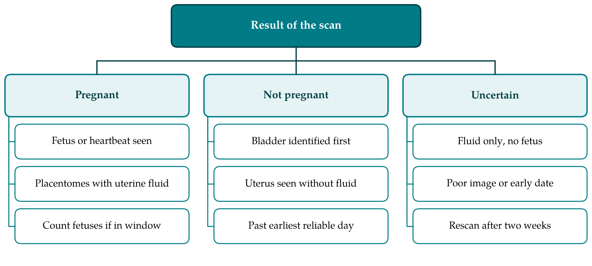
Figure 1 shows the decision. An animal is recorded as pregnant only when a fetus, a fetal heartbeat, or placentomes together with echo-free uterine fluid are seen. Placentomes appear as cup-shaped or ring-shaped echogenic bodies on the uterine wall and are specific to pregnancy. Echo-free fluid alone is not sufficient. An animal is recorded as not pregnant when the bladder and the non-gravid uterus or the region where it lies have been seen, no fluid or fetal structure is present, and at least six weeks have passed since the last possible mating. Every other case is uncertain. Fetal number is recorded only when the scan falls in the middle third of gestation by fetal size and the operator has been assessed for counting.
3.4. Rescanning and recording
Animals classed as uncertain, and those found negative when mating may have occurred less than six weeks earlier, are marked and scanned again after two weeks. A second uncertain result with fluid and no fetus in a doe should prompt veterinary examination for hydrometra. Each animal gets one line in a paper or phone record: identity, species, date, mating window, result, fetal number if counted, and the operator. Stored still images of each positive finding are worth keeping where the device allows, since they let a supervisor review the work.
4. Training and Quality Assurance
The protocol assumes an operator who has been taught on animals of known status. A reasonable scheme is supervised scanning of pregnant and non-pregnant animals at several stages until the trainee's classifications match those of the instructor, followed by periodic checks. The teaching should cover the false images as carefully as the true ones: the bladder, loops of intestine containing fluid, the full rumen on the left side, and the fluid-filled uterus without a fetus. Agreement between two operators scanning the same animals independently can be expressed with the kappa statistic and interpreted on the conventional scale of Landis and Koch (1977). The final test of any operator is the record of births, and services using the protocol should collect lambing and kidding outcomes from a sample of flocks each season and compare them with the scan sheets.
5. Fitting the Protocol to Field Conditions
Field use raises practical matters that clinic-based descriptions pass over. Screens are hard to read in sunlight, so scanning is done in shade or under a hood. Batteries, probe cables and phones fail in dust and heat, and a spare power source belongs in the kit. Probes are wiped and disinfected between flocks to avoid carrying infection on equipment. Keepers should be told beforehand what the three results mean, and in particular that an uncertain result is a request to return and not a failure of the test. Where a service is offered for a fee, the cost of the return visit should be included in the first.
The use made of the result should be agreed with the keeper before the visit. In a flock that lambs once a year the main gains are separating barren females for sale or remating and directing scarce supplementary feed to those carrying young, above all those carrying more than one. In a continuously mated village flock the gain is smaller for each animal but still real: a female sold for slaughter in the belief that she is barren may be pregnant, and a scan before sale prevents that loss. Handling should be calm and brief, with heavily pregnant animals never cast or lifted, and the keeper should restrain the animals personally wherever possible, since familiar handling reduces struggling and improves the image.
6. Limitations and What Remains to Be Shown
The protocol has been assembled from published work and has not been evaluated. Its accuracy under field conditions is unknown. A proper evaluation would scan a defined group of females once, according to the protocol, by operators unaware of mating records, and would take birth, abortion or slaughter findings as the reference standard. The design and reporting of such a study should follow the STARD statement for diagnostic accuracy research (Bossuyt et al., 2015), with sensitivity and specificity given separately for each stage of gestation and each operator, and with the uncertain results reported and not discarded.
Several limits are already clear. Pregnancies younger than about six weeks will be missed or deferred, which matters in systems where early rebreeding is the aim. Losses after a positive scan will appear as false positives against birth records. The gestational age cannot be estimated without fetal measurements and breed-specific growth curves, of the kind published for some dairy breeds (González de Bulnes et al., 1998), and these are not part of the protocol. Counting fetuses demands more skill than detecting pregnancy, and image quality from low-cost probes has not been compared formally with that of the cart-based machines used in the founding studies.
7. Conclusion
Portable scanners make pregnancy diagnosis possible in flocks that commercial scanning never reached. Their usefulness there depends on a method simple enough to teach and strict enough to keep errors visible. The protocol proposed in this article uses one transabdominal technique, fixed image criteria, an explicit uncertain category and a rescan rule, and it ties the operator's record to later births. A field accuracy study against birth outcomes is the necessary next step before it is recommended for routine use.
Conflict of interest
To be declared by the author(s) before publication.
Funding
To be declared by the author(s) before publication.