1. Introduction
People who have had a traumatic brain injury sleep badly. A meta-analysis of the prevalence studies estimated that about half report some form of sleep disturbance, with insomnia, hypersomnia and sleep apnoea all more frequent than in the general population (Mathias and Alvaro, 2012). The problems appear after mild as well as severe injury, they can last for years, and they are associated with slower recovery, worse mood and poorer cognition (Ouellet et al., 2015). For a long time they were treated as a consequence of injury that deserved management in its own right and little more.
A second observation has given them greater weight. Repeated head injury is associated with a distinctive tauopathy, chronic traumatic encephalopathy, in which the earliest deposits lie around small vessels at the depths of the cortical sulci (McKee et al., 2013). The perivascular position of those deposits invites a mechanical explanation, and the glymphatic model, proposed in 2012, supplies one. If the brain clears protein waste along perivascular routes, if that clearance depends on sleep, and if injury damages both, then disturbed sleep after injury is a candidate mechanism of later disease and not only a symptom. This review asks how much of that chain the evidence supports.
2. Approach of This Review
The article is a narrative review of published work. It takes the argument in stages: the glymphatic model and the objections to it, the nature of sleep disturbance after injury, the experimental and human evidence that connects them, and the implications for treatment and research. Animal and human findings are kept apart throughout, because most of the mechanistic evidence comes from anaesthetised rodents and most of the clinical evidence is indirect. Reviews by Rasmussen et al. (2018) on the glymphatic pathway in neurological disease and by Sandsmark et al. (2017) on sleep after injury cover the neighbouring ground in more detail.
3. The Glymphatic Model and Its Critics
The model has three parts, each resting on different evidence and each open to a different objection.
3.1. Perivascular exchange and aquaporin-4
Iliff et al. (2012) injected fluorescent tracers into the cerebrospinal fluid of mice and watched them enter the brain along the spaces surrounding penetrating arteries, spread into the interstitium and leave along veins. Mice lacking aquaporin-4, the water channel concentrated in astrocytic endfeet facing the vessels, showed much slower tracer influx and slower clearance of injected amyloid-β. The authors named the pathway glymphatic for its dependence on glia and its lymphatic-like function. The later description of lymphatic vessels in the dura gave the pathway a plausible outflow (Louveau et al., 2015).
3.2. Dependence on sleep
Xie et al. (2013) reported that tracer influx and amyloid-β clearance in mice were markedly greater during natural sleep and anaesthesia than in wakefulness, and that the interstitial space expanded in sleep. This fitted an earlier finding that interstitial amyloid-β rises in wakefulness and falls in sleep (Kang et al., 2009). In people, a night without sleep increases amyloid-β signal on positron emission tomography (Shokri-Kojori et al., 2018) and raises tau in cerebrospinal fluid (Holth et al., 2019). Recordings in sleeping volunteers show that electrical slow waves are followed by oscillations in blood volume and then by pulses of cerebrospinal fluid flow (Fultz et al., 2019), which offers a physical means by which deep sleep could promote fluid movement.
3.3. Points of dispute
None of this is settled. Hladky and Barrand (2014) argued on physical grounds that bulk flow through the narrow spaces of the neuropil is implausible and that diffusion accounts for solute movement within the parenchyma. Smith et al. (2017) failed to reproduce the dependence on aquaporin-4, although a subsequent report combining data from several laboratories did find it (Mestre et al., 2018). The sleep finding has also been challenged: a study using a different measurement approach concluded that clearance in mice was lower, not higher, during sleep and anaesthesia (Miao et al., 2024). Sleep loss might in addition raise amyloid-β and tau by increasing their release from active neurons, with no change in clearance at all. A fair summary is that perivascular tracer transport is well documented, its dependence on aquaporin-4 is probable, and the size and direction of the sleep effect are under active debate.
4. Sleep After Traumatic Brain Injury
Sleep disturbance after injury is not one condition. In the first weeks, increased sleep need and daytime sleepiness predominate. Later, insomnia becomes more prominent, along with circadian rhythm disorders and sleep-disordered breathing (Ouellet et al., 2015). Polysomnographic studies have reported reduced sleep efficiency and altered sleep architecture, though findings on slow-wave sleep are inconsistent across studies and between patients and animal models (Sandsmark et al., 2017). Pain, medication, mood disorder and the hospital environment all contribute, which makes it hard to isolate the effect of the injury itself.
Why injury disturbs sleep is only partly understood. Diffuse axonal injury affects the brainstem and hypothalamic nuclei that control arousal, and a post-mortem study found fewer hypocretin neurons in the hypothalamus of people who had died after severe head injury (Baumann et al., 2009). The observation is of interest here because the same peptide, also called orexin, influences interstitial amyloid-β in mice (Kang et al., 2009). Damage to one small group of cells could in principle affect wakefulness and protein dynamics together.
The inconsistency matters for the present argument. The glymphatic hypothesis is specific about which aspect of sleep counts: slow-wave activity, with its low noradrenergic tone and large slow oscillations. A patient who sleeps for more hours with less slow-wave activity, and a patient who sleeps for fewer hours with normal architecture, would be predicted to differ in clearance, yet both are recorded as having disturbed sleep in most clinical studies. Questionnaire measures, which dominate the literature, cannot distinguish them.
5. Evidence Linking Injury, Sleep and Clearance
The evidence divides by species. In rodents, perivascular transport can be measured directly, at the price of anaesthesia and invasive tracer delivery. In people it has to be inferred.
5.1. Experimental models
In a mouse model of closed-skull injury, Ren et al. (2013) found reactive astrogliosis with loss of the normal perivascular polarisation of aquaporin-4, which became spread across the astrocyte surface. Iliff et al. (2014) then showed that perivascular tracer influx and the clearance of interstitial solutes were reduced after injury and remained so for at least a month. In mice lacking aquaporin-4, injury produced more phosphorylated tau, more neuroinflammation and worse cognitive performance than in wild-type animals. These experiments are the strongest support for the idea that injury impairs clearance and that impaired clearance promotes tau pathology.
A related study bears on diagnosis. Plog et al. (2015) reported that the appearance in blood of injury markers such as S100β, glial fibrillary acidic protein and neuron-specific enolase depended on glymphatic transport, and that sleep deprivation, among other manipulations that suppress the pathway, lowered their serum levels after injury. If the same holds in people, a patient kept awake in an emergency department might have misleadingly low biomarker values. That possibility has not been tested clinically.
5.2. Human studies
Table 1 lists the methods available for assessing perivascular function in people. The most direct is magnetic resonance imaging after intrathecal injection of a gadolinium-based contrast agent, which showed delayed brain-wide enhancement and clearance in idiopathic normal pressure hydrocephalus (Ringstad et al., 2017). Using the same technique, Eide et al. (2021) found that one night of total sleep deprivation impaired clearance of the tracer from the brain. The method requires lumbar puncture and off-label use of contrast, and it has not been applied systematically after head injury.
| Method | What is measured | Main limitation |
|---|---|---|
| Intrathecal contrast MRI (Ringstad et al., 2017) | Entry and clearance of tracer in brain tissue over hours to days | Invasive; off-label contrast use; repeated scanning |
| Diffusion index along perivascular spaces (Taoka et al., 2017) | Water diffusivity in the direction of medullary veins | Indirect; influenced by white matter microstructure |
| Visible perivascular spaces on structural MRI | Number and volume of fluid-filled spaces around vessels | Structural marker; meaning for flow uncertain |
| Amyloid or tau PET (Shokri-Kojori et al., 2018) | Regional burden of deposited protein | Reflects production and clearance together |
| Cerebrospinal fluid sampling (Holth et al., 2019) | Concentrations of amyloid-β and tau over time | Invasive; cannot separate release from removal |
| Fast functional MRI of fluid inflow (Fultz et al., 2019) | Pulsatile cerebrospinal fluid movement at the fourth ventricle | Measures large-scale flow, not tissue exchange |
Non-invasive markers have been used more widely. The diffusion index introduced by Taoka et al. (2017) estimates water movement along the perivascular spaces of deep white matter, and enlarged perivascular spaces visible on structural imaging have been taken as a sign of stagnant fluid. In a study of military veterans, Opel et al. (2019) reported associations between a history of traumatic brain injury, the burden of enlarged perivascular spaces and poor sleep. Such findings are consistent with the hypothesis. They are also cross-sectional, and both markers are indirect: the diffusion index is sensitive to axonal injury, which head trauma produces independently of any change in fluid transport.
6. A Reciprocal Relationship
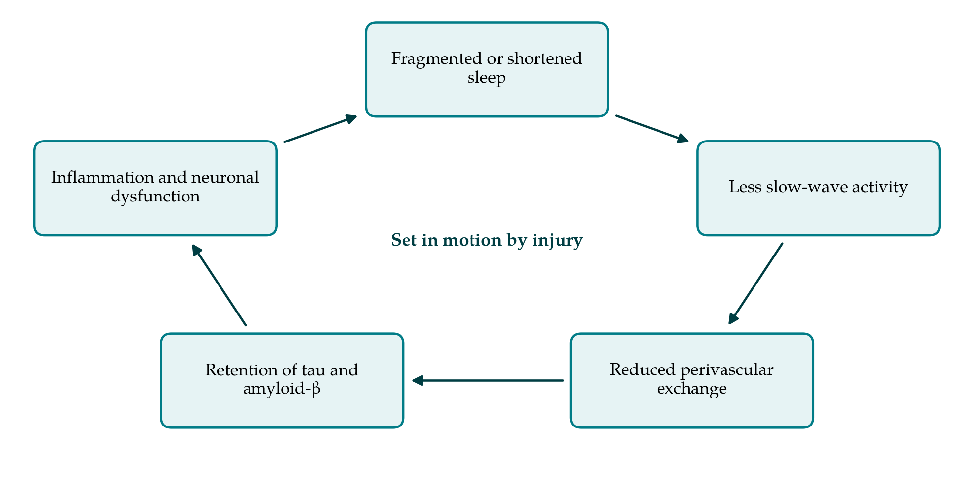
Several authors have drawn these strands into a self-reinforcing loop (Piantino et al., 2019), shown in Figure 1. Injury disturbs sleep and, separately, disrupts astrocytic water channels. Poorer sleep and damaged perivascular pathways both reduce the removal of solutes. Retained tau and amyloid-β, with the inflammatory response they provoke, impair neuronal function, including that of the brainstem and hypothalamic systems that regulate sleep. Worse sleep then feeds back on clearance.
The diagram leaves out influences that act on several steps at once. Age, vascular disease and sleep apnoea are each associated with poorer sleep, and each could plausibly alter fluid movement through effects on arterial pulsation or on astrocytes. All three are common among older adults admitted after falls, a group that makes up a growing share of head injury. A study that finds worse sleep and worse clearance markers in injured than in uninjured people has to show that these shared influences do not account for the association. Few of the available studies were designed to do so, and the groups in whom imaging markers have been examined after injury are small and mostly studied at a single time point.
The loop is coherent, and each arrow has some support. It should still be read as a hypothesis. The arrows from injury to reduced exchange and from reduced exchange to tau pathology rest on mouse experiments. The arrow from sleep loss to protein retention is supported in people, with the caveat about production noted above. The arrow from retained protein back to disturbed sleep is the least documented in the setting of injury. No longitudinal human study has yet followed sleep, a clearance measure and protein burden together after trauma.
7. Therapeutic Implications and Their Limits
If the loop is real, treating sleep after injury would protect the brain as well as relieve a symptom. That prospect has encouraged interest in sleep hygiene, cognitive behavioural therapy for insomnia, treatment of sleep apnoea and the timing of sedative drugs in this population. All of these have their own justification, since better sleep improves daytime function whatever happens to clearance (Ouellet et al., 2015). What cannot yet be claimed is that any of them alters glymphatic function or lowers the risk of later neurodegeneration. No trial has tested that outcome, and the choice of hypnotic drug on glymphatic grounds would be premature, given that the effects of different sedatives and anaesthetics on fluid transport are themselves disputed.
There is also a question of when to act. The mouse data suggest that perivascular function is depressed for weeks after a single injury. In patients this is the period in which sleep is most irregular and in which hospital routines, pain and noise interrupt it most often. If clearance matters, the early weeks would be the time to protect sleep, by reducing disturbance at night on the ward and treating pain adequately. Those measures are inexpensive and are justified on ordinary grounds of care. They deserve mention because they do not depend on the hypothesis being right.
8. Gaps and Priorities for Research
Three kinds of study would move the field. The first is measurement: agreement on which human marker of perivascular function to use, with validation of the non-invasive indices against intrathecal tracer studies in the same individuals. The second is longitudinal observation after injury that pairs objective sleep recording, including slow-wave activity, with repeated imaging and fluid biomarkers, so that the order of events can be established. The third is intervention: trials of sleep treatment after injury that include a clearance marker among their outcomes.
Work in animals has gaps of its own. Most experiments use young male mice under anaesthesia, whereas the clinical population is of both sexes and all ages, and anaesthetic agents differ in their effect on tracer movement. Whether repeated mild injury, the pattern linked to chronic traumatic encephalopathy (McKee et al., 2013), produces cumulative impairment of clearance is a question the existing models are suited to answer and have only begun to address.
9. Conclusion
The proposal that sleep disruption after traumatic brain injury impairs the clearance of waste proteins, and so contributes to later disease, draws on real findings: perivascular solute transport exists, injury damages the astrocytic machinery thought to support it in mice, and sleep loss raises amyloid-β and tau in people. The chain has not been demonstrated end to end in injured patients, and its central physiological claims are still argued over. Sleep problems after injury deserve treatment for the distress and disability they cause. Whether treating them also protects the brain is a question for studies that have yet to be done.
Conflict of interest
To be declared by the author(s) before publication.
Funding
To be declared by the author(s) before publication.