1. Introduction
Iron deficiency is the most common nutritional deficiency in pregnancy and the leading cause of anaemia in pregnant women worldwide. Antenatal programmes in most low- and middle-income countries respond by measuring haemoglobin and prescribing iron to those below a threshold, or to everyone (WHO, 2016). Haemoglobin, however, falls late. Iron stores can be exhausted while the haemoglobin concentration is still normal, and anaemia has causes other than iron lack, among them haemoglobinopathies, malaria, hookworm and chronic inflammation. Serum ferritin, which reflects storage iron, is the test recommended for identifying deficiency itself (WHO, 2020), but it requires a laboratory immunoassay that many antenatal clinics cannot reach.
Lateral flow immunoassays for ferritin have been developed to fill this gap. Like a pregnancy test, they need a drop of blood, a strip and some minutes of waiting, and their material cost is low. Some give a visual line for a yes or no answer at a chosen cut-off; others are read by a small optical reader or a telephone camera to produce a number. Before any such device is adopted in antenatal care it must be shown to agree with the laboratory method in the hands of the staff, on the specimens and under the conditions that will actually obtain.
This article proposes a protocol for that validation. It is written for a district health team, a non-governmental programme or a university department with access to a reference laboratory but without the resources of a diagnostics company. The protocol is assembled from published standards for method comparison and diagnostic accuracy research. It has not been carried out here, no device has been tested, and no performance data are reported. The aim is to set out what should be done, in what order, and why.
2. Background
Two bodies of knowledge shape the design: what ferritin does and does not reveal about iron status in pregnancy, and how lateral flow tests work and fail.
2.1. Ferritin as a measure of iron status in pregnancy
Ferritin is the principal iron storage protein, and the small amount that circulates in plasma is proportional to body stores in healthy people (Daru et al., 2017). The World Health Organization defines iron deficiency in apparently healthy adults, and in women in the first trimester of pregnancy, by a ferritin concentration below 15 μg/L (WHO, 2020). Some national guidelines use a higher cut-off in pregnancy: the British guideline treats a ferritin below 30 μg/L as indicating deficiency (Pavord et al., 2020). The choice of threshold matters for validation, because a point-of-care test should be most accurate near the value at which a decision changes.
Two features of ferritin complicate its use. It is an acute-phase protein, so infection or inflammation raises it and can mask deficiency; correction approaches based on C-reactive protein and α-1-acid glycoprotein have been proposed and compared (Thurnham et al., 2010; Namaste et al., 2017). And plasma volume expands as pregnancy advances, so that ferritin falls through the second and third trimesters for reasons that are partly dilutional. A validation study in antenatal clinics has to record gestational age and, where it can, a marker of inflammation.
2.2. How lateral flow ferritin tests work
A lateral flow strip carries the sample by capillary action along a membrane. Ferritin in the sample binds a labelled antibody, usually conjugated to colloidal gold or coloured latex, and the complex is captured at a test line by a second antibody; a control line confirms that the strip has run (Posthuma-Trumpie et al., 2009; Koczula and Gallotta, 2016). Signal at the test line increases with ferritin concentration over a limited range. Known sources of error include variable sample volume, the effect of haematocrit when whole blood is applied, degradation of antibodies in heat and humidity, reading at the wrong time, and subjective interpretation of faint lines. The criteria by which point-of-care tests for low-resource settings are judged, first summarised as ASSURED (Peeling et al., 2006) and later extended to REASSURED (Land et al., 2019), ask for affordability, ease of use and robustness as well as accuracy, and a validation should address each.
3. The Proposed Validation Protocol
The protocol has seven steps, listed in Table 1 with the output expected from each. Figure 1 shows their order, including the return to the manufacturer or to an earlier step if a stage fails. Steps 1 and 2 are completed before any pregnant woman is approached.
| Step | What is done | Output |
|---|---|---|
| 1. Define intended use | State the user, specimen, cut-off and the decision the result informs | Intended-use statement; acceptance criteria |
| 2. Laboratory verification | Check precision, lot variation, interference and stability on stored samples | Verification report; decision to proceed |
| 3. Clinic study design | Choose sites, consecutive enrolment and eligibility rules | Study protocol; ethics committee approval |
| 4. Reference testing | Collect a paired venous sample and run the laboratory immunoassay blind | Paired data set with quality control records |
| 5. Sample size | Calculate numbers from expected prevalence and the precision wanted | Justified enrolment target |
| 6. Analysis | Estimate sensitivity, specificity and agreement with confidence intervals | Accuracy tables; difference plot |
| 7. Usability and robustness | Examine reader agreement, invalid strips and storage in clinic conditions | Usability report; training needs |
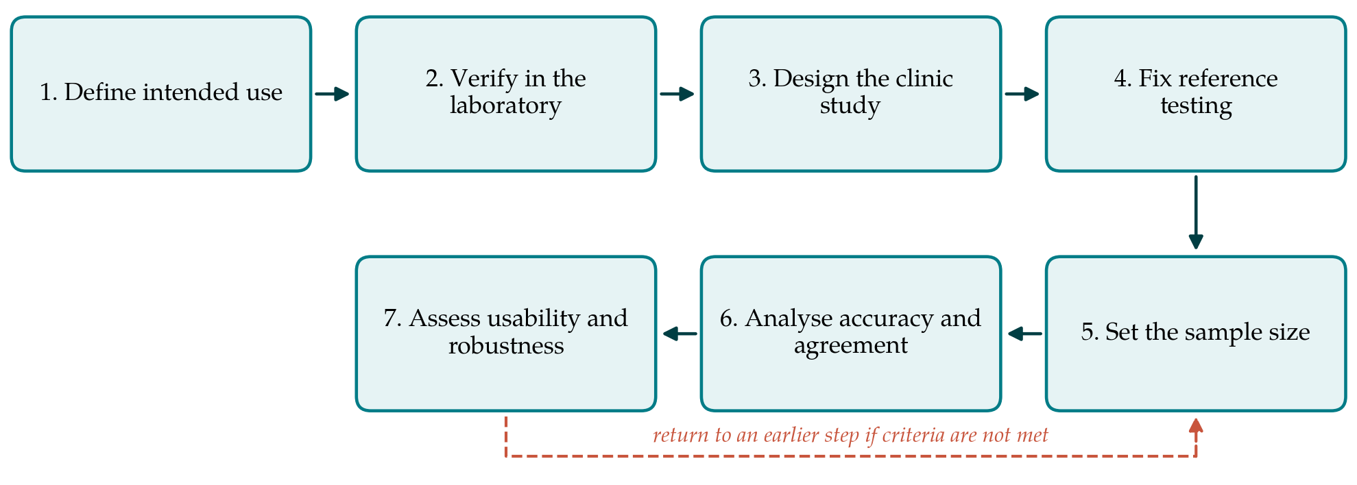
3.1. Step 1: Define the intended use and acceptance criteria
A validation can only succeed or fail against a stated purpose. The team should write down who will run the test (a midwife, a nurse, a laboratory technician), on what specimen (capillary whole blood from a fingerstick, most likely), at which visit, and what will be done differently on the strength of the result. A test meant to decide who receives iron is a different proposition from one meant to decide who is referred for further investigation. The clinically relevant cut-off follows from the guideline in force locally. Acceptance criteria are then set in advance: the lowest sensitivity and specificity, at that cut-off, that would make the test worth using, and the highest tolerable proportion of invalid strips. Fixing these before data are seen protects the study from the temptation to redefine success afterwards.
3.2. Step 2: Verify analytical performance in the laboratory
Before a clinic study is mounted, the device should be checked at the bench with stored or surplus samples. The points to cover are repeatability within a run and between days, agreement between strip lots, the range over which signal tracks concentration, and the effect of likely interferents such as haemolysis, lipaemia and extremes of haematocrit. For a test with a visual yes or no output, the relevant guidance is the Clinical and Laboratory Standards Institute protocol for qualitative tests, which describes how to characterise performance around the cut-off (CLSI, 2008). Strips should also be held for some weeks at the temperature and humidity of the intended clinics and retested. A device that fails here goes back to its maker, and no clinic time is spent on it.
3.3. Step 3: Design the clinic study
The clinical evaluation is a cross-sectional diagnostic accuracy study. Its credibility depends on avoiding the biases catalogued in QUADAS-2 (Whiting et al., 2011). Women should be enrolled consecutively, or by a random scheme, from those attending for routine antenatal care at the participating clinics, without selection on haemoglobin or symptoms; enrolling known anaemic women alongside healthy volunteers would exaggerate accuracy. Sites should span the range of settings in which the test is to be used. Gestational age, parity, recent iron supplementation, recent febrile illness and haemoglobin are recorded for each participant. Written consent and approval from the responsible ethics committee are prerequisites, and women found to be deficient by the reference method must be informed and treated under the usual local pathway.
3.4. Step 4: Fix the reference standard and the handling of specimens
Each participant gives a fingerstick sample for the lateral flow test, run on the spot by clinic staff, and a venous sample for the reference measurement. The reference should be a laboratory immunoassay calibrated against the international standard for ferritin and run in a laboratory with documented internal quality control and participation in external quality assessment, in line with the expectations of ISO 15189 (ISO, 2022). Serum is separated promptly and kept cold or frozen until analysis. The operator of the strip must not know the laboratory result, and the laboratory must not know the strip result. If resources allow, C-reactive protein is measured on the same serum so that the influence of inflammation on agreement can be examined.
3.5. Step 5: Set the sample size
Sample size follows from the acceptance criteria. For sensitivity, the number of women needed with deficiency by the reference standard is fixed by the expected sensitivity and the width of confidence interval judged acceptable; dividing by the expected prevalence of deficiency gives the total to enrol. The same reasoning applied to specificity gives a second total, and the larger of the two is used (Buderer, 1996). Prevalence should be taken from local surveys where they exist. Because the calculation is sensitive to that figure, a planned interim check of the observed prevalence, made without unblinding the comparison, allows the target to be adjusted.
3.6. Step 6: Analyse accuracy and agreement
For a qualitative strip, results are cross-tabulated against the reference classification at the pre-specified cut-off, and sensitivity, specificity and predictive values are reported with confidence intervals, together with the number of invalid and uninterpretable strips. For a reader-based quantitative device, agreement across the range is examined as well. A plot of the difference between methods against their mean, with limits of agreement, shows bias and its dependence on concentration (Bland and Altman, 1986); Passing-Bablok regression estimates proportional and constant differences without assuming that the reference is free of error (Passing and Bablok, 1983). A correlation coefficient alone is not evidence of agreement and should not be offered as such. Subgroup analyses by trimester and by inflammatory status are specified in advance and treated as exploratory. The report follows the STARD checklist, including a flow diagram of participants (Bossuyt et al., 2015).
3.7. Step 7: Assess usability and robustness
Accuracy in a supervised study overstates accuracy in routine use unless the people and conditions are the routine ones. A subset of strips should be read independently by two members of staff, and their agreement summarised with the kappa statistic (Landis and Koch, 1977). Staff are observed performing the test after the standard training and the errors noted: wrong volume, wrong timing, misread lines. Time per test, the proportion of invalid results, the storage conditions actually achieved in the clinic and the cost per valid result complete the picture that the REASSURED criteria ask for (Land et al., 2019).
4. Fitting the Protocol to Antenatal Practice
Several features of antenatal clinics shape how the protocol is applied. Clinics are busy, and a test that adds a quarter of an hour to each visit will be dropped whatever its accuracy, so workflow should be studied alongside performance. Many women attend first in the second trimester, when ferritin is already falling through haemodilution; the study population should mirror that pattern and not be restricted to early attenders. Where malaria or other infections are common, a share of women will have ferritin values raised by inflammation, and the laboratory reference will classify some of them as replete when they are not. The strip cannot be expected to do better than the analyte it measures, and this limitation is properly reported as a property of ferritin testing in that setting.
The consequences of each kind of error also differ. A false positive leads to iron being given to a woman who does not need it, which under a policy of universal supplementation changes little. A false negative withholds investigation or treatment from a woman who is deficient. Where universal supplementation is already in place, the test earns its cost mainly by identifying women who need more than the routine provision or who are anaemic for another reason, and the intended-use statement in Step 1 should say so.
5. Limitations and What Remains to Be Shown
The protocol is a proposal. It has not been piloted, and its feasibility in a real district, where staff turnover, stock-outs and competing priorities intervene, is untested. It deals with accuracy and usability and stops short of the question that finally matters, which is whether using the test improves maternal or newborn outcomes, or the efficiency of care, compared with haemoglobin screening alone. That question needs a comparative study, ideally randomised by clinic, and a favourable accuracy study is only the licence to conduct one.
Three technical uncertainties also remain. Capillary and venous ferritin may differ, and a discrepancy between strip and reference could reflect the specimen as much as the device; a nested comparison of capillary and venous samples on the reference method would settle this. Commercial laboratory immunoassays for ferritin do not agree perfectly among themselves, so the choice of reference method influences the apparent bias. And thresholds for deficiency in pregnancy are themselves under discussion (Daru et al., 2017), which means that a device validated at one cut-off may need re-evaluation if guidance changes.
6. Conclusion
A low-cost lateral flow ferritin test could bring the assessment of iron stores to clinics that at present measure only haemoglobin. Whether a given device is fit for that role is an empirical matter, and the seven-step protocol proposed here sets out how to find out: define the use, verify the chemistry, compare with a sound reference in consecutive attenders, size and analyse the study by accepted methods, and examine how the test behaves in ordinary hands. The protocol remains to be applied. Its purpose is to make sure that when a device is tested, the answer can be trusted and compared with others.
Conflict of interest
To be declared by the author(s) before publication.
Funding
To be declared by the author(s) before publication.