1. Introduction
Few drugs in rheumatology have as favourable a balance of benefit, safety and price as hydroxychloroquine. Withdrawal of the drug in a randomised trial led to more lupus flares (Canadian Hydroxychloroquine Study Group, 1991), a multiethnic cohort linked its use with better survival (Alarcón et al., 2007), and a systematic review found consistent evidence of reduced disease activity and damage with few serious adverse effects (Ruiz-Irastorza et al., 2010). Current European recommendations advise hydroxychloroquine for all patients with lupus, at a target dose of 5 mg per kilogram of real body weight per day, adjusted for the individual's risk of flare and of retinal toxicity (Fanouriakis et al., 2024).
The retina is the reason that dose is specified so carefully. Hydroxychloroquine can damage the photoreceptors and retinal pigment epithelium, and the damage is not reversible. Screening is meant to find it before the patient notices any loss of vision. In clinics where rheumatology itself is a scarce service and lupus presents late and severely (Tikly and Navarra, 2008), the eye examination is easily lost among more pressing matters. This communication examines that problem and what can reasonably be done about it.
2. What the Recommendations Ask For
The modern understanding of risk comes largely from a study of long-term users in a large American health plan, which showed that toxicity depends on daily dose by real body weight and on duration. At doses up to 5.0 mg per kilogram the risk was low during the first decade and rose substantially after twenty years; kidney disease and concurrent tamoxifen increased it (Melles and Marmor, 2014). On that basis the American Academy of Ophthalmology recommends a baseline fundus examination within the first year of treatment and annual screening from five years onward in patients on acceptable doses without major risk factors, earlier in those with them. The primary tests are automated visual fields and spectral-domain optical coherence tomography (Marmor et al., 2016).
Two refinements matter outside Europe and North America. In patients of Asian descent the earliest damage often lies in a pericentral ring and not beside the fovea, so a central 10-2 field can miss it and a wider test pattern is advised (Melles and Marmor, 2015). And because retinopathy found at a late stage can continue to progress after the drug is stopped (Marmor and Hu, 2014), older methods that detect only established disease, such as fundus inspection or the Amsler grid, are not regarded as adequate for screening. The United Kingdom guidance likewise relies on optical coherence tomography and fundus autofluorescence, performed annually after five years of treatment (Yusuf et al., 2021).
Chloroquine deserves a separate word, because in some countries it is cheaper or more reliably stocked than hydroxychloroquine and is used in its place. It is the more retinotoxic of the two, and the American recommendations set a correspondingly lower ceiling of 2.3 mg per kilogram of real body weight per day (Marmor et al., 2016). A clinic that prescribes chloroquine to patients with lupus has a stronger reason, and not a weaker one, to attend to dose and to screening.
3. Why Adherence Falters Where Resources Are Limited
Adherence to screening has two sides. One is whether the clinic refers at the recommended times; the other is whether the patient attends and the result returns to the prescriber. Figure 1 gathers the obstacles that stand between a prescription and a completed screen. They are described here from general knowledge of how such clinics work, since published measurements of screening uptake in low-resource lupus services are sparse.
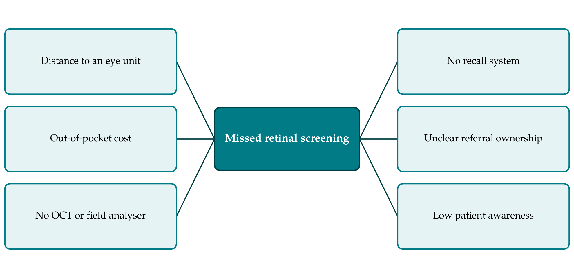
The health-system barriers are the hardest to shift. The global supply of ophthalmologists is very unevenly distributed, with the lowest numbers per head of population in low-income countries (Resnikoff et al., 2012), and those who are available are occupied with cataract, glaucoma, trauma and diabetic eye disease, which account for far more avoidable blindness. Optical coherence tomography and automated perimetry are concentrated in tertiary or private centres. A patient may face a day's travel and a fee that exceeds a month's supply of the drug in order to be screened for a complication that is unlikely in the first years of treatment.
Clinic-level barriers are more tractable. Many lupus clinics keep no record of when each patient started hydroxychloroquine, so the five-year point passes unnoticed. Patients are often not weighed, and a fixed daily tablet count is prescribed regardless of body size, which exposes small patients to doses well above the recommended ceiling. Responsibility for arranging screening is frequently unassigned between the rheumatologist, the general physician and the eye service. Patients, for their part, may never have been told why an eye test is needed when their sight seems normal. Written information in the patient's own language is seldom available, and a verbal explanation given once, during a consultation dominated by active disease, is unlikely to be remembered five years later when the first screen falls due.
4. A Minimum Workable Standard
Where the full recommendation cannot be met, the aim should be to keep the greatest share of its protective effect at the least cost. Table 1 pairs each barrier with a response that needs little or no equipment. The suggestions are drawn from the logic of the published guidance and have not been tested as a package.
| Level | Barrier | Low-cost response |
|---|---|---|
| Patient | Travel and fees for eye examination | Combine the eye visit with another appointment |
| Patient | Purpose of screening not understood | Explain early that damage is silent and permanent |
| Clinic | Start date of treatment unknown | Register of start date, weight and dose |
| Clinic | Fixed dosing regardless of weight | Weigh at each visit; prescribe by real body weight |
| Clinic | No one owns the referral | Named staff member reviews the register monthly |
| System | No tomography or field analyser | Agreed route to the nearest equipped centre |
| System | Too few eye specialists | Technician imaging with remote reading |
Correct dosing is the single most effective measure and costs nothing but a set of scales. Keeping the daily dose within the weight-based limit lowers risk for every patient whether or not a screen ever takes place, a point stressed in the joint statement of the American rheumatology, dermatology and ophthalmology societies (Rosenbaum et al., 2021). Renal function should be checked periodically for the same reason, since lupus nephritis is common and reduced clearance raises exposure.
Risk stratification allows scarce referrals to be used where they count. Patients beyond five years of treatment, those with kidney disease, those whose dose has exceeded the weight-based limit and those taking tamoxifen should be referred first. A patient in the first few years on a correct dose with normal kidneys is at low risk, and delaying that person's screen is a defensible compromise when capacity is short. Symptom questions and bedside tests should not be presented as equivalent to instrument-based screening, because they detect damage too late.
5. What Should Be Measured
The absence of local data is itself a barrier, because a problem that has not been counted is hard to argue for. A clinic register of the kind described makes a simple audit possible: the proportion of patients with a recorded weight and a dose within the limit, the proportion past five years who have had any instrument-based eye examination, and the interval since the last one. Such audits need no research funding and would establish, for the first time in many services, where the largest gaps lie. They would also show whether patients of African, South Asian or East Asian ancestry in these clinics display the pericentral pattern, on which evidence outside East Asian populations remains limited.
6. Conclusion
Retinal screening for hydroxychloroquine toxicity was designed around instruments that many lupus clinics cannot readily reach. The response should not be to stop a cheap and effective drug, nor to ignore a preventable cause of visual loss. Weight-based dosing, a register that records when treatment began, referral of the patients at highest risk first, and honest explanation to patients together make a standard that most clinics could meet now. Better evidence on actual screening uptake in these settings is needed, and collecting it is within the means of the clinics concerned.
Conflict of interest
To be declared by the author(s) before publication.
Funding
To be declared by the author(s) before publication.