1. Introduction
Carpal tunnel syndrome accounts for the great majority of median nerve entrapments, and the nerve is compressed far less often above the wrist. When it is, the cause is usually sought in the proximal forearm, where the nerve passes beneath the bicipital aponeurosis, between the two heads of pronator teres and under the fibrous arch of flexor digitorum superficialis (Johnson et al., 1979; Hartz et al., 1981). Compression in the arm itself is rarer still and is associated mainly with a supracondylar process and the ligament that may run from it to the medial epicondyle (Gessini et al., 1983).
Anatomical variation of the biceps brachii adds another candidate. The muscle quite often has a third head, and occasionally a fourth or more, arising from the humerus and joining the main belly or its insertion. Because the extra head arises on the medial side of the arm, close to the median nerve and the brachial artery, authors who describe it in the dissecting room commonly remark that it might compress them. The remark is repeated from paper to paper, often with the added thought that a labourer whose arm muscles are hypertrophied would be especially liable. This review asks what that claim rests on.
2. Scope and Approach
The article is a narrative review of the published literature and reports no new dissections or patients. It draws on the classical catalogues of muscular variation, on modern cadaveric series and anatomical encyclopaedias, on the surgical literature of proximal median nerve entrapment and on experimental work on nerve compression. No formal search protocol was used. The aim is to weigh the evidence and not to count it, and statements of frequency are given in words where series disagree.
3. Anatomy of the Accessory Head
Supernumerary heads of the biceps were recorded in detail by the nineteenth-century students of muscular anomaly, among them Macalister (1875), Testut (1884) and Le Double (1897), and the variation holds a regular place in modern compendia (Bergman et al., 1988; Tubbs et al., 2016). The usual form is a single extra head that takes origin from the anteromedial surface of the humerus, in the interval between the insertion of coracobrachialis and the upper part of the origin of brachialis. It passes downwards to join the deep surface of the common belly, the tendon of insertion or the bicipital aponeurosis. Like the rest of the muscle, it is supplied by the musculocutaneous nerve.
Rodríguez-Niedenführ et al. (2003) re-examined the variation in a large dissection series and proposed that supernumerary heads be named by their humeral origin. The infero-medial humeral head, as just described, was by far the commonest. A supero-medial head arising nearer the lesser tubercle and an infero-lateral head arising near the deltoid insertion were both much rarer. The scheme is useful because it draws attention to position: only the medial heads lie against the neurovascular bundle, and a lateral head has no relation to the median nerve at all.
How often a third head is present depends on who is dissected. Standard texts give a figure of about one arm in ten (Standring, 2016), but series from different populations range from a few per cent to around a fifth of arms. A study of South African cadavers found a notably high frequency (Asvat et al., 1993), and a Turkish series that included neonates showed that the head is already present at birth and is not an acquired response to use (Kopuz et al., 1999). It may be unilateral or bilateral, and some series find it more often in men. These differences are probably real in part, but small samples and differing definitions of what counts as a separate head also contribute.
4. Relation to the Median Nerve and Brachial Artery
In the lower half of the arm the median nerve descends in the groove on the medial side of the biceps, crossing in front of the brachial artery from lateral to medial, and both enter the cubital fossa deep to the bicipital aponeurosis. An infero-medial accessory head arises directly behind and lateral to this bundle. In most arms the extra belly simply lies alongside it, displacing nothing. The arrangement that draws comment is one in which the accessory head, or a slip detached from it, passes in front of the nerve and artery to reach the medial intermuscular septum, the brachial fascia or the aponeurosis, so that the bundle runs through a muscular or musculotendinous tunnel.
The clearest description is that of Nakatani et al. (1998), who reported a cadaver with four-headed biceps muscles on both sides in which the median nerve and brachial artery passed through a tunnel formed by a slip from the accessory head. The authors noted the potential for compression but, as in nearly all such reports, the specimen came without a clinical history. That is the general state of the evidence at this site. The anatomy that could produce entrapment has been shown repeatedly; whether it did so during life is not known for any of the dissected cases.
5. The Accessory Head Among Other Sites of Proximal Entrapment
An accessory biceps head has to be judged against the better documented causes of median neuropathy above the wrist, which are brought together in Figure 1 and compared in Table 1. The pronator syndrome was named by Seyffarth (1951), and the isolated palsy of the anterior interosseous nerve was described the following year by Kiloh and Nevin (1952). Operative series since then have shown that several structures can be responsible in the same limb and that fibrous bands within and around pronator teres and the superficialis arch are found far more often than any single named anomaly (Johnson et al., 1979; Hartz et al., 1981). Dellon and Mackinnon (1987) dissected the proximal forearm specifically to record such musculoaponeurotic variations and found them to be frequent in unselected cadaver limbs.
| Site | Compressing structure | Nature of the evidence |
|---|---|---|
| Lower arm | Accessory head of biceps or a slip from it | Cadaveric descriptions (Nakatani et al., 1998); few clinical reports |
| Distal humerus | Supracondylar process and its ligament | Operative cases and reviews (Gessini et al., 1983) |
| Cubital fossa | Bicipital aponeurosis | Operative series (Hagert, 2013) |
| Proximal forearm | Heads of pronator teres and fibrous bands | Operative series (Hartz et al., 1981) |
| Proximal forearm | Arch of flexor digitorum superficialis | Operative series (Johnson et al., 1979) |
| Anterior interosseous nerve | Accessory muscle heads and tendinous bands | Dissection studies (Dellon and Mackinnon, 1987) |
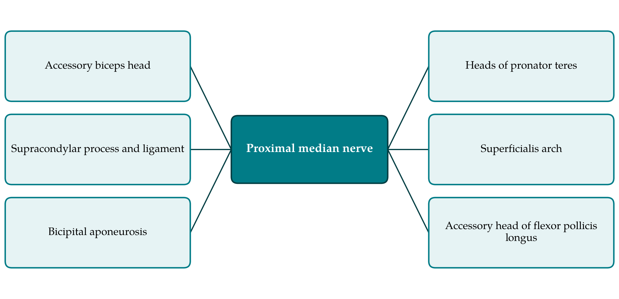
Two lessons carry over to the arm. The first is that an anatomical variant near a nerve is common and a symptomatic one is not, so the presence of an accessory head on imaging or at operation does not by itself explain a patient's complaint. The second concerns the bicipital aponeurosis. An accessory head that inserts into the aponeurosis may tighten it over the nerve during resisted flexion and supination, and compression beneath the aponeurosis has been described as a clinical entity that can be relieved by dividing it (Hagert, 2013). A variant head could thus act indirectly, at a recognised site, and need not form a tunnel of its own.
6. How Compression Injures a Nerve
Whether a muscular tunnel is likely to harm the nerve inside it depends on pressure and time. Experimental and clinical work has established that sustained external pressure first impairs venous outflow from the nerve, then produces oedema within the fascicles, and with longer exposure leads to focal demyelination and eventually to axonal loss (Mackinnon, 2002). The stages correspond to the lower grades of the classical schemes of nerve injury, in which conduction block with intact axons has a good prospect of recovery and axonal degeneration does not (Seddon, 1943; Sunderland, 1951). Early compression is characteristically intermittent and provoked by posture or effort, which fits an entrapment produced by a contracting muscle.
Repeated loading matters as well as steady pressure. Rempel et al. (1999) reviewed the response of peripheral nerves to loading and concluded that low pressures applied repeatedly or for long periods can produce the same sequence of changes as higher pressures applied briefly. A further idea relevant to manual workers is the double crush proposed by Upton and McComas (1973), in which a nerve already compromised at one level becomes more vulnerable at another. The hypothesis is still debated, but it offers one account of how a proximal variant that would otherwise be silent might add to the effect of compression at the carpal tunnel.
7. The Question of Manual Work
The proposed link with heavy work has an appealing logic. The accessory head contracts with the rest of the biceps in elbow flexion and forearm supination, movements that lifting, carrying and the use of hand tools demand forcefully and repeatedly. A muscle that is used in this way enlarges, and a tunnel bounded by muscle becomes tighter when the muscle swells or shortens. Intermittent pressure repeated over a working day is the pattern of loading that the experimental literature identifies as harmful.
Logic is not evidence, however, and several points weigh against a strong claim. The variant is present from birth in a substantial minority of people, most of whom do manual tasks at some time without trouble. No published study has compared the frequency of accessory heads in people with and without proximal median symptoms, or in manual and sedentary workers. The individual reports that exist cannot show causation, since a variant found at exploration may be incidental. Epidemiological work on occupation and median neuropathy concerns the carpal tunnel almost exclusively and cannot simply be transferred to the arm. An occupational contribution is therefore plausible but unproven.
8. Clinical Recognition
Certain features should lead a clinician to look above the wrist. Aching in the lower arm or proximal forearm that is brought on by resisted elbow flexion or supination, and sensory disturbance that includes the skin over the thenar eminence, both point proximally, because the palmar cutaneous branch leaves the median nerve before the carpal tunnel and is spared in carpal tunnel syndrome. Night waking is less typical than at the wrist. Weakness, when present, may involve pronator teres and the long flexors as well as the thenar muscles (Rodner et al., 2013).
Investigation has limits that are well recognised. Nerve conduction studies and electromyography are valuable for excluding carpal tunnel syndrome, cervical radiculopathy and plexus lesions, but in proximal median entrapment they are often normal or fail to localise the lesion (Preston and Shapiro, 2013). Imaging is better suited to showing the variant itself. High-resolution ultrasound can display an accessory muscle belly and follow the nerve through it during active contraction (Bianchi and Martinoli, 2007), and magnetic resonance imaging can show both the muscle and signal change in the nerve or in denervated muscles. Neither proves that the variant is the cause.
Management, as described for proximal median entrapment generally, begins with modification of the provoking activity and a period of observation, and surgical exploration is reserved for persistent or progressive cases (Rodner et al., 2013). There is no evidence base specific to the accessory biceps head on which to recommend one course over another, and this review does not offer treatment advice. What can be said is that a surgeon exploring the median nerve in the lower arm should know that an accessory head may be present and should record what is found.
9. Gaps and Future Work
The gap between the anatomical and the clinical record could be narrowed in three ways. Cadaveric series should report not only how often an accessory head occurs but how often it or its slips cross the neurovascular bundle, using an agreed classification such as that of Rodríguez-Niedenführ et al. (2003). Ultrasound surveys of the arm in symptomatic and symptom-free people, including groups defined by occupation, would give the comparison that is missing and could examine the nerve during contraction. And operative reports of proximal median decompression should state whether the arm was inspected and describe any variant muscle with photographs and follow-up, so that single cases can eventually be pooled. Until such data exist, the frequency of clinically significant compression by an accessory head cannot be estimated.
10. Conclusion
Accessory heads of the biceps brachii are common, usually arise from the medial side of the humerus and lie next to the median nerve and brachial artery. In a small proportion of dissections they enclose the bundle in a tunnel that could plausibly compress it, particularly during forceful flexion and supination. That is as far as the evidence goes. The clinical literature has not shown how often symptoms result, nor that manual workers are at greater risk, and the variant should be regarded as one possible and uncommon explanation for median neuropathy that does not fit the pattern of carpal tunnel syndrome. Better imaging and more careful operative documentation are the means by which the old anatomical suggestion can be tested.
Conflict of interest
To be declared by the author(s) before publication.
Funding
To be declared by the author(s) before publication.