1. Introduction
Surgery for a glioma of the dominant hemisphere is governed by two aims that pull against each other. Removing more tumour is associated with longer survival in low-grade and high-grade disease alike (Smith et al., 2008), and the diffuse gliomas now defined by their molecular profile (Louis et al., 2021) infiltrate well beyond the margin a surgeon can see. At the same time a resection that costs the patient fluent speech is a poor exchange. Awake craniotomy with direct electrical stimulation was developed to reconcile the two. The patient names pictures, counts or reads while small currents are applied to the exposed cortex, and sites where stimulation reliably disturbs performance are left in place.
The method rests on the observation, made in a classic series by Ojemann et al. (1989), that the cortical sites essential for naming are small, sharply bounded and placed differently from one person to the next. Anatomical landmarks therefore do not identify them. Later work showed that a tailored exposure in which no positive site is found can also guide a safe resection (Sanai et al., 2008), and a meta-analysis of published series associated stimulation mapping with fewer lasting severe deficits and with more extensive removal of tumour (De Witt Hamer et al., 2012).
Nearly all of that evidence concerns one language per patient. Bilingualism is common across the world, and in many neurosurgical units it is the usual case. A patient who uses one language at home and another at work has two competences to lose, and the surgeon has to decide whether mapping one of them protects the other. This review examines what the literature offers on that decision and on its consequences.
2. Scope and Approach
This is a narrative review of the published literature. It draws on the stimulation mapping studies that included bilingual or multilingual patients, on functional imaging and aphasia research that bears on how languages are represented, and on the large surgical series and reviews that describe awake mapping in general. No formal search protocol was applied and no pooled estimate is offered. The term bilingual is used broadly, for anyone who uses two or more languages in daily life, and the first-acquired and later-acquired languages are called L1 and L2 as in the source literature. Studies of sign language and of childhood surgery fall outside the scope.
3. Language Representation in the Bilingual Brain
Whether two languages occupy the same cortex is an old question. Penfield and Roberts (1959) held that a single cortical mechanism served all of a speaker's languages. The clinical study of aphasia in bilingual speakers complicated that view, because recovery does not always run in parallel. One language may return before the other, or return alone, or the two may alternate, patterns catalogued by Fabbro (2001), who noted that parallel recovery is nonetheless the most frequent. Paradis (2004) argued that such dissociations need not imply separate anatomical sites and could arise from differences in the activation threshold of each language within a common system.
3.1. Functional imaging
Imaging gave the first picture in healthy speakers. Kim et al. (1997) reported that people who learned their second language in adulthood activated spatially distinct parts of the inferior frontal gyrus for each language, whereas early bilinguals did not, and that posterior temporal activation overlapped in both groups. Perani et al. (1998) found that when proficiency was high the patterns for L1 and L2 converged regardless of the age of acquisition, and concluded that proficiency mattered more. The two results are often set against each other, though they used different tasks, one based on production and the other on comprehension. The broad conclusion of later imaging work is one of substantial overlap, with additional recruitment of control regions when the weaker language is used (Abutalebi and Green, 2007).
Imaging has a known limit for surgical purposes. It shows the regions that take part in a task and not the regions the task cannot do without. Speech depends on a distributed system with considerable redundancy, and only an intervention that briefly disables a site can show whether that site is indispensable.
3.2. Direct stimulation
Stimulation mapping supplies that test, and its findings in bilingual patients are summarised in Table 1. The earliest report, by Ojemann and Whitaker (1978), described two patients in whom naming was disturbed in both languages at some sites and in only one language at others. Roux and Trémoulet (2002) tested naming, reading and counting in a larger group and found language-specific sites in a proportion of patients, in frontal as well as temporoparietal cortex. The most detailed comparison is that of Lucas et al. (2004), who mapped bilingual patients and set them beside monolingual controls. They found shared sites and sites specific to each language, reported that L2-specific sites lay mainly in posterior temporal and parietal cortex, and observed that the layout of L1 sites did not differ from that seen in people with one language.
| Source | Patients and tasks | Main observation | Surgical implication |
|---|---|---|---|
| Ojemann and Whitaker, 1978 | Two bilingual patients; object naming | Some sites disturbed both languages, others only one | One language cannot stand in for the other |
| Roux and Trémoulet, 2002 | Bilingual patients; naming, reading, counting | Language-specific and task-specific sites in some patients | Test more than one task per language |
| Lucas et al., 2004 | Bilingual patients with monolingual controls; naming | Shared and specific sites; L2 sites mainly posterior | L1 mapping does not locate L2 sites |
| Walker et al., 2004 | Bilingual patients with mass lesions; speech mapping | Distinct and overlapping sites in the same patients | Map every language the patient relies on |
| Bello et al., 2006 | Multilingual glioma patients; cortical and subcortical mapping | Separate cortical sites and subcortical tracts for different languages | Continue testing below the cortex |
| Moritz-Gasser and Duffau, 2009 | Bilingual glioma patients; naming during resection | Stimulation provoked involuntary switching between languages | Record switching as a distinct error |
Other groups have reached similar conclusions. Walker et al. (2004) found distinct and overlapping sites in patients operated on for mass lesions, and Bello et al. (2006) extended the observation to multilingual patients with gliomas and to the white matter beneath the cortex. A review of this literature by Giussani et al. (2007) concluded that most stimulation studies had found language-specific sites and that mapping all languages in which the patient is fluent was the prudent course. The studies are small, they differ in the tasks used and in how proficiency was judged, and none can say how often a language-specific site lies within a planned resection. What they establish is that such sites exist and that their position is not predictable in advance.
3.3. Control and switching
Speaking one language while knowing two requires the unused one to be held back. The inhibitory control model of Green (1998) treats this as an executive function, and imaging has associated it with prefrontal, anterior cingulate, inferior parietal and caudate regions (Abutalebi and Green, 2007). Stimulation has given the idea some anatomical support. Moritz-Gasser and Duffau (2009) described involuntary switching into the other language when particular cortical and subcortical sites were stimulated, and argued for a large-scale network serving language selection. The clinical point is that a patient may come through surgery able to speak each language and still be unable to keep them apart, a deficit that naming tests in a single language will not detect.
4. Mapping Practice in Bilingual Patients
The technical conduct of awake surgery has been described at length, including anaesthetic regimens, stimulation settings and the handling of intraoperative seizures (Hervey-Jumper et al., 2015). What changes for the bilingual patient is mainly the neurolinguistic part of the procedure. Figure 1 sets out the points at which the patient's languages enter the pathway, from the first clinic visit to follow-up.
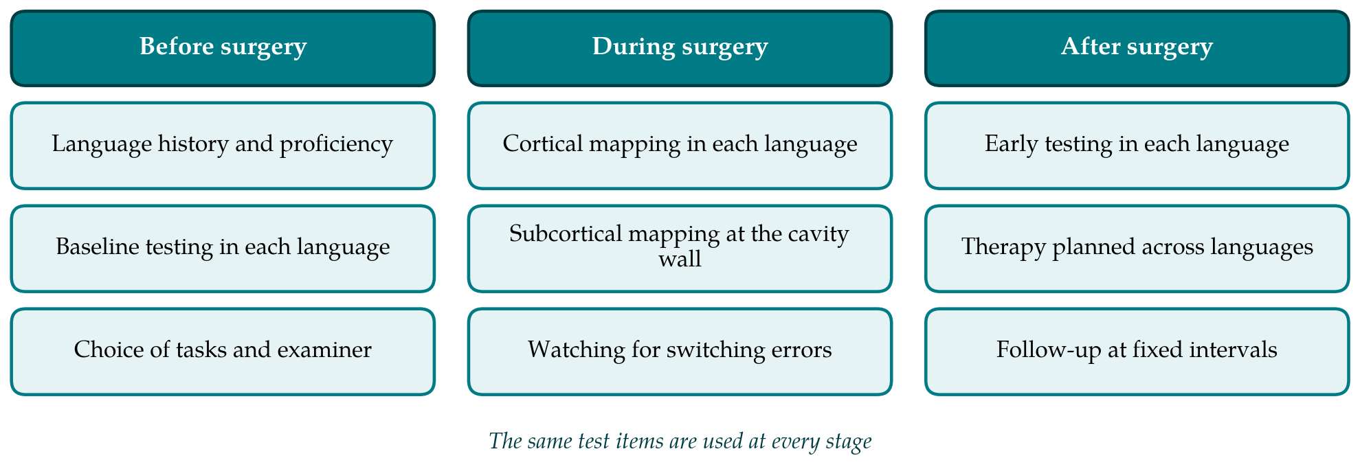
Preparation begins with a language history: which languages are used, when each was acquired, how well each is spoken and read, and which the patient needs for work and for family life. Baseline testing should then be done in each language. The Bilingual Aphasia Test of Paradis and Libben (1987) was designed to give equivalent assessments across many languages, and it remains the instrument most often named for this purpose. Items the patient cannot name reliably before surgery are removed from the intraoperative set, since an error on the table must be attributable to the stimulus and not to the tumour or to an unfamiliar word.
Which languages to map is itself a judgement. A patient may list four languages and rely on two. Testing a language the patient barely uses consumes time that could be given to the ones that matter, and a weak language yields unreliable baseline naming, which makes errors under stimulation hard to interpret. The stimulation studies offer no rule on this point. A reasonable reading of them is that the choice should follow the patient's own account of which languages daily life depends on, recorded before surgery and discussed with the family, and that the operation note should state which languages were tested and which were not.
During mapping, each stimulation site has to be tested in each language, which lengthens a procedure already limited by the patient's stamina. Published protocols handle this in different ways. Some map the cortex fully in one language and then the other. Others alternate. A reviewer of neurolinguistic practice in awake surgery found wide variation in tasks and little standardisation (De Witte and Mariën, 2013), and that variation is greater still where two languages are involved. Object naming is the common core. Counting, reading and tasks that require the patient to switch languages on cue have been added by individual groups.
Mapping should not stop at the cortical surface. The principle that subcortical pathways must be identified by stimulation as the resection deepens was established for language in general by Duffau et al. (2003), and the multilingual series of Bello et al. (2006) suggests it applies to each language separately. An examiner who speaks the patient's languages, or a trained interpreter working with the speech therapist, is needed throughout, because the classification of an error as anomia, paraphasia, speech arrest or a switch depends on understanding what was said.
5. Outcomes: What Is and Is Not Known
For awake mapping as a whole the evidence on outcome is consistent, though it comes from observational series and not from randomised comparison. In the large single-centre experience of Sanai et al. (2008), language function worsened in a minority of patients in the first days after surgery and had recovered in nearly all of them by six months. Transient deterioration followed by recovery is the expected course, and it is usually attributed to oedema and to the reorganisation that slow growing tumours induce in surrounding networks (Duffau, 2005).
For bilingual patients specifically, three statements can be supported. First, the mapping studies listed in Table 1 report that resections guided by testing in every language were completed without lasting loss of either language in most patients, but they were designed to describe localisation and their follow-up of language was brief and informal. Second, differential effects on the two languages after surgery have been described, in keeping with the recovery patterns known from stroke (Fabbro, 2001), yet their frequency is unknown. Third, no published comparison shows that mapping two languages gives a better result than mapping one. The recommendation to map all languages follows from the localisation data and from caution, and it is reasonable on those grounds.
Assessment after surgery has the same weakness as testing during it when only one language is used. A patient examined on the ward in the working language of the hospital may appear intact while having lost fluency in the language spoken at home, and the reverse can happen. Because early deficits after awake surgery usually improve, a single examination in the first week also says little about the final result. Testing each language with the instrument used before surgery, and repeating it after some months, is the least that is needed to describe outcome. Few published reports meet that standard.
There is also an interpretive difficulty. Stimulation interrupts a site and the network connected to it, so a positive response does not prove that the site alone is essential, and a negative response at one current does not prove that it is dispensable (Mandonnet et al., 2010). A language-specific site may therefore mark a point where one language is more vulnerable to disruption, for instance because it is less practised, and need not be a place where that language is stored. The account given by Paradis (2004) predicts exactly this. Whether sparing such a site matters for long-term outcome is the open question.
6. Gaps and Future Work
The first need is for outcome data reported by language. Large awake series could record the languages each patient uses, state which were mapped, and give standardised scores for each before surgery, in the first week and at later follow-up. Even without a comparison group this would show how often the languages diverge after surgery and whether divergence persists.
The second need is for agreed descriptors. Age of acquisition, proficiency and daily use are measured in different ways from one report to the next, or are not given, which makes it impossible to test the competing claims from imaging about what shapes the representation of L2. A short common dataset, agreed between centres, would cost little.
Third, the testing of language control deserves attention. Switching tasks are easy to add to a mapping protocol, and involuntary switching is easy to record once the team is looking for it. Whether deficits of control after surgery affect daily life in people who move between languages many times a day has not been studied. Finally, most of the literature comes from centres in Europe and North America and from a limited set of language pairs. Pairs that differ in script, in tonal structure or in word order may behave differently, and the centres best placed to study them are those where such patients are the majority.
7. Conclusion
The evidence that some cortical and subcortical sites are specific to one of a bilingual patient's languages is consistent across several small stimulation studies, and it justifies testing every language the patient depends on during awake surgery for a glioma near language cortex. The evidence that doing so improves outcome has not yet been gathered. Published series describe good preservation of both languages when both are mapped, but they do not report outcomes in a form that allows firm conclusions, and the effect of surgery on the ability to select and switch between languages is largely undocumented. Reporting language history, mapping strategy and outcome for each language would turn a sensible precaution into a tested practice.
Conflict of interest
To be declared by the author(s) before publication.
Funding
To be declared by the author(s) before publication.