1. Introduction
The extent to which a glioblastoma is removed is associated with how long the patient lives. That relationship was documented in large retrospective series before modern adjuvant treatment (Lacroix et al., 2001) and it has persisted since radiotherapy with temozolomide became standard (Stupp et al., 2005). The difficulty is that the infiltrating edge of the tumour often cannot be told from oedematous brain by eye or by touch. Fluorescence guidance addresses this by making tumour, or tissue closely associated with it, emit visible light during the operation.
In well-equipped centres the technique is routine. Elsewhere it is rare. The shortage of neurosurgical capacity in low and middle income countries is well described (Dewan et al., 2019), and the Lancet Commission on Global Surgery made the wider case that surgical care depends as much on infrastructure, supply chains and trained teams as on individual operators (Meara et al., 2015). An operating microscope with an integrated fluorescence module and a reliable supply of a licensed fluorophore are exactly the kind of resources that are missing.
This article sets out a workflow intended for such theatres. It does not describe a new technique. It organises what is already published into a sequence that a small team can follow with basic equipment, and it marks the points where a shortage of resources calls for a specific adaptation. The workflow is proposed for discussion and local testing and has not been applied by the author in the form given here.
2. Background: Agents and Optics
Three fluorophores are in clinical neurosurgical use. They differ in what they label, in the light needed to see them and in cost, and Table 1 compares them from the standpoint of a theatre with few resources.
| Agent | Source of contrast | Light and equipment | Main constraint |
|---|---|---|---|
| 5-aminolevulinic acid (Stummer et al., 2006) | Protoporphyrin IX formed within tumour cells | Violet-blue excitation; red emission; dedicated microscope module | Drug cost and supply; darkened field |
| Sodium fluorescein (Acerbi et al., 2018) | Leakage where the blood-brain barrier is disrupted | Blue excitation; yellow-green emission; filter or module | Not tumour-specific; off-label; timing matters |
| Fluorescein at high dose under white light (Shinoda et al., 2003) | As above, visible to the unaided eye | No special optics | Stains normal tissue over time; skin and urine discolouration |
| Indocyanine green (Raabe et al., 2003) | Intravascular dye showing blood flow | Near-infrared camera | Shows vessels, not tumour margin |
| No fluorophore | Colour, texture, anatomy | White light only | Infiltrated margin often invisible |
2.1. 5-aminolevulinic acid
5-aminolevulinic acid is a natural precursor in haem synthesis. Given by mouth before surgery, it leads to accumulation of protoporphyrin IX in malignant glioma cells, which emit red light when illuminated in the violet-blue range (Stummer et al., 1998). After a prospective series showed that residual fluorescence predicted residual tumour (Stummer et al., 2000), a randomised multicentre trial found that fluorescence guided surgery produced more complete resections of contrast-enhancing tumour and better progression-free survival at six months than white light surgery (Stummer et al., 2006). The drug was subsequently licensed in Europe and later in the United States (Hadjipanayis and Stummer, 2019). Its positive predictive value for tumour is high, while weakly infiltrated tissue may not fluoresce visibly (Hadjipanayis et al., 2015).
2.2. Sodium fluorescein
Fluorescein was used to localise brain tumours as long ago as the 1940s (Moore et al., 1948). It is given intravenously, stays largely outside cells, and collects where the blood-brain barrier is defective, which in a high-grade glioma corresponds broadly to the region that enhances on imaging (Diaz et al., 2015). Shinoda et al. (2003) used a high dose that could be seen without any filter. The introduction of a microscope filter matched to the dye allowed much lower doses (Schebesch et al., 2013), and a multicentre phase II study reported a high rate of complete resection of enhancing tumour with this approach (Acerbi et al., 2018). No randomised comparison with white light surgery or with 5-aminolevulinic acid has established an effect on survival. The dye is cheap, widely available and has a long safety record in ophthalmology, though rare severe allergic reactions occur.
2.3. What limits adoption
The barriers are practical. The licensed oral agent is costly per case and must be imported in many countries. Commercial fluorescence modules are tied to particular microscopes. Visual judgement of fluorescence is subjective and depends on ambient light and on the distance and angle of the microscope, as work on quantitative measurement has made clear (Valdés et al., 2011). Simple detection systems built from a light source and inexpensive filters have been described for fluorescein (Bongetta et al., 2016), which suggests that the optical requirement can be met cheaply. What such reports do not supply is the surrounding procedure that makes the technique safe and repeatable, and that is the gap the present workflow aims to fill.
3. The Proposed Workflow
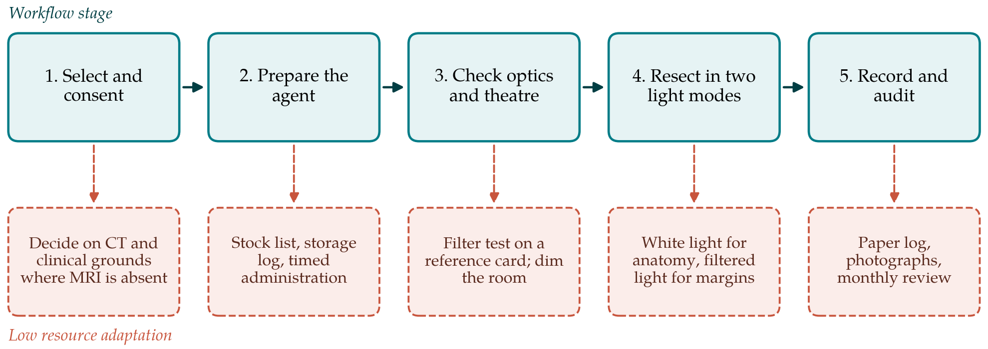
The workflow has five stages. Figure 1 shows them in order, each paired with the adaptation proposed for a setting in which the usual resource is absent. The description assumes fluorescein as the agent, since it is the one most likely to be obtainable, and notes where 5-aminolevulinic acid would differ.
3.1. Stage 1: Case selection and consent
Fluorescence guidance is most useful for intrinsic tumours that are expected to be high grade and for which a maximal safe resection is the aim. Where magnetic resonance imaging is not available, that judgement rests on contrast-enhanced computed tomography and the clinical picture, and the team should accept that some cases will prove to be other lesions. Fluorescein also accumulates in metastases, abscesses and other lesions with a disrupted barrier, so fluorescence does not confirm the diagnosis. Consent should cover the off-label status of the dye for this purpose, the possibility of allergic reaction, and the temporary yellow colouring of skin and urine. A history of reaction to fluorescein is a reason not to use it.
3.2. Stage 2: Preparing the agent
The dye, its expiry date and the resuscitation drugs needed to treat anaphylaxis are checked the day before. Dose and timing follow a single written local protocol based on a published regimen, agreed with the anaesthetist and the pharmacist, and are not varied from case to case. This article does not recommend a dose. The point of the stage is that timing determines contrast: fluorescein given too long before the margin is inspected spreads into oedematous brain around the tumour and loses its discrimination. The time of injection is therefore written on the theatre board. For 5-aminolevulinic acid the equivalent step is oral administration some hours before anaesthesia in line with the product licence, followed by protection of the patient from strong light for the period the licence specifies.
3.3. Stage 3: Checking optics and theatre
Before the patient is draped, the light source and filters are tested on a reference target, for example a card marked with a dilute drop of the dye beside an unmarked area. If the mark cannot be distinguished clearly at working distance, the case proceeds under white light alone. The theatre lights are arranged so that they can be dimmed or switched off during inspection, and windows are covered. The check takes a few minutes and can be added to the equipment section of the surgical safety checklist, a tool whose value in varied settings is well supported (Haynes et al., 2009).
3.4. Stage 4: Resection in two light modes
The bulk of the tumour is removed under white light using ordinary microsurgical judgement. Filtered light is used at defined moments: on first exposure, to confirm that the lesion fluoresces; as the resection approaches its planned limit; and at the end, to inspect the cavity wall systematically. Fluorescence is treated as a prompt to look again and never as an instruction to resect. Anatomy, the proximity of eloquent cortex and tracts, and vascular territory set the limit, and fluorescent tissue in a functionally critical area is left. Blood and cottonoids are cleared before each inspection because both alter the signal.
3.5. Stage 5: Recording and audit
For each case the team records the agent, the time of administration, the result of the optics check, whether residual fluorescence was seen at the end and why any was left, and any adverse event. A paper form is sufficient. Where the microscope has no camera, a photograph through the eyepiece is an acceptable record. Postoperative imaging, of whatever kind is available, is compared with the surgeon's impression. A monthly review of the forms shows whether the technique is being used consistently and whether the surgeon's reading of fluorescence agrees with imaging and histology.
4. Fitting the Workflow to Local Conditions
A unit will need to make three decisions of its own. The first concerns the optical system: whether to use a commercial module if one can be obtained, an improvised filter set, or high-dose dye under white light, each of which changes Stage 3 and the interpretation at Stage 4. The second concerns regulation and ethics, since off-label use of a drug is governed differently from one jurisdiction to another and may require approval by a hospital committee. The third concerns people. The workflow assumes that the scrub nurse, the anaesthetist and the surgeon each own a part of it, and it will work best where it is introduced by the whole team in a few supervised cases before becoming routine.
5. Limitations and What Remains to Be Shown
The workflow is a proposal built from published protocols and has not been validated. Several of its premises are uncertain. The evidence that fluorescein improves the extent of resection comes from uncontrolled studies performed with commercial filters in well-resourced centres, and it may not carry over to improvised optics. Because the dye marks barrier disruption and not tumour cells, it can label surgically injured brain and can miss non-enhancing infiltrated tissue. Subjective reading of weak fluorescence varies between observers (Valdés et al., 2011).
A unit adopting the workflow should therefore treat its first cases as an evaluation. The questions to answer are whether the optics check reliably predicts usable contrast in the operative field, whether the surgeon's judgement of complete resection agrees with postoperative imaging more often than before, whether adverse reactions occur, and whether the added steps lengthen operations to a degree that matters. A comparison of extent of resection before and after introduction would be a reasonable first study, and a multicentre registry among units in similar circumstances would be more informative still. Until such data exist, fluorescence in this setting should be regarded as an aid to the surgeon's eye and nothing more.
6. Conclusion
Fluorescence guided resection need not be confined to hospitals that can afford a licensed drug and a dedicated microscope. Fluorescein with simple filters offers a route to it, at the cost of lower specificity and thinner evidence. The five-stage workflow proposed here places that technique inside a routine of selection, preparation, checking, disciplined use and audit. Whether it improves resection or outcome in the theatres for which it is intended is not yet known, and finding out is the necessary next step.
Conflict of interest
To be declared by the author(s) before publication.
Funding
To be declared by the author(s) before publication.