1. Introduction
In the normal dog, blood draining the stomach, intestines, pancreas and spleen reaches the liver through the portal vein before it enters the caudal vena cava. A congenital portosystemic shunt is a single large vessel that connects the portal system directly to the systemic veins. Ammonia, bile acids and other substances absorbed from the gut escape hepatic clearance, and the liver, deprived of portal flow and the trophic factors it carries, remains small. The clinical result is a young dog that grows poorly and shows episodes of dullness, ataxia, circling, head pressing or seizures, often worse after a protein-rich meal, together with vomiting, polyuria or signs of ammonium urate urolithiasis (Berent and Tobias, 2009).
Dietary and medical management reduces the signs but leaves the anomaly in place. Surgical attenuation of the shunt aims to restore portal perfusion of the liver, and since the 1990s a series of devices has made gradual closure possible. A practitioner faced with a juvenile dog and a confirmed shunt has to judge whether to operate, when, and by which method. This review sets out what the published literature offers in answer, and where it falls short.
2. Scope and Approach
The article is a narrative review of the veterinary literature on single congenital shunts in dogs. It relies on the large retrospective series from referral hospitals, the small number of prospective and comparative studies, the systematic reviews of treatment, and standard surgical texts. Sources were selected for their bearing on technique, complications and outcome; no formal search protocol was followed. Multiple acquired shunts, which arise from portal hypertension and are not treated by attenuation, are mentioned only as a complication. Cats are excluded.
3. Anatomy, Breed and Presentation
Shunts are classified by their position relative to the liver. Extrahepatic shunts arise from the portal vein or one of its tributaries, commonly the left gastric, splenic or right gastric vein, and end in the caudal vena cava, the azygos vein or the phrenic vein. Computed tomographic angiography has shown that a small number of anatomical patterns account for the great majority (Nelson and Nelson, 2011). Intrahepatic shunts lie within the liver and are described as left, central or right divisional; the left divisional form corresponds to a ductus venosus that failed to close after birth.
Breed predicts the type. Extrahepatic shunts are typical of small and toy breeds such as the Yorkshire terrier, Maltese, miniature schnauzer and Cairn terrier, and intrahepatic shunts of large breeds including the Irish wolfhound, Labrador retriever and Australian cattle dog (Hunt, 2004; Tobias and Rohrbach, 2003). Pedigree studies in the Cairn terrier support an inherited basis (van Straten et al., 2005). Most dogs are presented before two years of age, though some reach adulthood with few signs.
4. Confirming and Mapping the Shunt
Routine blood tests suggest the diagnosis: microcytosis, low urea, low albumin, low cholesterol and mild increases in liver enzymes are common. The tests of liver function in general use are fasting and post-prandial serum bile acids and fasting plasma ammonia. In a study designed to compare them, ammonia was the more specific indicator of shunting, while bile acids were sensitive but raised in other liver diseases as well (Gerritzen-Bruning et al., 2006). Neither test separates a congenital shunt from acquired shunting or from portal vein hypoplasia without imaging.
The distinction matters for prognosis. Primary hypoplasia of the portal vein, also described as microvascular dysplasia, produces similar laboratory changes and occurs in the same small breeds, sometimes together with a macroscopic shunt. It has no surgical treatment. A liver biopsy taken at the time of attenuation is therefore standard practice, although the histological changes of the two conditions overlap and the biopsy cannot by itself predict how well the liver will recover once portal flow is restored (Berent and Tobias, 2009).
Abdominal ultrasonography can identify the vessel in experienced hands. Computed tomographic angiography has become the preferred method where it is available, because it displays the origin, course and termination of the shunt and lets the surgeon plan the approach (Nelson and Nelson, 2011). Intraoperative mesenteric portovenography remains useful. Images taken before and after temporary occlusion show how well the intrahepatic portal branches are developed, and poorer arborisation has been associated with a worse outcome (Lee et al., 2006).
5. Medical Stabilisation and the Decision to Operate
Dogs with signs of hepatic encephalopathy are stabilised before anaesthesia. The established measures are a diet with restricted but high-quality protein, lactulose to acidify the colon and speed transit, and an antimicrobial to reduce urease-producing gut bacteria (Lidbury et al., 2016). Some dogs do well for years on this regimen alone (Watson and Herrtage, 1998).
The argument for surgery rests mainly on one prospective comparison, in which owners chose between treatments and dogs were followed over time. Dogs that had surgery survived longer than those managed medically, and follow-up of the same group confirmed the difference along with better owner-reported quality of life (Greenhalgh et al., 2010; Greenhalgh et al., 2014). Treatment was not randomly allocated, and dogs considered poor candidates may have been steered towards medical care. Age at treatment did not influence survival in that work, which undermines the idea that surgery must be done as early as possible. Medical management is a legitimate course when surgery is declined or unaffordable.
6. Techniques of Attenuation
The surgical problem is that the hypoplastic portal vasculature of many dogs cannot at once accept the whole of the portal flow. Sudden complete closure can produce fatal portal hypertension. The techniques differ chiefly in how they manage this risk, as Table 1 summarises.
| Technique | Principle | Advantages | Limitations |
|---|---|---|---|
| Complete suture ligation | Shunt tied off fully at one operation | Immediate, definitive closure | Tolerated by only a proportion of dogs |
| Partial suture ligation | Shunt narrowed as far as portal pressure allows | Controlled by the surgeon | Residual flow; second operation often needed |
| Ameroid constrictor | Casein ring in a steel collar swells and incites fibrosis | Gradual closure; short operation | Rate of closure unpredictable; kinking of vessel |
| Cellophane banding | Thin film band provokes inflammatory closure | Cheap; suits larger vessels | Material and closure vary; may remain patent |
| Hydraulic occluder | Inflatable cuff filled through a subcutaneous port | Closure adjustable after surgery | Repeated visits; device failure |
| Transvenous coil embolisation | Coils placed in shunt behind a caval stent | Avoids liver dissection for intrahepatic shunts | Specialist equipment; repeat procedures |
6.1. Suture ligation
The original operation places a silk or polypropylene ligature around the shunt as close to its termination as possible. The surgeon occludes the vessel temporarily and watches for signs of portal hypertension: congestion and cyanosis of the intestine and pancreas, increased intestinal motility, a rise in measured portal pressure and a fall in arterial pressure. If these appear, the ligature is tied only partly. Dogs that tolerate complete ligation tend to have good long-term results, but many do not tolerate it, and partially ligated dogs may continue to shunt and need a second operation. The degree of narrowing achieved and the diameter of the portal vein were both related to prognosis in one series (Kummeling et al., 2004).
6.2. Devices for gradual occlusion
The ameroid constrictor is a C-shaped ring of compressed casein inside a stainless steel collar. Casein absorbs fluid and swells, and because the collar prevents outward expansion the lumen narrows. Its use for extrahepatic shunts was described by Vogt et al. (1996), and the largest series reported low perioperative mortality and a good or excellent outcome in most dogs (Mehl et al., 2005). Closure appears to depend as much on fibrous tissue reaction and thrombosis as on swelling of the casein, which explains why it is sometimes faster than intended and sometimes incomplete.
Cellophane banding, introduced by Youmans and Hunt (1998), wraps a folded strip of thin film around the vessel and secures it with clips. The band induces a slow inflammatory reaction. Results in a large series were comparable with those of the constrictor (Hunt et al., 2004). The hydraulic occluder, an inflatable silicone cuff connected to an injection port under the skin, allows closure to be advanced in steps after the operation and has been used mainly for intrahepatic shunts (Adin et al., 2006).
6.3. Intrahepatic shunts and endovascular treatment
Intrahepatic shunts are harder to treat by open surgery. The vessel may be buried in liver tissue, dissection risks haemorrhage, and reported mortality has been higher than for extrahepatic shunts. Percutaneous transvenous coil embolisation was developed as an alternative. Through the jugular vein a stent is placed in the caudal vena cava across the mouth of the shunt, and thrombogenic coils are released into the shunt through the mesh of the stent, which stops them migrating. A series of one hundred dogs treated in this way recorded low procedural mortality and outcomes at least as good as historical surgical results (Weisse et al., 2014). Gastrointestinal ulceration, a recognised late problem in dogs with intrahepatic shunts, led the authors to recommend lifelong gastroprotectant treatment.
6.4. What the comparative evidence shows
Two systematic reviews from the same group assessed the evidence for extrahepatic and for intrahepatic shunts and reached the same conclusion: the literature consists overwhelmingly of retrospective case series with differing definitions of outcome, and it does not allow one technique to be recommended over another (Tivers et al., 2012; Tivers et al., 2017). A later systematic review with meta-analysis reported broadly the same position for extrahepatic shunts (Serrano et al., 2019). In practice the choice follows the surgeon's training, the size and site of the vessel and the equipment to hand.
7. Complications and Perioperative Care
The hazards change as the dog passes through treatment, and Figure 1 sets the main one against each stage. Juvenile and toy-breed dogs have small glycogen reserves and are prone to hypoglycaemia and hypothermia under anaesthesia. Low albumin and reduced production of clotting factors add to the anaesthetic risk, and drugs that depend on hepatic metabolism act for longer than expected (Johnston and Tobias, 2018).
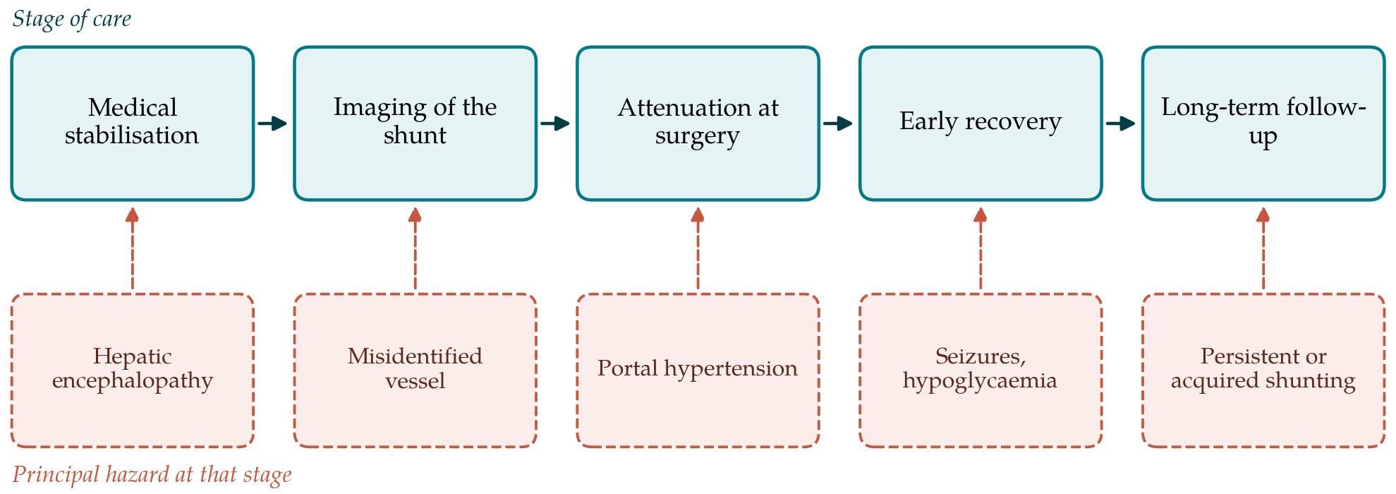
Care in the first days after surgery is largely a matter of observation. Blood glucose is checked repeatedly in small dogs, body temperature is supported, and intravenous fluids are continued until the dog eats. Abdominal girth, comfort, mucous membrane colour and mental state are assessed at short intervals, since the two serious complications both declare themselves through these simple signs. The diet and lactulose started before surgery are continued through recovery and withdrawn only when follow-up tests show that liver function has improved (Johnston and Tobias, 2018).
Portal hypertension shows itself in the first day or two as abdominal pain, distension with ascites, bloody diarrhoea and circulatory collapse. It is uncommon after gradual occlusion but remains the reason for close observation after any attenuation. Severe cases require removal of the ligature or device. Milder, sustained hypertension can cause multiple acquired shunts to open over the following weeks, which returns the dog to a shunting state that surgery cannot correct (Berent and Tobias, 2009).
Neurological deterioration after surgery is the complication surgeons fear most. It ranges from tremors and ataxia to generalised seizures and status epilepticus, typically arises within the first few days, and is not explained by hyperammonaemia or hypoglycaemia (Tisdall et al., 2000). It affects a small minority of dogs, but mortality among those that develop generalised seizures is high. Its cause is unknown. Prophylactic levetiracetam was associated with fewer seizures in one retrospective study (Fryer et al., 2011), yet a larger multicentre study found no protective effect (Mullins et al., 2019). The practice is common, and its value is unproven.
8. Outcomes After Surgery
Most dogs that survive the perioperative period improve clinically: neurological signs resolve, growth resumes and medical treatment can often be withdrawn. Clinical improvement does not prove that the shunt has closed. Studies that re-examined dogs with bile acid tests, scintigraphy or angiography found continued shunting in a meaningful share of animals that appeared well, through a patent original vessel or through acquired collaterals (Mehl et al., 2005; Hunt et al., 2004). Low preoperative albumin, a high white cell count and occurrence of postoperative complications have each been linked with poorer results in individual series. Long-term survival after successful surgery is good, and the available comparison favours surgery over medical care (Greenhalgh et al., 2014). Follow-up testing at intervals after the operation is advisable so that persistent shunting is recognised and diet adjusted accordingly.
9. Gaps and Future Work
The weaknesses of the evidence are plain. No randomised trial compares techniques. Definitions of a good outcome differ between reports, some relying on owner opinion and others on liver function tests or imaging, so pooled analysis is unreliable. The mechanism of post-attenuation seizures is unknown, which leaves prevention to guesswork. Predictors that would tell a surgeon before the operation which dog will tolerate complete closure are lacking. Agreement on a core set of outcome measures, prospective registries shared between referral centres and trials of seizure prophylaxis with adequate numbers would each move the subject forward. Breeding studies that identify the genetic basis in predisposed breeds could in time reduce the number of affected puppies.
10. Conclusion
Surgical attenuation offers young dogs with a single congenital portosystemic shunt the prospect of a normal life, and the published record supports it over indefinite medical management in dogs fit for anaesthesia. Gradual occlusion with an ameroid constrictor or cellophane band is now the usual approach to extrahepatic shunts and coil embolisation an established option for intrahepatic ones, but the evidence does not rank the methods. Careful stabilisation, accurate imaging, vigilant observation in the first days and objective follow-up matter at least as much as the device chosen.
Conflict of interest
To be declared by the author(s) before publication.
Funding
To be declared by the author(s) before publication.