1. Introduction
Children make up a small share of the patients who undergo computed tomography (CT) and a larger share of the concern about its risks. Early modelling suggested that the lifetime cancer risk attributable to a CT examination was appreciably higher for a young child than for an adult, partly because settings designed for adults were then applied to children without adjustment (Brenner et al., 2001). Large record-linkage studies in the United Kingdom and Australia later reported associations between CT in childhood and subsequent leukaemia and brain tumours (Pearce et al., 2012; Mathews et al., 2013). The size of the effect remains debated, since the conditions that prompt a scan may themselves be linked to cancer, but the findings reinforced a principle that was never in dispute: a justified examination should use no more radiation than the diagnostic task needs.
Head CT is the examination to which that principle most often applies in children. It is the standard investigation for significant head injury, where validated decision rules identify those who do not need imaging (Kuppermann et al., 2009), and it is used in the follow-up of hydrocephalus and in suspected intracranial infection. Campaigns such as Image Gently have promoted child-sized protocols for many years (Goske et al., 2008; Strauss et al., 2010). Whether the protocols of a given department are in fact well adjusted can be judged only by comparison, and the instrument for comparison is the diagnostic reference level (DRL).
This article sets out a protocol for a multi-centre regional audit leading to DRLs for paediatric head CT. The protocol is proposed on the basis of published recommendations. It has not been run, and the article contains no dose data.
2. Background
Three matters need to be settled before an audit is designed: what a DRL is and how it is derived, which dose quantities it is expressed in, and how children are to be grouped.
2.1. The reference level concept
A DRL is a level of a readily measured dose quantity, set for a defined examination and patient group, against which a facility compares the median of its own practice. It is an investigation level and not a limit: it does not apply to individual patients, and exceeding it calls for review, not for sanction (ICRP, 2017). The International Commission on Radiological Protection recommends that a national or regional DRL be set at the third quartile of the distribution of facility medians, so that by construction about a quarter of facilities lie above it and are prompted to examine their practice. The median of the same distribution, often called the achievable dose, gives a further target for departments already below the DRL (Kanal et al., 2022). In the European Union the establishment, regular review and use of DRLs is a legal obligation on member states (Council of the European Union, 2013), and international safety standards carry the same expectation (IAEA, 2018).
2.2. Dose quantities for CT
Two quantities are displayed by every modern scanner, as the product standard requires (IEC, 2009): the volume CT dose index (CTDIvol, in mGy) and the dose-length product (DLP, in mGy cm). Both describe the output of the scanner as measured in a standard acrylic phantom, 16 cm in diameter for head examinations, and neither is the dose to the patient (McCollough et al., 2011). Because they are recorded automatically and depend directly on the protocol, they are the quantities recommended for DRLs. The size-specific dose estimate corrects CTDIvol for the attenuation of the individual patient, and a formulation for the head based on water-equivalent diameter is available (AAPM, 2011; AAPM, 2019). It is a useful secondary quantity.
2.3. Grouping children
Adults are treated as one group for DRL purposes; children cannot be. For examinations of the trunk, body weight is the preferred grouping variable. For the head, where attenuation depends on skull size and that tracks age more closely than weight, age bands are recommended (ICRP, 2017). The European guidelines propose four bands for head CT: from birth to under 3 months, 3 months to under 1 year, 1 year to under 6 years, and 6 years and above (European Commission, 2018). Earlier surveys used other bands, which is one reason their values are hard to compare (Shrimpton et al., 2006; Vassileva et al., 2015). Adopting the European bands makes a regional result directly comparable with the European reference values.
3. The Proposed Audit Protocol
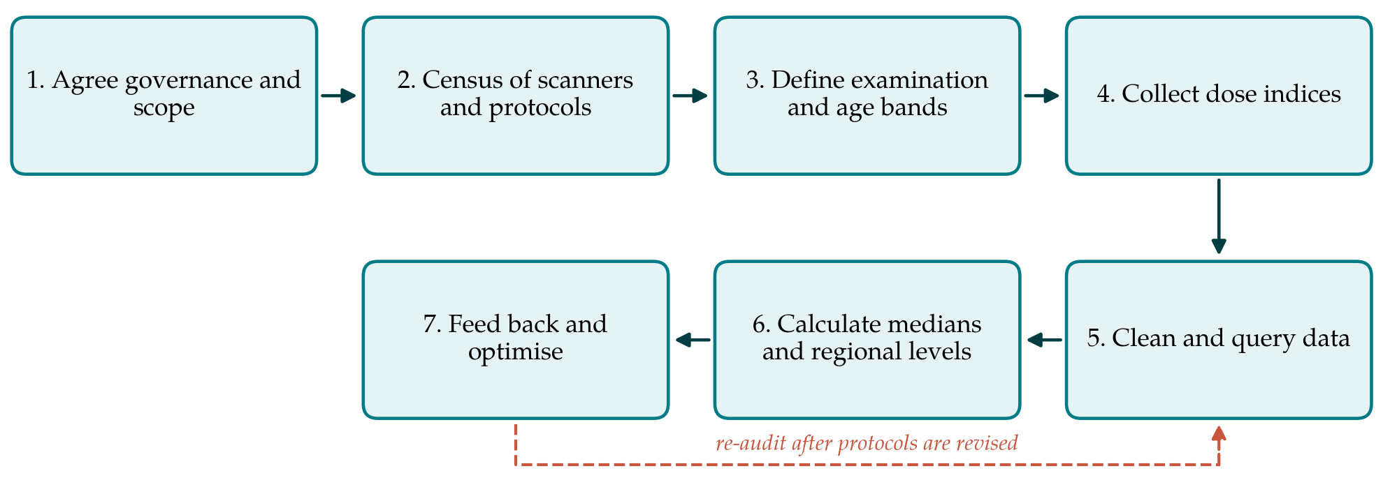
The protocol has seven steps. Table 1 lists each with its main activity and the output it should produce, and Figure 1 shows the sequence, including the return from feedback to a later round of data collection. The steps are described in order.
| Step | Main activity | Output |
|---|---|---|
| 1. Governance and scope | Form a steering group and invite every site that scans children | Agreed audit plan |
| 2. Scanner and protocol census | Record scanners and export current head protocols | Census table for every scanner |
| 3. Examination and age bands | Define examination, indications, exclusions and age bands | Written inclusion criteria |
| 4. Data collection | Record dose indices for consecutive eligible examinations | Site data in a common format |
| 5. Cleaning | Query implausible records; correct or exclude by written rules | Clean data set and log of queries |
| 6. Calculation | Compute site medians, then regional third quartile and median | Regional levels by age band |
| 7. Feedback and re-audit | Return results, review high protocols, schedule next round | Site reports and re-audit date |
3.1. Step 1: Governance and scope
A regional audit needs a small steering group with a named lead, normally a medical physicist, together with a radiographer and a radiologist who have paediatric experience. The group invites every facility in the region that scans children, including general hospitals where paediatric head CT is occasional, because these are the sites where adult-derived settings are most likely to persist. The group agrees in advance the examination to be audited, the collection period, the data fields and the rule by which results will be shared: this protocol recommends that each site sees its own results against the anonymised regional distribution. The audit is registered under local clinical audit and information governance arrangements.
3.2. Step 2: Scanner and protocol census
Before any patient data are gathered, each site lists its scanners with manufacturer, model, year of installation and reconstruction options, and exports the paediatric head protocols as they stand: tube voltage, tube current or image quality setting, automatic exposure control mode, rotation time, pitch, slice thickness and reconstruction algorithm. The census serves two purposes. It explains differences found later, since a site with high indices can be matched at once to the protocol that produced them. It also confirms that the displayed CTDIvol refers to the 16 cm phantom on every scanner and that dose displays have been checked at routine quality control, without which the pooled values would not be comparable (McCollough et al., 2011).
3.3. Step 3: Examination and age bands
The audited examination is unenhanced CT of the brain as a single acquisition from skull base to vertex. Head CT is requested for different purposes that tolerate different image noise: the search for a small haemorrhage after trauma needs more than a check of ventricular size in a child with a shunt. Indication should therefore be recorded, and where numbers allow, the common indications should be analysed separately, in line with the move towards indication-based reference levels (European Commission, 2018). Patients are assigned to the four European age bands by age on the day of the scan. Examinations combined with the cervical spine or facial bones in one acquisition, contrast-enhanced studies and angiography are excluded or recorded as separate categories.
3.4. Step 4: Data collection
Each site collects consecutive eligible examinations over the agreed period, without selecting cases thought to be typical, since selection hides the outliers the audit exists to find. The Commission asks for at least 20 patients in each group at each facility where that is practicable and accepts that fewer may have to be used in paediatric work (ICRP, 2017). A collection period of twelve months is proposed so that smaller sites can contribute to the youngest bands. For each examination the record holds an audit identifier, site and scanner, date, age, indication, protocol name, CTDIvol, total DLP, number of acquisitions and scan length. Where a dose management system or structured dose reports are available the fields are extracted automatically; where they are not, they are transcribed from the dose page, and a second person checks a sample.
3.5. Step 5: Cleaning the data
Pooled dose data contain predictable errors. Adult protocols applied to adolescents, repeated acquisitions after movement summed into one DLP, localiser doses added to the total, examinations labelled as head that include the neck, and transcription slips all occur. The steering group applies written rules: records with implausible combinations of CTDIvol, DLP and scan length are returned to the site for checking, and are excluded only if they prove to be errors of recording. Genuine high values stay in. Repeated acquisitions are kept and flagged, because the repeat rate is itself a finding that bears on immobilisation and sedation practice. The number of records received, queried, corrected and excluded is reported for each site.
3.6. Step 6: Calculating the regional levels
For each site, age band and quantity the median is calculated. The median is used in preference to the mean because dose distributions are skewed and a few high values would otherwise dominate (ICRP, 2017). The regional DRL for each band is the third quartile of the site medians, and the regional achievable dose is their median. Where a band has data from only a handful of sites, a quartile of so few points means little; the result should then be reported as a regional typical value, the median of the site medians, and labelled as provisional. Results are set beside the European values and any national values for context (European Commission, 2018; Kanal et al., 2022).
3.7. Step 7: Feedback, optimisation and re-audit
Each site receives its medians plotted against the regional distribution, with the protocol settings from the census alongside. A site above the regional DRL in any band reviews the relevant protocol with its physicist and radiologist. The usual causes are well described: tube current or noise settings not scaled to head size, tube voltage left at the adult value, scan length extending beyond the region of interest, and failure to use iterative reconstruction where it is installed (Strauss et al., 2010). A site far below the median is asked to confirm that image quality is adequate, since a low dose that yields a non-diagnostic scan is not optimised. Changes are documented, and data collection is repeated after an interval. The Commission suggests that reference levels be reviewed every three to five years, and a shorter interval is reasonable after a first round (ICRP, 2017).
4. Fitting the Protocol to Regional Practice
The protocol was designed around three practical constraints. Small numbers are the first. A general hospital may scan very few infants in a year, and no adjustment of method creates data that do not exist. Pooling across a region is the remedy for the DRL itself, but it does not give the small site a stable median of its own. Such sites should compare their protocol settings, and not only their few dose values, with those of the regional paediatric centre.
Image quality is the second. Dose indices say nothing about whether the images answered the clinical question, and a DRL process that rewards low numbers alone will drift towards under-exposure. The guidelines ask that image quality be assessed alongside dose (European Commission, 2018). A proportionate approach is for a radiologist at each site to grade a small random sample of audited examinations against agreed criteria, such as visibility of the boundary between grey and white matter and of the posterior fossa, and for the grades to be reported with the dose results.
The third is effort. Manual transcription from dose pages is slow and prone to error, and the audit will be repeated. Regions that expect to maintain DRLs should plan for automatic extraction from structured dose reports, which also allows size-specific estimates to be computed for the head (AAPM, 2019).
5. Limitations and What Remains to Be Shown
This is a proposed protocol that has not been carried out, and its workability is untested. The twelve-month collection period, the rule for labelling values as provisional and the image quality sampling are suggestions that a pilot in two or three sites would refine. The protocol also assumes that displayed dose indices are accurate, which depends on the quality control performed at each site.
Some limits belong to the DRL method itself. CTDIvol and DLP describe scanner output, not patient dose or risk, and a regional value at the third quartile reflects what local practice is, not what it ought to be (McCollough et al., 2011). A region in which every site uses poorly adjusted protocols will produce a comfortable-looking DRL. Comparison with external values guards against this only in part, because scanner generations differ. Whether an audit of this kind lowers doses in a region without loss of diagnostic quality is an empirical question. National programmes that have repeated their surveys give grounds for expecting so (Shrimpton et al., 2006), but the protocol proposed here would have to demonstrate it for itself through its second round.
6. Conclusion
Regional diagnostic reference levels for paediatric head CT can be established with resources that most hospital groups already have: scanners that display standard dose indices, a physicist to lead, and agreement to share results. The seven-step protocol described here applies the international and European recommendations to that setting, with age bands that allow direct comparison with European values, explicit rules for cleaning and for small numbers, and a feedback stage that ties the results of each site to its protocol settings. Its value lies less in the numbers it would produce than in the cycle of comparison, review and re-audit that those numbers start. The protocol awaits application, and its first use should be reported with its difficulties as well as its results.
Conflict of interest
To be declared by the author(s) before publication.
Funding
To be declared by the author(s) before publication.