1. Introduction
Men with localised prostate cancer have long been offered a choice between treating the whole gland and treating nothing. The ProtecT trial showed that, at ten years, deaths from prostate cancer were few whether men were assigned to active monitoring, radical prostatectomy or radiotherapy, although progression and metastasis were more frequent with monitoring (Hamdy et al., 2016). The companion report on patient-reported outcomes documented the price of radical treatment: urinary leakage and loss of erectile function were worst after surgery, and bowel symptoms after radiotherapy (Donovan et al., 2016). Many men, particularly those with intermediate-risk disease, find neither option comfortable.
Focal therapy was proposed as a third course. Onik et al. (2002) described cryoablation confined to the side of the gland containing tumour, with the explicit aim of preserving potency, and likened the idea to lumpectomy for breast cancer. Since then high-intensity focused ultrasound, cryotherapy, irreversible electroporation, vascular-targeted photodynamic therapy, focal laser ablation and focal brachytherapy have all been used to ablate part of the prostate (Valerio et al., 2017). The technologies differ, but the clinical questions they raise are the same.
This review addresses two of those questions: how patients should be selected and how they should be followed. Both have been the subject of formal consensus exercises, and both remain less settled than the volume of publication might suggest.
2. Approach of This Review
The article is a narrative review of the published literature. It relies on the diagnostic trials that underpin modern lesion localisation, on prospective and registry series of focal therapy, on systematic reviews of those series, and on Delphi consensus statements and guidelines. No formal search strategy was applied, and the sources were chosen for their influence on current practice. Salvage focal therapy after radiotherapy is not covered.
3. Rationale: The Index Lesion
Prostate cancer is usually multifocal. Whole-mount studies of prostatectomy specimens found several separate tumours in most glands, but the largest focus accounted for most of the tumour volume and the secondary foci were typically small and of low grade (Wise et al., 2002). Genomic analysis of men who died of metastatic disease indicated that their metastases shared a single clonal origin (Liu et al., 2009). From these observations came the index lesion hypothesis: that one dominant focus drives the natural history of the disease, and that destroying it may be sufficient while small low-grade foci elsewhere are observed (Ahmed, 2009).
The hypothesis is reasonable and not proven. The lethal clone is not always the largest lesion, and the behaviour of untreated tissue over decades is unknown. Focal therapy is therefore best understood as treatment combined with continued surveillance of the remaining gland, a point that shapes both selection and follow-up.
4. Patient Selection
Selection has three components: the grade and extent of the cancer, the confidence with which it has been localised, and the characteristics of the man himself. Table 1 summarises the criteria on which consensus statements broadly agree and the points that remain open.
| Domain | Generally accepted | Still debated | Sources |
|---|---|---|---|
| Risk group | Intermediate-risk disease, chiefly grade group 2 | Grade group 3; small high-grade lesions | Donaldson et al., 2015; Tay et al., 2017 |
| Low-risk disease | Active surveillance is the preferred strategy | Treatment of large-volume grade group 1 | Mottet et al., 2021 |
| Imaging | Multiparametric MRI before biopsy and before treatment | Role of PSMA PET and ultrasound variants | Ahmed et al., 2017; Turkbey et al., 2019 |
| Biopsy | Targeted cores plus systematic sampling of the untreated gland | Need for transperineal template mapping | Kasivisvanathan et al., 2018; Tay et al., 2017 |
| Untreated foci | Small low-grade foci outside the field may be observed | Size limit of acceptable untreated disease | Donaldson et al., 2015 |
| Patient factors | Sufficient life expectancy; willingness to attend follow-up | Gland size, calcification, prior surgery | Valerio et al., 2017 |
4.1. Risk group and grade
Early series enrolled many men with low-risk cancer, who would now be advised to choose active surveillance. Expert panels have since shifted the indication towards intermediate-risk disease. The consensus meeting reported by Donaldson et al. (2015) concluded that focal therapy should be directed mainly at men with intermediate-risk cancer and that low-grade, low-volume disease outside the treatment field could be left untreated. The international Delphi project led by Tay et al. (2017) reached a similar position. In the grade group system adopted by the International Society of Urological Pathology (Epstein et al., 2016), the typical candidate has a localised grade group 2 lesion; opinion is divided on grade group 3, and high-risk disease is generally regarded as unsuitable outside research.
4.2. Localising the cancer
Focal treatment is only as good as the map on which it is based. The PROMIS study showed that multiparametric magnetic resonance imaging (mpMRI) was considerably more sensitive than transrectal ultrasound-guided systematic biopsy for clinically significant cancer, using transperineal template mapping as the reference (Ahmed et al., 2017). The PRECISION trial then showed that an MRI-targeted biopsy pathway detected more significant cancers and fewer insignificant ones than standard systematic biopsy (Kasivisvanathan et al., 2018). Reporting is standardised by the Prostate Imaging Reporting and Data System (Turkbey et al., 2019).
Imaging does not see everything. Small and low-grade foci are often invisible, and some significant tumours are missed. For that reason consensus panels ask for systematic sampling of the areas that will not be treated in addition to targeted cores from the visible lesion (Tay et al., 2017). Transperineal template mapping gives the most complete picture but at the cost of anaesthesia and morbidity, and with good-quality mpMRI most panels no longer consider it mandatory.
4.3. Matching the patient and the energy
The systematic review by Valerio et al. (2017) found that high-intensity focused ultrasound and cryotherapy had the largest evidence base, with other sources at an earlier stage of evaluation. Choice of energy is influenced by lesion position. Transrectal ultrasound energy reaches posterior tumours readily, whereas anterior lesions in large glands may be better suited to cryotherapy or to needle-based techniques. Men should also understand before treatment that focal therapy commits them to a schedule of tests, and that a proportion will need a second focal procedure or a later radical treatment. A man who is unlikely to attend surveillance is not a good candidate.
5. What Published Series Show
The prospective development study of focal high-intensity focused ultrasound by Ahmed et al. (2012) established that unifocal and multifocal lesions could be ablated with a low rate of genitourinary side effects. Focal cryotherapy has been reported from a large national registry (Ward and Jones, 2012). A multicentre series from the United Kingdom reported five-year outcomes in men of whom most had intermediate- or high-risk cancer (Guillaumier et al., 2018), later extended to a larger cohort with longer follow-up (Reddy et al., 2022). A phase 2b study of MRI-guided focused ultrasound enrolled men with intermediate-risk disease (Ehdaie et al., 2022). Across these reports continence was preserved in the great majority and erectile function in most, while a minority of men required retreatment or went on to radical therapy.
The limits of this evidence are plain. Almost all of it is single-arm. The one large randomised trial, of padeliporfin vascular-targeted photodynamic therapy, compared treatment with active surveillance in men with low-risk cancer, a group for whom intervention is now seldom recommended (Azzouzi et al., 2017). An updated systematic review found that the quality of the evidence had improved little over five years and that definitions of success still varied between studies (Hopstaken et al., 2022). European guidelines accordingly advise that focal therapy be offered within a clinical trial or a well-designed prospective registry (Mottet et al., 2021).
6. Follow-Up Protocols
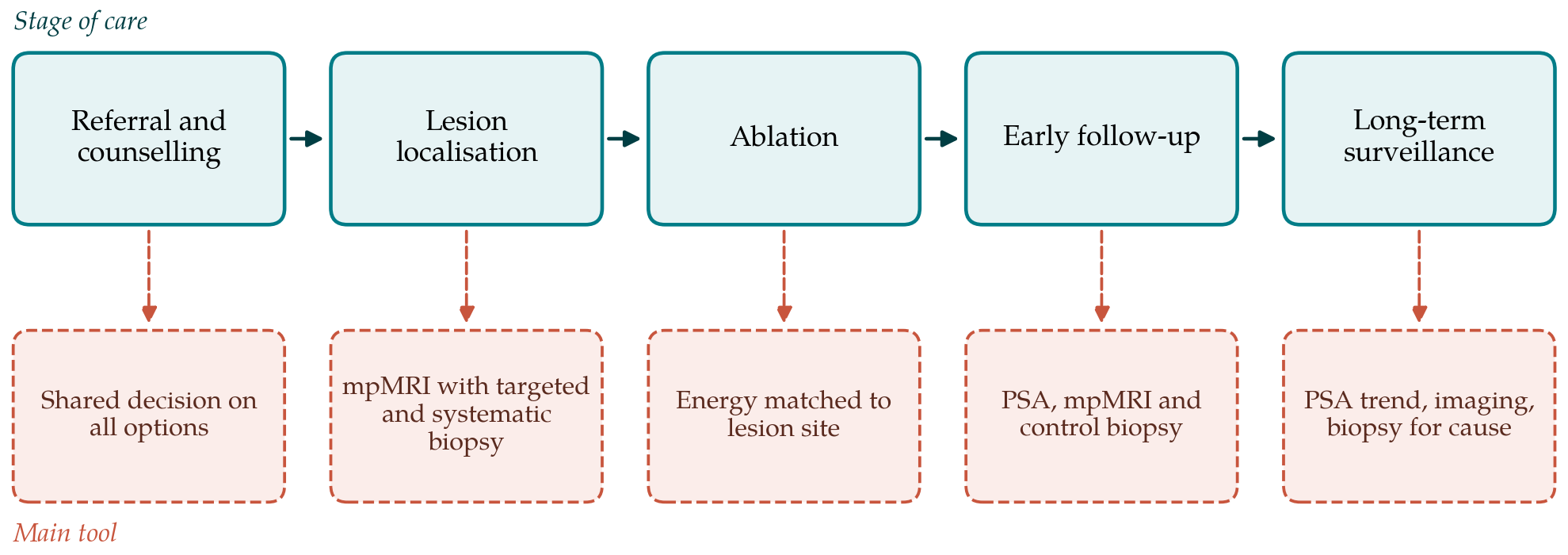
Follow-up after focal therapy has to answer two questions at once: whether the ablation destroyed the target, and whether significant cancer has appeared in the untreated gland. Figure 1 places follow-up in the pathway of care, with the main tool used at each stage.
6.1. Prostate-specific antigen
Prostate-specific antigen (PSA) falls after ablation but does not become undetectable, because viable prostate tissue remains. No threshold has been validated to define failure. The Phoenix definition of nadir plus 2 ng/mL was derived for radiotherapy (Roach et al., 2006) and has been borrowed by some focal therapy series without evidence that it predicts clinical outcomes in this setting. Consensus statements therefore treat PSA as a trend to be watched and a trigger for further tests, not as an endpoint in itself (Muller et al., 2015; Lebastchi et al., 2020).
6.2. Imaging the treated gland
Multiparametric MRI is the imaging standard after treatment as before it, but the ablated zone looks different from untreated prostate. Scarring, loss of volume and altered enhancement make the pre-treatment scoring rules unreliable. Early contrast-enhanced imaging can confirm that the intended zone has been devascularised; later scans look for focal early enhancement and restricted diffusion at the margin of the cavity. A dedicated scoring proposal for the treated prostate, Prostate Imaging after Focal Ablation, has been published to make reporting more consistent (Giganti et al., 2023). Its performance against biopsy has still to be established in independent cohorts.
6.3. Control biopsy
Histology remains the reference. The Delphi project on follow-up recommended that men be followed for at least five years, with PSA at regular intervals, mpMRI and a biopsy of the treated area within about the first year (Muller et al., 2015). The later multidisciplinary consensus endorsed a protocol biopsy combining targeted cores from the ablation zone with systematic sampling of the rest of the gland, and proposed standard terms that separate in-field recurrence, which reflects failure of ablation, from out-of-field disease, which reflects selection or new tumour (Lebastchi et al., 2020). The distinction matters because the two call for different responses and say different things about a programme's quality.
6.4. Defining failure and reporting function
There is broad agreement that small amounts of grade group 1 cancer on control biopsy need not be counted as failure, and that clinically significant cancer in the treated field should be. Beyond that, endpoints differ: some series report any positive biopsy, others freedom from radical or systemic treatment. Comparison between centres is unreliable until a common definition is used. Functional outcomes should be collected with validated patient-completed questionnaires at baseline and at fixed intervals, since preservation of continence and erectile function is the reason for choosing focal therapy in the first place.
How long and how intensively to continue surveillance is unresolved. After a negative control biopsy, most protocols rely on PSA at intervals of several months and periodic mpMRI, with further biopsy prompted by a rising PSA or a new imaging finding. The untreated gland remains at risk indefinitely, so follow-up in effect merges with active surveillance of the remaining prostate. Men in whom significant cancer is found may be offered repeat focal ablation, surgery or radiotherapy, and the feasibility and morbidity of these salvage treatments form part of what a programme should report.
7. Gaps and Future Work
The central gap is comparative evidence. Randomised trials against radical treatment have proved difficult to recruit, because men who seek focal therapy are often unwilling to be allocated to surgery. Prospective registries with agreed selection criteria, a fixed biopsy schedule and standard definitions are the practical alternative, and their value depends on completeness of follow-up. Long-term data are also needed: prostate cancer evolves over decades, and most series report medians of only a few years. Further questions concern the place of prostate-specific membrane antigen imaging in selection, the validation of post-ablation MRI scoring, and whether a safe schedule can be defined that spares men repeated biopsy when imaging and PSA are reassuring.
8. Conclusion
Focal therapy offers men with localised, well-characterised, intermediate-risk prostate cancer a means of treating the dominant lesion with a low risk of incontinence and a moderate risk to erectile function. Its results depend on accurate localisation with mpMRI and combined biopsy, on restricting treatment to men whose disease fits the index lesion model, and on disciplined surveillance in which a control biopsy, not PSA alone, determines success. The evidence is still largely uncontrolled. Until comparative and long-term data are available, treatment within prospective registries or trials, with candid counselling about retreatment, is the defensible standard.
Conflict of interest
To be declared by the author(s) before publication.
Funding
To be declared by the author(s) before publication.