1. Introduction
An adolescent who complains of a lump in the throat, a change in the voice or difficulty swallowing solids will usually be examined with a flexible endoscope, and now and then the examiner sees a rounded, reddish mass sitting in the midline of the tongue base, between the circumvallate papillae and the epiglottis. The appearance is distinctive enough to suggest the diagnosis of lingual thyroid at once to a clinician who has seen one before. Many have not. Ectopic thyroid tissue is uncommon, the tongue base is much its most frequent site, and the literature on it is made up mainly of single cases and series collected over decades (Neinas et al., 1973; Noussios et al., 2011).
The rarity matters because the natural surgical response to a tongue base mass, to biopsy it or take it out, can do lasting harm. In most patients with a lingual thyroid no gland is present in the neck, and the ectopic tissue is the sole source of thyroid hormone (Yoon et al., 2007). Removing it without knowing this leaves a growing young person abruptly and permanently hypothyroid. Incisional biopsy of a vascular gland at the back of the tongue brings its own risk of bleeding into the airway.
This article sets out a pathway intended to prevent those errors. It is a proposal, built from the published descriptions of the condition and from established principles of paediatric thyroid care. It does not report patients, and it has not been tested prospectively. Its purpose is to give a department that may see such a case once in several years an ordered way of thinking about it.
2. Background
The thyroid begins as a thickening of endoderm in the floor of the primitive pharynx, at the point later marked by the foramen caecum of the tongue. It descends in front of the hyoid and larynx, remaining attached for a time by the thyroglossal duct, and reaches its position in front of the trachea early in gestation. Arrest of this migration at its starting point produces a lingual thyroid, and arrest lower down produces sublingual or prelaryngeal ectopy (Batsakis et al., 1996). The transcription factors NKX2-1, FOXE1 and PAX8 direct the formation and movement of the gland, and their disruption causes thyroid dysgenesis in animal models, although mutations in these genes are found in only a small minority of people with an ectopic gland (De Felice and Di Lauro, 2004; Van Vliet, 2003).
Ectopy is the commonest form of thyroid dysgenesis underlying congenital hypothyroidism, and many affected children are identified by newborn screening (Léger et al., 2014). Others have enough hormone production to pass the screen and remain well through childhood. They present later, and the literature consistently notes two features of that presentation: a clear female preponderance and a clustering around puberty and pregnancy, when physiological demand rises and thyroid-stimulating hormone (TSH) drives the ectopic tissue to enlarge (Ibrahim and Fadeyibi, 2011; Guerra et al., 2014). Symptoms are those of a mass in the oropharynx: a sensation of a foreign body, dysphagia, a muffled voice, snoring or obstructed breathing during sleep, and occasionally bleeding. A substantial proportion of patients are hypothyroid when first tested (Toso et al., 2009).
Lingual thyroid is not the only midline lesion at this site. Hypertrophy of the lingual tonsil, vallecular cyst, thyroglossal duct cyst, vascular malformation, dermoid cyst, lymphoma and, rarely, sarcoma or minor salivary gland tumour may look similar on a brief examination. Lingual tonsil tissue is usually lobulated and spread across the tongue base, a vallecular cyst is translucent and sits to one side, and a thyroglossal duct cyst is more often felt in the neck at the level of the hyoid than seen in the pharynx. None of these distinctions is reliable enough to act on alone. The pathway therefore begins with the mass and not with the diagnosis.
3. The Proposed Pathway
The pathway has six steps, listed in Table 1 with the question each is meant to answer. Its governing rule is simple: no tissue is taken from the mass until steps one to four are complete.
| Step | Action | Question answered |
|---|---|---|
| 1. Clinical assessment | History, neck palpation, flexible endoscopy with photograph | Is the airway safe and is the mass midline |
| 2. Thyroid function | Serum TSH and free thyroxine | Is the patient euthyroid |
| 3. Neck ultrasound | Scan of the thyroid bed and the mass | Is a normally sited gland present |
| 4. Scintigraphy | Radionuclide thyroid scan | Is the mass functioning thyroid, and the only one |
| 5. Cross-sectional imaging | MRI or CT when surgery is considered | What is the extent and relation to the airway |
| 6. Joint decision | Otolaryngology and endocrine review with family | Observe, treat medically or operate |
3.1. Step 1: Clinical assessment
The history should cover the duration and progression of symptoms, breathing during sleep, bleeding, and features of hypothyroidism such as fatigue, cold intolerance, constipation and slowed growth or delayed puberty. Newborn screening results and any family history of thyroid disease are recorded. Examination includes palpation of the neck for a thyroid in its usual place, though a small normal gland in a slim adolescent is easily missed and its apparent absence proves little. Flexible endoscopy documents the site, surface and size of the mass and the space left in the oropharyngeal airway. A lingual thyroid is typically smooth, firm and covered by intact mucosa with visible vessels (Williams et al., 1996). Where the history suggests obstructed breathing during sleep, a sleep study gives an objective measure of its severity and a baseline against which treatment can be judged. Stridor, marked obstruction or active bleeding take the patient out of the elective pathway and into urgent airway management.
3.2. Step 2: Thyroid function
Serum TSH and free thyroxine are measured at the first visit. A raised TSH with a low or low-normal free thyroxine both supports the diagnosis and identifies a treatable cause of enlargement. Normal results do not exclude ectopy, since many ectopic glands maintain adequate output until demand increases. Where hypothyroidism is found, treatment is begun by the endocrine team and not held back until imaging is complete, since correcting the deficiency is of benefit whatever the final diagnosis.
3.3. Step 3: Neck ultrasound
Ultrasound of the neck is quick, free of radiation and well tolerated by young people. Its main use here is to show whether thyroid tissue occupies the thyroid bed. An empty bed in the presence of a tongue base mass makes lingual thyroid very likely. Ultrasound can also demonstrate the mass itself through a submental window and may reveal further ectopic tissue along the line of the thyroglossal tract. The request should say plainly what is being asked, since a sonographer told only of a throat lump may not examine the thyroid bed with that question in mind.
3.4. Step 4: Scintigraphy
Radionuclide scanning with technetium-99m pertechnetate or radioiodine is the investigation that settles the matter. Uptake at the tongue base confirms that the mass is functioning thyroid tissue, and the absence of uptake in the neck confirms that it is the only such tissue (Kansal et al., 1987; Rahbar et al., 2008). The radiation dose is small, but in a young patient the scan should be requested once, with a clear question, and not repeated without reason.
3.5. Step 5: Cross-sectional imaging
Magnetic resonance imaging, which involves no ionising radiation, defines the depth of the mass in the tongue musculature and its relation to the vallecula and epiglottis. Computed tomography without contrast shows thyroid tissue as dense because of its iodine content and is an alternative where MRI is not available. Imaging at this level is needed mainly when surgery is being planned or when the endoscopic appearance is atypical.
3.6. Step 6: Joint decision
With the diagnosis secure, management is decided jointly by the otolaryngologist and a paediatric endocrinologist, with the patient and family. Figure 1 shows the three broad courses. An adolescent with no symptoms and normal thyroid function can be observed, with thyroid tests repeated at intervals through puberty. One who is hypothyroid, or whose gland is enlarging, is offered levothyroxine under endocrine supervision. Replacement corrects the deficiency and, by lowering TSH, removes the stimulus to growth; a reduction in the size of the mass and in symptoms is commonly described, though it is neither rapid nor guaranteed (Kansal et al., 1987; Toso et al., 2009).
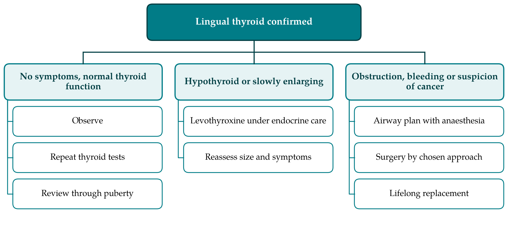
Surgery is reserved for airway obstruction, significant dysphagia, recurrent bleeding, failure of medical treatment or suspicion of malignancy. Carcinoma arising in a lingual thyroid is rare (Massine et al., 2001), and where it is suspected, assessment should follow the principles laid down for thyroid nodules in children (Francis et al., 2015), with needle sampling preferred to incisional biopsy. Transoral excision suits most glands, and external approaches through the hyoid or lateral pharynx have been described for larger ones (Prasad and Bhat, 2000; Rahbar et al., 2008). Whatever the route, the anaesthetist needs warning that a mass lies above the larynx, and the family needs to understand before the operation that lifelong hormone replacement will follow if no other thyroid tissue exists. Radioiodine ablation, used in some older adults who are unfit for surgery, is generally avoided in the young.
The position is different in the minority who have a normally sited gland as well as lingual tissue. Excision of the ectopic tissue then carries no threat of permanent hypothyroidism, and the threshold for surgery in a symptomatic adolescent can reasonably be lower. This is one reason for completing the imaging steps in every case: the same endoscopic appearance can call for opposite advice depending on what lies in the neck.
4. Putting the Pathway to Use
Nothing in the pathway calls for unusual resources. Thyroid function tests and ultrasound are available in any hospital, scintigraphy in most regional centres, and the order of investigations is chosen so that the cheapest and least invasive come first. What the pathway does require is that the clinician who first sees the mass recognise the possibility and resist the reflex to sample it. A short written protocol in the endoscopy clinic, stating that a midline tongue base mass in a child or adolescent is investigated with thyroid tests and neck ultrasound before any biopsy, would meet that need.
Follow-up is part of the pathway. Patients managed by observation need periodic thyroid function tests, because hypothyroidism may develop later, and women should be advised to seek review when planning or early in pregnancy. Those treated surgically need endocrine follow-up for life. Transition from paediatric to adult services is a point at which such patients are easily lost.
Communication deserves the same care as investigation. The diagnosis should be written prominently in the record and in letters to the family doctor, in words that a clinician unfamiliar with the condition cannot misread: the tongue base mass is thyroid tissue, it may be the only thyroid the patient has, and it is not to be biopsied or removed without endocrine advice. Young people move, change doctors and attend emergency departments, and an endoscopist meeting the mass for the first time years later will face the same temptation as the first.
5. Limitations and What Remains to Be Shown
The pathway rests on a literature of case reports and retrospective series, the weakest forms of clinical evidence, and on the judgement of the authors of those reports. No comparative study of observation, suppression and surgery exists, and none is likely given the rarity of the condition. The indications for surgery proposed here are therefore conventional and not evidence-based in any strict sense. The pathway has not been applied to a series of patients, and its effect on the outcomes that matter, namely avoidance of inadvertent removal of the only thyroid tissue, time to diagnosis and exposure to radiation, has not been measured.
Several specific points are uncertain. How often suppressive treatment produces a worthwhile reduction in size is unknown. The need for scintigraphy when ultrasound has already shown an empty thyroid bed could be questioned, and some centres may reasonably omit it in a typical case. The best surgical approach has been debated for decades (Batsakis et al., 1996; Williams et al., 1996) and newer transoral techniques have yet to be compared with older ones. A multicentre registry would be the practical means of addressing these questions. The pathway is also written for the adolescent. In infants identified by newborn screening the questions and the investigations differ and are covered by endocrine guidance (Léger et al., 2014), and in adults the range of tumours that may arise in the tongue base is wider.
6. Conclusion
Lingual thyroid is rare enough that a clinician may never see a case and common enough among tongue base masses in the young that it must always be considered. The proposed pathway places thyroid function tests, neck ultrasound and scintigraphy ahead of any tissue sampling, brings endocrinology into the decision from the start, and reserves surgery for defined indications. It is offered as a framework for departments to adapt and, ideally, to audit, and not as a validated standard.
Conflict of interest
To be declared by the author(s) before publication.
Funding
To be declared by the author(s) before publication.