1. Introduction
Leptospirosis is a bacterial zoonosis caused by pathogenic spirochaetes of the genus Leptospira, which colonise the renal tubules of mammals and are shed in urine. People and animals are infected through broken skin or mucous membranes in contact with urine or with water and soil contaminated by it (Levett, 2001). The disease is most frequent in the humid tropics, and estimates of its global burden in people place most cases there (Costa et al., 2015). Dry regions receive less attention, on the reasonable ground that leptospires survive poorly when desiccated.
Working horses, donkeys and mules are of interest in this context for two reasons. They live in close contact with households that depend on them and have limited access to veterinary care, as welfare assessments of these populations have documented (Pritchard et al., 2005). And they drink daily from communal troughs, ponds and canals, the places where exposure in a dry landscape would be expected. A serological survey that finds antibody in such animals therefore invites strong conclusions. This note considers how far those conclusions can go.
2. What the Microscopic Agglutination Test Shows
The reference serological method is the microscopic agglutination test, in which dilutions of serum are mixed with live cultures of selected serovars and examined under dark-field microscopy for agglutination (WHO, 2003). The result is a titre against each antigen in the panel. Several properties of the test shape what a survey can claim, and Figure 1 gathers them. The test detects antibody, and so indicates exposure at some time and not current infection or shedding. Agglutinating antibody may persist for months or years. A single titre at or above 100 is the conventional evidence of past exposure, whereas diagnosis of acute disease requires a rising titre in paired samples or demonstration of the organism (Picardeau, 2013).
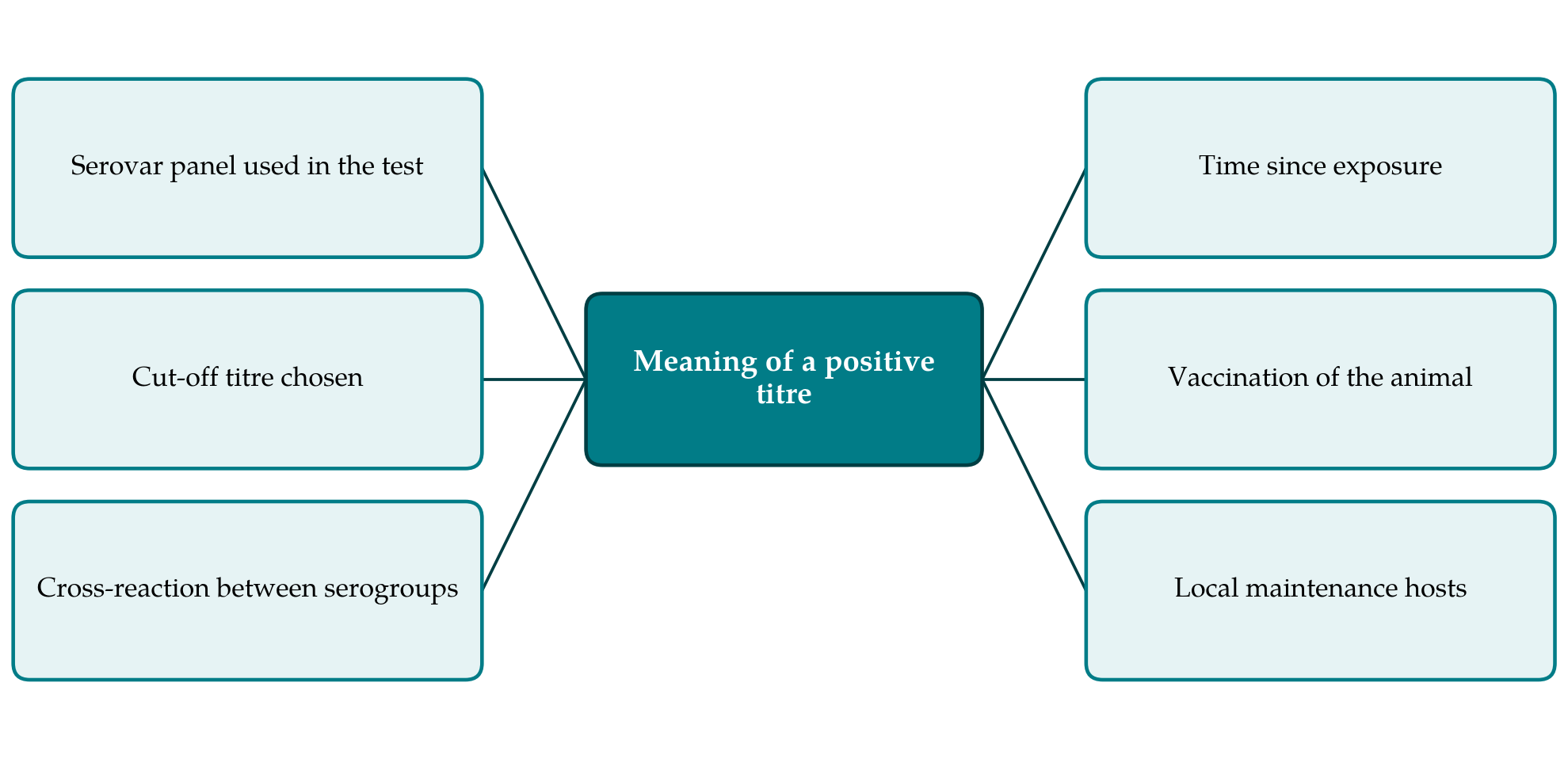
The panel matters as much as the cut-off. Antibody is directed mainly against the lipopolysaccharide that defines the serogroup, so a serum is recorded as negative if the infecting serogroup is not represented. Panels borrowed from another country or from a cattle laboratory may miss locally circulating strains. Cross-reactions between serogroups are common, and the antigen giving the highest titre is not a dependable guide to the infecting serovar (Levett, 2001). Statements that a particular serovar "predominates" in a surveyed population should be read as statements about serogroup reactivity. Table 1 summarises what each of the main laboratory approaches contributes.
| Approach | What it shows | Main limitation |
|---|---|---|
| Microscopic agglutination test | Antibody to serogroups in the panel; past exposure | Needs live cultures; depends on panel and cut-off |
| Paired-sample serology | Rising titre; recent infection | Second visit rarely feasible in field surveys |
| Enzyme immunoassay | Genus-level antibody | Few assays validated for equids |
| Polymerase chain reaction on urine | Leptospiral DNA; current shedding | Shedding intermittent; no serovar identity |
| Culture and typing | The local strain itself | Slow, demanding and often unsuccessful |
3. Leptospira and the Equine Host
Horses are susceptible to a range of serovars. Most infections pass unnoticed. Clinical consequences that are well documented include abortion and stillbirth, acute renal disease in foals and, most characteristically, equine recurrent uveitis, an immune-mediated inflammation of the eye that can follow infection by months and lead to blindness (Verma et al., 2013; Divers et al., 2019). Serovars of the Pomona serogroup are those most often linked with abortion and uveitis in North America, and Grippotyphosa with uveitis in Europe, while serovar Bratislava is widely regarded as adapted to the horse, though its role in disease is disputed (Ellis, 2015).
Almost all of this knowledge comes from horses in temperate, high-income settings. Donkeys and mules have been studied far less, and it cannot be assumed that their response to infection matches that of the horse. For working equids the relevant clinical questions, whether exposure contributes to eye disease, reproductive loss or poor condition in these populations, have scarcely been asked. A horse can also excrete leptospires in urine, which gives seropositivity a possible public health meaning (Hamond et al., 2013), but antibody alone does not identify a shedder.
4. Exposure in Semi-Arid Environments
Aridity limits the survival of leptospires in open ground. It does not remove transmission. Reviews of the environmental determinants of leptospirosis consistently identify contact with surface water, flooding and rainfall as risk factors (Mwachui et al., 2015), and in a dry landscape those conditions are confined in space and time. Animals of many species converge on the few available wells, troughs, reservoirs and irrigation channels, whose muddy margins stay moist and are contaminated repeatedly with urine. Short rainy seasons produce standing water and a pulse of rodent breeding. Irrigated cropping creates permanently wet ground inside an otherwise dry district.
The maintenance hosts in such places are the usual ones: rodents for serogroups such as Icterohaemorrhagiae, and cattle, pigs and dogs for the serovars adapted to them (Adler and de la Peña Moctezuma, 2010). Working equids are typically stabled at night in or beside the household compound, where feed stores attract rats, and are tethered by day near other livestock. Antibody in these animals is therefore plausible on ecological grounds and need not indicate anything unusual about the region. Equally, a low figure from a survey carried out late in a dry season, with a small panel, says little about the risk after rain.
5. Points for the Design and Reporting of Surveys
Several modest measures would make serological reports on working equids more useful. The sampled population should be described, with the way animals were selected, since animals brought to a free clinic differ from those that are not. The season of sampling and recent rainfall should be stated. The full antigen panel, the strains used and the cut-off titre belong in the methods, and results should be tabulated by titre and serogroup and not reduced to a single percentage. Species should be reported separately for horses, donkeys and mules. Vaccination against leptospirosis is unusual in working equids, but its absence should be confirmed.
Where resources allow, two additions change what can be concluded. Testing urine by polymerase chain reaction in a subset shows whether seropositive animals are shedding. Sampling the species that share the water point, particularly cattle, dogs and trapped rodents, and attempting isolation, indicates where the equids' exposure originates (Ellis, 2015). An eye examination and a brief reproductive history for each animal cost little and would begin to connect serology with the conditions known to follow infection in horses.
6. Conclusion
Antibody to Leptospira in working equids of a semi-arid region is evidence that the animals have met the organism, most likely at shared water and in contact with other hosts. It is not, taken alone, evidence of disease in the animal or of risk to its handlers, and the prevalence reported depends on the panel, the cut-off and the season. Surveys that state these choices clearly and add even limited clinical and molecular information would allow the significance of exposure in these neglected animals to be judged.
Conflict of interest
To be declared by the author(s) before publication.
Funding
To be declared by the author(s) before publication.