1. Introduction
The assessment of acute chest pain is organised around a small number of conditions that kill quickly: acute coronary syndrome, pulmonary embolism, aortic dissection, cardiac tamponade and tension pneumothorax. The established tools are the history, the examination, a 12-lead electrocardiogram recorded within ten minutes of arrival, and serial measurement of high-sensitivity cardiac troponin (Gulati et al., 2021; Collet et al., 2021). These tools work well for myocardial infarction. They are less informative about the other conditions on the list, and troponin results take time to return.
Ultrasound machines small enough to be carried to the bedside have changed what a clinician can learn in those early minutes (Moore and Copel, 2011). A focused cardiac examination can show a pericardial effusion, a severely impaired left ventricle or a dilated right ventricle within a few minutes, findings that redirect the work-up before any laboratory result is available. Professional bodies have defined the scope of such examinations and the training they require (Labovitz et al., 2010; Spencer et al., 2013; Via et al., 2014).
What those documents do not provide is an operational protocol for triage: a fixed sequence that says which patients, which views, which questions, and what to do with each answer. Without one, practice depends on the habits of whoever is on duty. This article proposes such a protocol. It is assembled from published recommendations and has not been tested in patients by the authors; the aim is to give departments a defined procedure that can be adopted, audited and formally evaluated.
2. Background
Focused cardiac ultrasound differs from comprehensive echocardiography in purpose and in method. A comprehensive study follows a long standard sequence of views and measurements and is reported by a specialist (Mitchell et al., 2019). A focused study answers a few predefined questions, mostly by visual judgement, and is interpreted at once by the person holding the probe. The international recommendations limit its targets to findings that are gross and reliably recognised after modest training: global left ventricular systolic function, right ventricular size, pericardial effusion, and the size and respiratory variation of the inferior vena cava (Via et al., 2014). Regional wall motion and valve lesions are regarded as needing more expertise.
European recommendations on emergency echocardiography make the same distinction and stress that a focused study should lead to a comprehensive one whenever it is abnormal or inconclusive (Neskovic et al., 2013). Emergency medicine has also produced multi-organ sequences for particular presentations, of which the best known are the RUSH examination for undifferentiated shock (Perera et al., 2010) and the BLUE protocol for acute respiratory failure (Lichtenstein and Mezière, 2008). No equivalent sequence has become standard for the stable patient with chest pain at triage, which is the gap the present proposal addresses.
3. Design Principles
Four principles governed the design. First, the scan must not delay any time-critical step. The electrocardiogram comes first, and a patient whose tracing shows ST-segment elevation proceeds to reperfusion without waiting for ultrasound. Second, the questions are binary and limited to findings within the accepted scope of focused ultrasound, so that operators with basic accreditation can answer them. Third, the protocol is used to rule in and never to rule out: a positive finding accelerates care, and a negative scan leaves the standard pathway unchanged. Fourth, every scan leaves a stored record that another person can review.
4. The Protocol
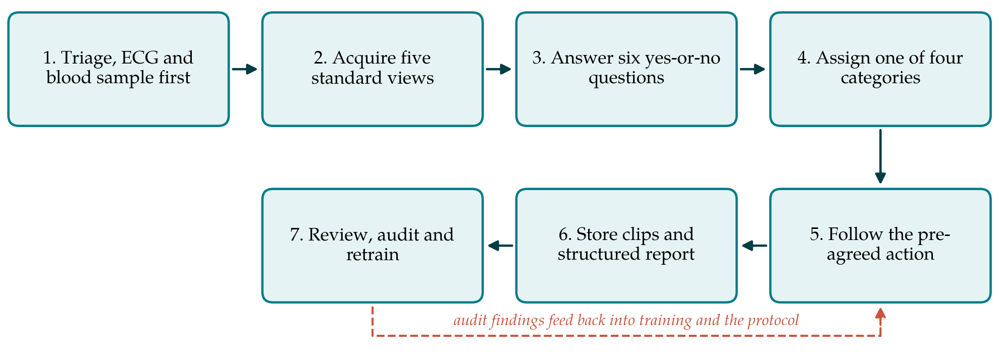
The protocol has seven steps, shown in Figure 1. The first five take place at the bedside and the last two afterwards.
4.1. Step 1: Selection and timing
The protocol applies to adults presenting with non-traumatic chest pain or an equivalent symptom such as unexplained breathlessness or syncope with chest discomfort. The scan is performed after the first electrocardiogram has been recorded and read and after blood has been drawn, during the interval in which the patient would otherwise be waiting. A fixed upper limit on scanning time is set locally; five minutes is suggested as a starting point. If adequate images cannot be obtained in that time, the scan is abandoned and recorded as non-diagnostic.
4.2. Step 2: Five views
The operator acquires, in a fixed order, the parasternal long-axis view, the parasternal short-axis view at the level of the papillary muscles, the apical four-chamber view, the subcostal four-chamber view and the subcostal view of the inferior vena cava. These are the views named in the focused ultrasound recommendations (Spencer et al., 2013) and form the core of European training syllabuses (Neskovic et al., 2018). Where the operator is also competent in lung ultrasound, one anterior view on each side of the chest may be added to look for lung sliding and for diffuse B-lines, using the definitions of the international lung ultrasound recommendations (Volpicelli et al., 2012). The lung views are optional and do not count towards the core record.
4.3. Step 3: Six questions
From those views the operator answers six questions, each as yes, no or cannot assess. Table 1 lists them with the view that best shows each finding and the diagnosis it should bring to mind. All are judged by eye. The right ventricle is called dilated when it appears as large as or larger than the left in the apical four-chamber view, in line with the qualitative sign used in the European guideline on pulmonary embolism (Konstantinides et al., 2020). The vena cava is called plethoric when it is distended and fails to collapse on inspiration, the pattern that Kircher et al. (1990) related to raised right atrial pressure.
| Question | Best view | A positive answer prompts consideration of |
|---|---|---|
| Is there a pericardial effusion? | Subcostal; parasternal long axis | Tamponade; pericarditis; dissection with haemopericardium |
| Is there chamber collapse with an effusion? | Subcostal; apical four-chamber | Tamponade requiring urgent senior review |
| Is left ventricular function severely reduced? | Parasternal long and short axis | Extensive infarction; acute heart failure; myocarditis |
| Is the right ventricle dilated? | Apical four-chamber | Pulmonary embolism; right ventricular infarction |
| Is the aortic root visibly dilated, or a flap seen? | Parasternal long axis | Acute aortic syndrome; urgent computed tomography |
| Is the inferior vena cava plethoric? | Subcostal | Raised right-sided pressure from any of the above |
Two deliberate omissions should be noted. The protocol does not ask about regional wall motion, because the consensus documents place it beyond basic focused competence and because an apparently normal wall does not exclude ischaemia. Nor does it ask for any measurement. An operator with advanced training may record additional observations as free text, but they do not alter the category assigned.
4.4. Step 4: Four categories
The answers are reduced to one of four categories. Category A, critical, is assigned when there is an effusion with chamber collapse, or a dilated root or flap in a patient with compatible pain. Category B, abnormal, covers any other positive answer. Category C is assigned when all six answers are no; it is worded as no focused abnormality seen and never as a normal echocardiogram. Category D records that one or more questions could not be assessed. Fixing the categories in advance removes the temptation to soften or embellish a report at the bedside.
4.5. Step 5: Action attached to each category
Each category carries an action agreed beforehand between emergency medicine, cardiology and radiology. Category A triggers immediate review by a senior clinician and transfer to a resuscitation area. Category B moves the patient up the queue for senior review and for definitive imaging appropriate to the finding, for instance computed tomographic pulmonary angiography when the right ventricle is dilated and the clinical probability of embolism supports it. Categories C and D change nothing: the patient continues on the department's standard chest pain pathway, with serial troponin and risk stratification as the guidelines direct (Gulati et al., 2021). In particular, suspected dissection is investigated by computed tomography whatever the ultrasound shows.
4.6. Step 6: Record
A short clip of each view is stored against the patient's record, together with a structured report giving the six answers, the category, the time of the scan and the operator's name. Emergency ultrasound guidelines treat archiving and documentation as part of the examination and not as an optional extra (American College of Emergency Physicians, 2017). A scan that is not stored cannot be reviewed, cannot be used for teaching, and gives the next clinician nothing to compare against.
4.7. Step 7: Training and quality review
Operators should hold whatever basic focused ultrasound accreditation the department recognises and should complete a short induction on this protocol specifically. A named lead reviews a regular sample of stored scans, together with every Category A and B scan, for image adequacy and for agreement with the recorded answers. Disagreements are fed back to the operator. Where a comprehensive echocardiogram or other definitive test followed, its result is compared with the triage category, and the comparisons are collated for audit.
5. Adapting the Protocol to a Department
Departments differ in staffing, equipment and patient mix, and three elements are meant to be set locally. The time limit can be lengthened where operators are less experienced or shortened in a very busy unit. The optional lung views can be made part of the core set where pneumothorax and pulmonary oedema are frequent diagnostic problems. The actions in Step 5 depend on what imaging is available out of hours and must be written with the services that provide it. The six questions and four categories should be left unchanged, since their value lies in being the same for every operator and every shift.
Handheld devices make the protocol feasible in the triage bay itself, although their small screens and limited penetration will increase the proportion of Category D scans in some patients. Infection control, charging and secure image transfer need to be arranged before the first scan is done. A department that cannot store images reliably should resolve that problem before introducing the protocol.
6. Limitations and What Remains to Be Shown
The protocol is proposed and has not been validated here. It rests on consensus statements whose own evidence is largely observational, and on the assumption that findings recognisable in general focused ultrasound are recognised equally well at triage, under time pressure, by the operators a department actually has. That assumption needs testing. The first requirement is a diagnostic accuracy study, designed and reported according to the STARD statement (Bossuyt et al., 2015), in which triage categories are compared with final adjudicated diagnoses in consecutive patients. The second is a study of agreement between operators, and between operators and expert review of the stored clips, expressed with a chance-corrected statistic and interpreted with the usual caution (Landis and Koch, 1977).
Accuracy alone would not justify adoption. It would also have to be shown that the protocol shortens the time to diagnosis of the non-coronary emergencies without lengthening the time to electrocardiogram or to reperfusion, and that Category C results do not lead clinicians to discharge patients earlier than the standard pathway allows. False reassurance is the principal hazard of any bedside test with limited sensitivity, and a prospective evaluation should measure it directly. Incidental findings and the downstream testing they generate are a further cost that should be counted.
7. Conclusion
Focused cardiac ultrasound is already used in the early assessment of chest pain, but mostly without a fixed method. The protocol proposed here specifies five views, six questions, four categories and the action tied to each, places the scan after the electrocardiogram and beside the standard pathway, and requires a stored record and regular review. It gives departments something definite to implement and investigators something definite to test. Its accuracy, reproducibility and effect on patient flow are unknown and should be established before it is treated as routine care.
Conflict of interest
To be declared by the author(s) before publication.
Funding
To be declared by the author(s) before publication.