1. Introduction
Fluorescence in situ hybridization grew out of the demonstration that nucleic acid probes carrying a hapten could be detected on chromosomes without radioactivity, and that such probes allowed quantitative analysis of interphase nuclei as well as metaphases (Pinkel et al., 1986). Its application to archival pathology material made it a routine tool for detecting gene amplification and rearrangement in solid tumors. Guidelines for HER2 testing in breast cancer and for ALK testing in lung cancer brought the method into almost every diagnostic laboratory (Wolff et al., 2018; Lindeman et al., 2013).
Many patients who need these tests never have a surgical specimen. Their diagnosis is made on a pleural fluid or an aspirate, and the material for ancillary testing is a cell block: a pellet of cells fixed, embedded in paraffin and sectioned like tissue. Guidance on thoracic small samples accepts cell blocks for molecular and in situ studies and stresses that their handling must be known and controlled (Roy-Chowdhuri et al., 2020). In practice a cell block is a less uniform substrate than a tissue block, and protocols written for tissue sections do not always transfer.
The commonest technical failure is a slide with weak or absent signals in nuclei that otherwise look intact, or bright signals in nuclei so damaged that they cannot be counted. Both are failures of probe penetration and its control. This article proposes a stepwise procedure by which a laboratory can optimize penetration for its own cell blocks. It describes a method and its rationale. No experimental results are presented, and the procedure has not been validated here.
2. Barriers Between Probe and Target
Formaldehyde fixes by forming methylene bridges between reactive groups on proteins and between proteins and nucleic acids, and the reaction continues for many hours after the fixative has penetrated (Fox et al., 1985). The result is a dense network around the chromatin that preserves morphology and excludes large molecules. Prolonged fixation deepens the network and also damages nucleic acids (Srinivasan et al., 2002), while delay before fixation allows degradation that no pretreatment can repair (Hewitt et al., 2008). Labeled probe fragments, typically a few hundred nucleotides long, must cross this network to reach denatured target DNA. Figure 1 shows the barriers in the order in which a probe meets them and the step that deals with each.
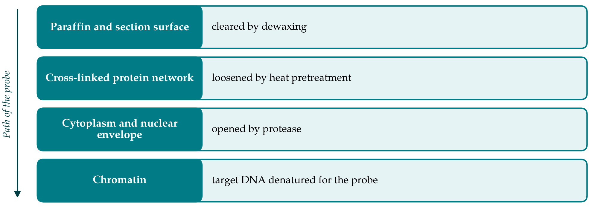
Pretreatment reverses enough of the fixation to let the probe in. After dewaxing, sections are heated in a buffer or a chaotropic solution, a step that owes its logic to the heat-induced retrieval introduced for immunohistochemistry (Shi et al., 1991), and are then digested with a protease, usually pepsin at acid pH or proteinase K. Published protocols for paraffin sections and tissue microarrays agree that these two steps, and above all their duration, decide the outcome (Chin et al., 2003; Summersgill et al., 2008). Too little treatment leaves the target covered. Too much dissolves the nuclear proteins that hold chromatin in place, and the DNA is lost or the nucleus collapses.
3. Why Cell Blocks Behave Differently
Three features set cell blocks apart from tissue. The first is the variety of preparation methods. Cells may be clotted with plasma and thrombin, suspended in agar or a commercial gel, collected in a collodion bag or processed by an automated system, and each matrix has its own permeability (Jain et al., 2014; Krogerus and Kholová, 2018). The second is pre-fixation. Samples often reach the laboratory in an alcohol-based transport or liquid-based cytology medium and meet formalin only afterwards. Alcohols coagulate proteins instead of cross-linking them, so a block of this kind may need a different pretreatment from one fixed in formalin alone (Shidham, 2019).
The third feature is the absence of stroma. Cells in a block lie singly or in small groups, with little collagen around them, and are reached by reagents faster than cells deep in a tissue section. A digestion time that suits a core biopsy can over-digest a cell block. Cellularity is also uneven, and good blocks depend on concentrating the cells in one plane of section (Nathan et al., 2000). The consequence for the laboratory is that conditions must be established on its own blocks, made by its own method, and cannot be copied from a tissue protocol or from another institution.
4. The Proposed Optimization Procedure
The procedure has six stages. It assumes a laboratory that already performs the assay on tissue sections and wishes to extend it to cell blocks, or one whose cell block failure rate is unacceptable.
4.1. Stage 1: Define the material and build control blocks
The laboratory first writes down how its cell blocks are made: collection medium, any alcohol exposure, fixative, fixation time, matrix and processing schedule. It then prepares control blocks by the same route from residual effusion specimens or cultured cells with a known status for the target of interest, in sufficient number to supply sections for all later stages. Sections are cut at a single stated thickness, four to five micrometers being usual for interphase analysis, and mounted on positively charged slides. Optimization on control material protects scarce patient blocks and makes runs comparable.
4.2. Stage 2: Fix a scoring rubric
Before any variable is changed, the criteria for judging a slide are fixed in writing. Four attributes are scored on a simple ordinal scale by two observers: signal intensity relative to background, the proportion of nuclei showing the expected signals, preservation of nuclear outline and counterstain, and non-specific or autofluorescent background. Table 1 lists the appearances that indicate insufficient and excessive treatment at each step, which the rubric is designed to capture. A written rubric prevents the common drift in which conditions are judged by the brightest field on the slide.
| Step | Variable to tune | Too little | Too much |
|---|---|---|---|
| Dewaxing | Solvent changes and time | Patchy hybridization; hazy film | Seldom a problem |
| Heat pretreatment | Buffer, temperature, time | Weak signal; strong autofluorescence | Sections lift; nuclei swell |
| Protease digestion | Enzyme, concentration, time | Cytoplasm persists; signals faint or absent | Ghost nuclei; pale counterstain; signal lost |
| Denaturation | Temperature and time | No signal despite good morphology | Blurred chromatin; diffuse signals |
| Hybridization | Duration and humidity | Low proportion of nuclei with signals | Drying artifact at edges |
| Stringency wash | Salt, temperature, time | High background; cross-hybridization | Specific signal weakened |
4.3. Stage 3: Tune heat pretreatment and digestion together
Heat pretreatment and protease digestion interact: a longer heat step shortens the digestion needed, and the best combination cannot be found by varying one while the other is held at an arbitrary value. The proposal is to test them jointly in a small factorial layout, for example three durations of heat pretreatment crossed with three durations of digestion at fixed temperature and enzyme concentration, giving nine slides from serial sections of one control block, all hybridized with the same probe lot in the same run. Factorial designs of this kind are standard practice for locating an optimum when two factors interact (Montgomery, 2017). The starting ranges should bracket the manufacturer's recommendation for the pretreatment kit in use, extending further toward shorter digestion than a tissue protocol would suggest.
Each slide is scored with the rubric. The condition chosen is not the one with the brightest signal but the one that sits in the middle of the acceptable region, so that ordinary variation between blocks does not push a case into failure. If no combination is acceptable, the buffer or the enzyme is changed and the grid repeated. Published protocols differ in the heating solution and the enzyme they recommend (Summersgill et al., 2008), which gives a ready list of alternatives.
4.4. Stage 4: Adjust denaturation, hybridization and washing
With pretreatment settled, the remaining steps are checked one at a time. Co-denaturation temperature and time are confirmed with a calibrated hot plate or hybridizer, since a deviation of a few degrees changes the result. Hybridization time is set according to the buffer: conventional formamide-based buffers are usually left overnight, whereas buffers based on other solvents were designed to shorten the step considerably (Matthiesen and Hansen, 2012). The stringency wash is the last control on background. Changes at this stage should be small, and any alteration that degrades the rubric scores sends the laboratory back to the previous setting.
4.5. Stage 5: Verify across preparation types
The chosen protocol is then run on a set of blocks that represents the range received in practice: formalin-only and alcohol-prefixed, bloody and clean, sparse and cellular, recent and archived. The aim is to learn where the protocol holds and where it does not. If alcohol-prefixed blocks behave consistently differently, a second validated protocol with its own digestion time is preferable to a single compromise that serves neither. A decision rule for the bench should follow, stating which protocol is applied to which type of block and what is done when the history is unknown.
4.6. Stage 6: Set cut-offs and monitor
Sectioning truncates nuclei, so some normal cells will show fewer signals than they contain, and overlapping nuclei can mimic gain. Cut-off values for calling a deletion, gain or rearrangement must therefore be derived on cell block sections processed by the final protocol, following the statistical approach set out in professional standards (Mascarello et al., 2011; Wolff et al., 2007). Thereafter each run carries a control section, the failure rate is recorded by block type, and a rise prompts a return to stage 3.
5. Fitting the Procedure to Practice
A laboratory with few cases can compress the scheme. The factorial grid can be reduced to two levels of each factor, and stage 5 can be met by accumulating cases over time, provided each new block type is run alongside a control. Laboratories using automated stainers have fewer free variables, and for them the procedure reduces to selecting among the pretreatment programs the instrument offers, scored by the same rubric. Where the assay is a regulated companion diagnostic, changes to the validated method may not be permitted, and the proper use of the procedure is then to define which cell blocks are acceptable for the unmodified assay. Pre-analytical control remains the most effective intervention of all: prompt fixation in neutral buffered formalin for a duration within the range recommended for the test (Wolff et al., 2018) removes much of the variation that optimization would otherwise have to absorb.
6. Limitations and What Remains to Be Shown
The procedure is a proposal, and its benefit over the informal trial and error that most laboratories use has not been measured. A fair test would compare failure rates and inter-observer agreement before and after its adoption across several laboratories that use different cell block methods. The rubric is ordinal and partly subjective; image analysis of signal-to-background ratio would be more objective but is not widely available. Control blocks made from cell lines or pooled effusions may not reproduce the behavior of scanty aspirates. And the scheme optimizes for one probe at a time, whereas probes differ in size and labeling, so a condition that suits a centromeric probe may be marginal for a small locus-specific one.
7. Conclusion
Probe penetration in formalin-fixed cell blocks is governed by a balance between unmasking the target and preserving the nucleus, and the balance point depends on how the block was made. The procedure proposed here asks a laboratory to characterize its own blocks, to judge slides by fixed written criteria, to tune heat pretreatment and digestion jointly, and to verify the outcome across the block types it receives before setting cut-offs. Each element is taken from established practice. Their combination into a defined sequence remains to be evaluated.
Conflict of interest
To be declared by the author(s) before publication.
Funding
To be declared by the author(s) before publication.