1. Introduction
Hormone therapy is the most effective treatment for vasomotor symptoms, and most women who use it start within a few years of their final menstrual period. For this group the absolute risks of treatment are low, but they are not nil, and venous thromboembolism is the harm that appears earliest. In the Women's Health Initiative trial of conjugated equine estrogens with medroxyprogesterone acetate, venous thrombosis was roughly twice as frequent in the treated group as in the placebo group (Rossouw et al., 2002), and a detailed analysis showed that the excess was greatest in the first year and was amplified by older age, obesity and factor V Leiden (Cushman et al., 2004).
Those trials used oral estrogen. Whether estradiol given through the skin carries the same risk has been asked since patches became widely available, and the question matters most at the start of treatment, when a prescriber chooses a route for a woman who may use it for years. Because thrombosis is uncommon in women in their early fifties, the question has been approached largely through laboratory markers of coagulation, which respond within weeks and can be measured in trials of modest size.
This review brings together the evidence on how oral and transdermal estradiol affect such markers, with attention to women in early postmenopause, and considers how far the laboratory findings can be read as evidence about clinical thrombosis. It reports no new data.
2. Scope and Approach
The article is a narrative review of the published literature. It draws on randomized comparisons of oral and transdermal estrogen that measured hemostatic variables, on the case-control and cohort studies that examined route of administration and venous thromboembolism, and on the position statements of professional societies. No formal search protocol was applied and no pooled estimate is offered. The aim is to describe the shape of the evidence and its limits, not to replace the published meta-analyses, which are cited where relevant.
3. Why the Route of Administration Matters
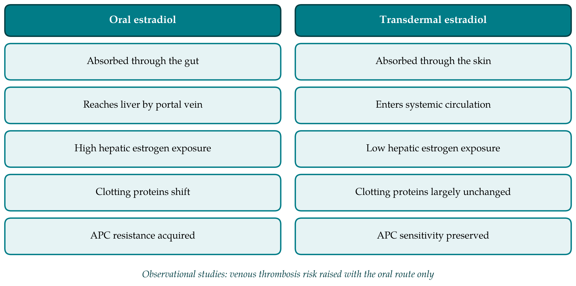
Estradiol swallowed as a tablet is absorbed into the portal circulation and reaches the liver before it reaches any other organ. A large fraction is converted to estrone and to conjugates in the gut wall and liver, so that the hepatocyte is exposed to estrogen concentrations several times higher than those in peripheral blood, and circulating estrone exceeds estradiol. Estradiol absorbed through the skin enters the systemic circulation directly, and the ratio of estradiol to estrone stays close to that of the premenopausal state (Kuhl, 2005).
The liver synthesizes most coagulation factors and their inhibitors, together with sex hormone-binding globulin, angiotensinogen and C-reactive protein, and the output of all of these is sensitive to estrogen. The hepatic first pass therefore offers a plausible mechanism by which the same hormone could have different hemostatic effects depending on how it is given. Figure 1 sets the two pathways side by side. The mechanism is an argument from pharmacology; the sections that follow examine whether measurement bears it out.
4. Laboratory Markers of Coagulation
No single test captures thrombotic tendency, and trials have reported a mixture of activation markers, inhibitor levels, global assays and fibrinolytic proteins. Table 1 summarizes the direction of change reported for the markers most often studied. The findings are taken up in three groups below.
| Marker | Oral estrogen | Transdermal estradiol | Representative sources |
|---|---|---|---|
| Prothrombin fragment 1+2 | Raised | Little or no change | Scarabin et al., 1997; Oger et al., 2003 |
| Antithrombin | Lowered | Little or no change | Scarabin et al., 1997 |
| Resistance to activated protein C | Increased | Slight or no increase | Post et al., 2003; Oger et al., 2003 |
| Plasminogen activator inhibitor-1 | Lowered | Little change | Vehkavaara et al., 2001 |
| C-reactive protein | Raised | Unchanged | Vehkavaara et al., 2001; Lowe et al., 2001 |
| Sex hormone-binding globulin | Raised markedly | Small rise | Kuhl, 2005 |
4.1. Markers of thrombin formation
Prothrombin fragment 1+2 is released when prothrombin is cleaved to thrombin and is the most widely used index of coagulation activation in this literature. In a randomized trial that assigned postmenopausal women to oral estradiol, transdermal estradiol, each with micronized progesterone, or no treatment, Scarabin et al. (1997) found that the oral regimen raised fragment 1+2 and lowered antithrombin activity, while the transdermal regimen produced no significant change in either. The same contrast was reported by the SARAH investigators in a placebo-controlled trial (Oger et al., 2003). Fragment 1+2 is a sensitive marker, but its concentration reflects clearance as well as formation, and the increases seen with oral estrogen are modest in absolute terms.
4.2. Natural anticoagulants and resistance to activated protein C
Activated protein C limits thrombin generation by inactivating factors Va and VIIIa. A poor plasma response to it is the phenotype of factor V Leiden, and an acquired form occurs with estrogen exposure. Work on oral contraceptives showed that a test based on thrombin generation detects this acquired resistance far better than the older clotting-time test (Rosing et al., 1999). Applied to menopausal therapy, the method showed a clear increase in resistance with oral estradiol and a much smaller one with the transdermal route (Post et al., 2003). Oger et al. (2003) likewise found that oral estradiol induced resistance relative to both placebo and transdermal estradiol, and that transdermal estradiol did not differ significantly from placebo.
These findings are the strongest laboratory support for a difference between routes, because acquired resistance to activated protein C has a recognized inherited counterpart with an established thrombotic risk. They remain surrogate measurements. The degree of resistance that translates into a given clinical risk in a postmenopausal woman has not been defined.
4.3. Fibrinolysis and inflammation
Oral estrogen does not move every variable in a prothrombotic direction. It lowers plasminogen activator inhibitor-1, which would be expected to favor fibrinolysis, and it raises C-reactive protein, an effect now generally attributed to hepatic synthesis and not to systemic inflammation. In a randomized comparison, Vehkavaara et al. (2001) recorded both changes with oral estradiol and neither with transdermal estradiol. A population survey that compared users of the two routes reached a similar picture for factor IX, resistance to activated protein C and C-reactive protein (Lowe et al., 2001). The net result of oral treatment is thus a mixed profile in which procoagulant changes coexist with enhanced fibrinolytic potential, and the balance cannot be read from any one marker.
5. From Markers to Clinical Events
Whether the laboratory contrast corresponds to a clinical one has been tested only in observational studies. The first was the French ESTHER case-control study, which found that current use of oral estrogen was associated with a several-fold increase in the odds of idiopathic venous thromboembolism whereas transdermal estrogen was not associated with any increase (Scarabin et al., 2003; Canonico et al., 2007). The E3N cohort of French women gave a concordant result (Canonico et al., 2010b), as did the Million Women Study in the United Kingdom (Sweetland et al., 2012).
A meta-analysis of the studies available in 2008 concluded that oral estrogen increased the risk of venous thromboembolism and that transdermal estrogen did not appear to (Canonico et al., 2008), and a later synthesis reached the same conclusion with more data (Rovinski et al., 2018). The largest single analysis, nested in two British primary care databases, again found raised risk with oral preparations and none with transdermal ones, and it added that among oral products conjugated equine estrogens carried a higher risk than estradiol (Vinogradova et al., 2019).
An informative detail comes from women with inherited thrombophilia. In ESTHER, carriers of factor V Leiden or the prothrombin G20210A variant who took oral estrogen had a risk far above that conferred by either exposure alone, while carriers who used transdermal estrogen had a risk similar to that of carriers who used none (Straczek et al., 2005). This is what would be expected if oral estrogen adds an acquired resistance to activated protein C to an inherited one.
The limits of this evidence should be stated plainly. Route was not assigned at random. In several countries clinicians prefer patches for women they regard as being at higher vascular risk, which would bias against the transdermal route, but confounding in the opposite direction cannot be excluded. The studies are consistent across designs and populations, and the association shows the specificity and biological coherence that support a causal reading, yet it remains an association (Olié et al., 2010).
6. Evidence Specific to Early Postmenopause
Most of the randomized marker studies enrolled women across a broad postmenopausal age range, and few analyzed results by time since menopause. The trial designed for recently menopausal women is the Kronos Early Estrogen Prevention Study, which compared low-dose oral conjugated equine estrogens, transdermal estradiol and placebo, each active arm with cyclic micronized progesterone, in women within three years of their final period. Its primary outcome was progression of carotid intima-media thickness, which did not differ between groups (Harman et al., 2014). The oral and transdermal arms differed in their effects on lipids, insulin sensitivity and other hepatic proteins in the directions pharmacology predicts, and venous thrombotic events were too few to compare (Miller et al., 2019).
Two cautions follow. The oral comparator in that trial was conjugated equine estrogens and not estradiol, so it is not a pure test of route for one molecule. And the ELITE trial, which did stratify women by time since menopause, used oral estradiol only and addressed atherosclerosis, not hemostasis (Hodis et al., 2016). The long-term follow-up of the Women's Health Initiative indicates that absolute risks of treatment are lower in women in their fifties than in older women (Manson et al., 2013), which is reassuring for either route but also means that any difference between routes will be small in absolute terms and hard to demonstrate in this age group.
7. The Contribution of the Progestogen
Women with a uterus take a progestogen with their estrogen, and the choice is not neutral. In the E3N cohort, norpregnane derivatives were associated with venous thrombosis while micronized progesterone and pregnane derivatives were not (Canonico et al., 2010b). A laboratory study in users of transdermal estrogen found resistance to activated protein C in those taking norpregnanes and not in those taking micronized progesterone (Canonico et al., 2010a). A comparison of routes that ignores the accompanying progestogen can therefore mislead, and marker studies should report it.
8. Gaps and Future Work
Professional societies have drawn practical conclusions from this body of work while acknowledging its weaknesses. The Endocrine Society suggests a non-oral route for women at increased risk of venous thromboembolism (Stuenkel et al., 2015), and the North American Menopause Society notes that observational data indicate lower risk with transdermal therapy while pointing out that no randomized trial has compared the routes for this outcome (NAMS, 2022).
Several questions are open. A trial powered for thrombosis in healthy women in their early fifties would need to be very large, and it is unlikely to be funded; well-designed emulations of such a trial in linked health records are the realistic alternative. Head-to-head marker studies of oral and transdermal estradiol at doses matched for symptom relief, restricted to women within a few years of menopause and using a standardized thrombin generation assay, would address the gap this review has identified. The relation between the size of a change in resistance to activated protein C and subsequent thrombosis needs prospective study. And the newer routes and products, including gels, sprays and vaginal rings delivering systemic doses, have far less hemostatic evidence behind them than patches do.
9. Conclusion
Randomized trials show that oral estrogen raises markers of thrombin formation, lowers antithrombin and induces resistance to activated protein C, and that transdermal estradiol has little or no effect on these variables. Observational studies in several countries find venous thromboembolism associated with oral but not with transdermal treatment. The two lines of evidence support each other, and the hepatic first pass explains them. Direct evidence in recently menopausal women is thin, no trial has tested the routes against a thrombotic endpoint, and the markers are surrogates whose predictive value is not quantified. A preference for the transdermal route in women with additional risk factors for thrombosis is a reasonable reading of the literature, and it should be presented as such: probable, well supported and unproven.
Conflict of interest
To be declared by the author(s) before publication.
Funding
To be declared by the author(s) before publication.