1. Introduction
Hemophagocytic lymphohistiocytosis (HLH) is a syndrome of uncontrolled immune activation in which cytotoxic lymphocytes and macrophages, failing to switch off after a stimulus, release cytokines in excess and infiltrate the marrow, liver, spleen and central nervous system. Untreated it is frequently fatal. It was first characterised in infants with inherited defects of lymphocyte cytotoxicity, but it is now recognised across the whole span of life, and in adults it most often arises as a complication of infection, malignancy or autoimmune disease (Ramos-Casals et al., 2014).
Young adults occupy an awkward position in this picture. They are past the age at which paediatricians think of familial disease and younger than most people with lymphoma-associated HLH. When a previously well person in the late teens to thirties develops persistent fever, falling blood counts and deranged liver tests in the days or weeks after a viral illness, the working diagnoses are usually severe viral infection itself, bacterial sepsis or a drug reaction. HLH is considered late, sometimes only once several organs are failing, although the laboratory clues are inexpensive and available in almost any hospital.
This article proposes a stepwise diagnostic approach for that scenario. It is addressed to general physicians, infectious disease specialists and intensivists, who are likelier than haematologists to meet such patients first. The approach is a synthesis of published diagnostic criteria, a validated probability score and expert consensus statements. It is offered as a structured proposal: it has not been tested prospectively, the article describes no individual cases, and the sequence is intended to support specialist judgement and not to replace it. Treatment is mentioned only so far as the diagnostic steps bear on it.
2. Background
The design of the approach rests on three points: the overlap between inherited and acquired disease, the range of viral triggers, and the reasons the diagnosis is so often made late.
2.1. Primary and secondary forms
The inherited forms of HLH result from mutations that disable the granule-mediated killing pathway of natural killer (NK) cells and cytotoxic T cells; the first to be identified was in the perforin gene (Stepp et al., 1999). A lymphocyte that cannot kill an infected or antigen-presenting cell goes on being stimulated by it, and the resulting cytokine output activates macrophages. Secondary, or acquired, HLH reaches the same end state without a known single-gene cause. The division is less sharp than the names suggest. Zhang et al. (2011) found hypomorphic variants in the genes responsible for familial disease in a proportion of adults with apparently acquired HLH, which implies that some young adults carry a partial defect that declares itself only under the stress of a strong infectious trigger.
2.2. Viral triggers
Virus-associated hemophagocytic syndrome was described as a distinct entity by Risdall et al. (1979). Herpesviruses dominate the list of triggers, Epstein-Barr virus (EBV) above all, followed by cytomegalovirus (CMV); HIV, influenza, dengue and parvovirus B19 are also well documented (Rouphael et al., 2007). EBV-associated HLH ranges from a self-limiting illness to a fulminant one and has been studied most closely in East Asia (Imashuku, 2002). In young men, fulminant EBV infection with HLH may be the first sign of X-linked lymphoproliferative disease. The hyperinflammation seen in some people with severe COVID-19 renewed general interest in screening for the syndrome (Mehta et al., 2020).
2.3. Why the diagnosis is missed
No single feature of HLH is specific. Fever, cytopenias, raised ferritin, abnormal liver function and coagulopathy all occur in severe sepsis, and in HLH they accumulate over days, so that the full pattern may not be present at any one assessment. Hemophagocytosis in the marrow, the finding that gives the syndrome its name, is often absent on the first aspirate (Gupta et al., 2008) and can be seen in other conditions. The diagnosis therefore depends on someone deciding to look for a combination of findings and to keep looking.
3. The Proposed Approach
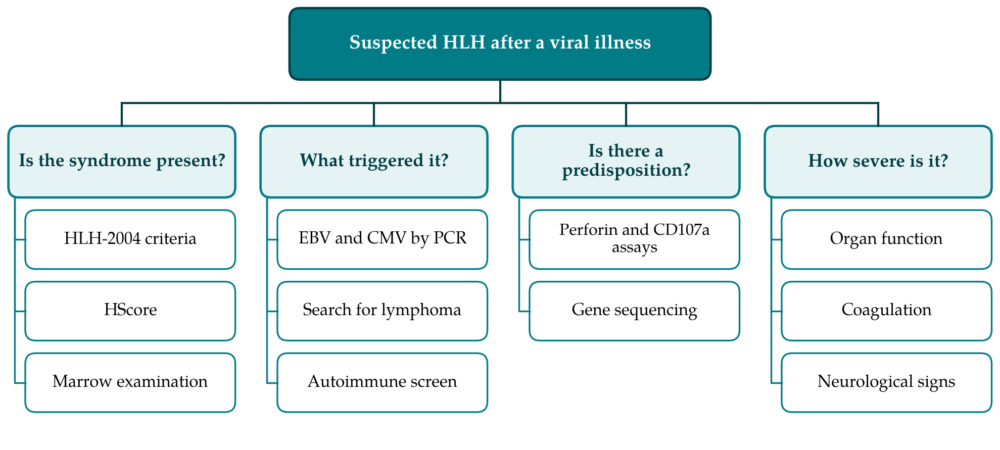
The approach has seven steps. They are set out below in the order in which they would usually be started, though in a seriously ill person several run in parallel. Table 1 compares the two instruments on which Step 3 relies, and Figure 1 shows the four questions that the work-up as a whole is meant to answer.
| Feature | HLH-2004 criteria | HScore | Practical note |
|---|---|---|---|
| Fever | One criterion | Graded by peak temperature | Often persistent despite antibiotics |
| Organomegaly | Splenomegaly | Liver, spleen or both | Imaging is more sensitive than palpation |
| Cytopenias | Two or more lineages affected | Graded by number of lineages | The trend matters as much as the value |
| Ferritin | 500 μg/L or above | Graded in three bands | Widely available; not specific in adults |
| Triglycerides, fibrinogen | Raised fasting triglycerides or low fibrinogen | Scored separately | Fibrinogen falls despite inflammation |
| Hemophagocytosis | In marrow, spleen or lymph node | On marrow aspirate | Neither required nor sufficient |
| NK-cell activity, soluble CD25 | Two separate criteria | Not included | Specialised laboratories only |
| AST, known immunosuppression | Not included | Both included | Available at first assessment |
3.1. Step 1: Recognise the pattern
The prompt for the work-up is a clinical pattern and not a single result. HLH should enter the differential diagnosis when fever continues beyond the expected course of a viral illness, or returns after initial improvement, together with any two of the following: a fall in two or more blood cell lines, a palpable or imaged spleen, liver enzymes rising without another explanation, unexplained coagulopathy, or new neurological symptoms. Failure to respond to appropriate antibiotics strengthens the suspicion. The threshold is deliberately low at this point, since what follows is a set of inexpensive blood tests.
3.2. Step 2: Request the first-line screen
The first-line screen consists of a full blood count with film, ferritin, fasting triglycerides, fibrinogen with a coagulation profile, liver enzymes, lactate dehydrogenase and renal function. Ferritin is the test most often omitted and among the most informative. In children a very high ferritin is strongly suggestive of HLH (Allen et al., 2008). In adults the same cannot be assumed: Schram et al. (2015) reviewed adults with extreme hyperferritinemia at a general hospital and found that most had renal failure, liver injury, infection or haematological malignancy, with HLH accounting for a minority. A normal ferritin makes HLH much less likely; a raised one is a reason to proceed and not a diagnosis. Because the abnormalities evolve, the screen is repeated every day or two while suspicion persists, and the direction of travel is noted. A falling fibrinogen in a febrile person is a particular warning, since fibrinogen ordinarily rises with inflammation.
3.3. Step 3: Apply the criteria and the score formally
Two instruments should be completed in writing. The HLH-2004 criteria, devised for a paediatric treatment trial, are met by a molecular diagnosis or by five of eight findings: fever; splenomegaly; cytopenias in at least two lineages; raised fasting triglycerides or low fibrinogen; hemophagocytosis in tissue; low or absent NK-cell activity; ferritin of 500 μg/L or more; and raised soluble CD25 (Henter et al., 2007). The HScore was derived and validated in adults and converts nine weighted variables into a probability of reactive hemophagocytic syndrome (Fardet et al., 2014). The two overlap, as Table 1 shows, but they differ in purpose. The criteria classify; the score estimates likelihood and can be calculated before specialised tests return. Consensus recommendations for adults endorse the use of both and stress that failure to meet five criteria on a given day does not exclude the diagnosis (La Rosée et al., 2019).
3.4. Step 4: Obtain tissue and specialised tests
If Steps 2 and 3 leave HLH probable, a bone marrow aspirate and trephine are indicated. Their purpose is twofold: to look for hemophagocytosis and, more importantly, to exclude lymphoma, leukaemia and marrow infection. The absence of hemophagocytosis should not stop the work-up. Soluble CD25, the shed α chain of the interleukin-2 receptor, reflects T-cell activation and should be requested where a laboratory offers it, as should NK-cell function. These results take days, and experienced authors advise against waiting for them when the clinical picture is deteriorating (Jordan et al., 2011).
3.5. Step 5: Identify the trigger
The antecedent viral illness should be defined by testing and not presumed. Quantitative polymerase chain reaction (PCR) for EBV and CMV in blood is the core of this step, since serology can mislead when immunity is disturbed. It is accompanied by HIV testing and, according to exposure and geography, tests for dengue, influenza, parvovirus B19, the hepatitis viruses and others (Rouphael et al., 2007). Finding a virus does not close the search. Lymphoma, of T-cell and NK-cell types especially, can present as HLH with EBV in the blood, so cross-sectional imaging, with biopsy of any accessible abnormal tissue, forms part of the same step. An autoimmune screen and a review of recent medication complete it. In people with a known rheumatic disease the same syndrome is termed macrophage activation syndrome and has its own classification criteria (Ravelli et al., 2016).
3.6. Step 6: Consider an inherited predisposition
In a young adult the possibility of a late-presenting genetic form deserves explicit thought, particularly when the episode is severe, when EBV is the trigger, when it recurs, or when there is parental consanguinity or a family history of early death from infection. Flow cytometric screening for perforin expression, for degranulation measured by surface CD107a and, in males, for the SAP and XIAP proteins can be followed by sequencing of the relevant genes (Canna and Marsh, 2020). The result matters beyond the acute episode, because confirmed primary disease is an indication to consider haematopoietic stem cell transplantation and has implications for relatives.
3.7. Step 7: Refer early and reassess
Haematology should be involved from the moment HLH becomes probable, and critical care early if organ function is declining. Published regimens derive from the HLH-94 paediatric protocol, built on etoposide and dexamethasone (Henter et al., 2002), and adult practice adapts them to the trigger and to the condition of the individual (Schram and Berliner, 2015; La Rosée et al., 2019). The choice and intensity of treatment lie outside this guide. What the diagnostic approach contributes is a dated record of which criteria were met and when, a search for the trigger that continues after treatment starts, and a planned reassessment of the diagnosis if the expected response does not follow.
4. Fitting the Approach to Different Settings
Not every hospital can perform every step. Steps 1 to 3 need only routine tests, with the HScore calculable from them and a marrow aspirate, and can be completed in a district hospital within a day or two. Soluble CD25, NK-cell function, degranulation assays and genetic testing are typically sent to reference centres. Where they are unavailable, the consensus view is that a decision can rest on the clinical criteria and the score, provided malignancy and uncontrolled infection have been sought actively (La Rosée et al., 2019). In regions where dengue, malaria, tuberculosis, leishmaniasis or rickettsial infection are endemic, the search in Step 5 must be widened accordingly, since several of these have specific treatments that take priority.
5. Limitations and What Remains to Be Shown
The approach is proposed and has not been validated. Its components have been studied separately, but the sequence as a whole has not been tested for whether it shortens time to diagnosis, alters treatment or improves survival. The HLH-2004 criteria were developed in children, and their thresholds, that for ferritin in particular, are of uncertain fitness for adults. The HScore was derived from a retrospective cohort in which malignancy was a common trigger, and its calibration in young adults with viral triggers, and in intensive care populations where sepsis mimics HLH, is less well established. A low entry threshold at Step 1 will generate negative work-ups, and the balance between earlier recognition and unnecessary marrow examinations is unknown.
A further caution concerns overdiagnosis. Treatment for HLH is immunosuppressive and, in its fuller forms, cytotoxic. Applying it to someone who has sepsis or an occult lymphoma, on the strength of a score, could do harm. For that reason the approach places the search for a trigger and for malignancy alongside the confirmation of the syndrome and not after it. Prospective registries that record the timing of each step and the eventual diagnosis would show whether the proposed order is the right one.
6. Conclusion
HLH after a viral infection in a young adult is uncommon, dangerous and recognisable with tests that most hospitals already run. The difficulty is one of sequence and attention: thinking of the syndrome, measuring ferritin, triglycerides and fibrinogen repeatedly, applying the HLH-2004 criteria and the HScore formally, and pursuing the trigger and any inherited predisposition in parallel. The seven-step approach set out here organises those actions. It awaits prospective evaluation, and it should be used as an aid to early referral and structured thinking, never as a substitute for specialist assessment.
Conflict of interest
To be declared by the author(s) before publication.
Funding
To be declared by the author(s) before publication.