1. Introduction
Developmental neurotoxicity is among the most demanding endpoints in regulatory toxicology. The rodent study described in OECD Test No. 426 requires dosing through gestation and lactation, with behavioural and neuropathological assessment of the offspring (OECD, 2007). It consumes large numbers of animals and takes years, and in consequence it has been conducted for only a minority of registered pesticide active ingredients. For formulated products it is essentially never conducted at all.
That omission matters because formulations are not simply dilute active ingredient. They contain surfactants, solvents, adjuvants and stabilisers chosen to improve wetting, penetration and stability, and those co-formulants have their own biological activity and can alter the uptake of the active ingredient. The regulatory treatment of such components as inert has been criticised for many years (Cox and Surgan, 2006), and comparisons of formulations with their declared active principles in cell systems have reported differences that are difficult to attribute to the active ingredient alone (Mesnage et al., 2014). Whatever view is taken of individual studies, the general point stands: the tested entity and the marketed entity are not the same thing.
The zebrafish embryo offers a tractable screen. Development is external and rapid, the embryo is transparent through the period when the primary neurons, axonal scaffold and neurotransmitter systems are laid down, the staging series is standardised (Kimmel et al., 1995), and the genome is sequenced and well annotated, with orthologues for the great majority of human disease genes (Howe et al., 2013). Locomotor responses to light transitions appear within the first week and provide a quantitative behavioural read-out (Irons et al., 2010). This article sets out a protocol for using that system on formulations. The protocol is proposed and has not been validated here.
2. Background to the Design Choices
Two existing guidelines bound the design. OECD Test No. 236, the fish embryo acute toxicity test, fixes husbandry, medium composition, plate format and the four apical endpoints of lethality (OECD, 2013). Its history and its extension beyond acute testing have been described by Braunbeck et al. (2015). OECD Test No. 426 defines what a developmental neurotoxicity study is expected to cover and is the standard against which any screen will eventually be judged (OECD, 2007).
The regulatory status of the embryo also shapes the design. Under the European directive on the protection of animals used for scientific purposes, the free-feeding larval stage marks the beginning of protection, and work before that point falls outside the authorisation requirements (European Parliament and Council, 2010). The reasoning and the boundaries have been discussed by Strähle et al. (2012). A protocol intended as a replacement screen should therefore terminate before independent feeding begins, which in practice means ending at five days after fertilisation.
A third consideration is what the zebrafish screen is for. It is not a substitute for the rodent study and will not generate a point of departure for risk assessment. Its value is in ranking, in comparing a formulation with its active ingredient, and in flagging products that warrant closer work. Large screening exercises have shown that the system can process chemical libraries at that level (Padilla et al., 2012; Truong et al., 2014), and reviews of neurotoxicity assessment in the model have set out both its reach and its limits (Bailey et al., 2013; Fitzgerald et al., 2021).
3. The Proposed Protocol
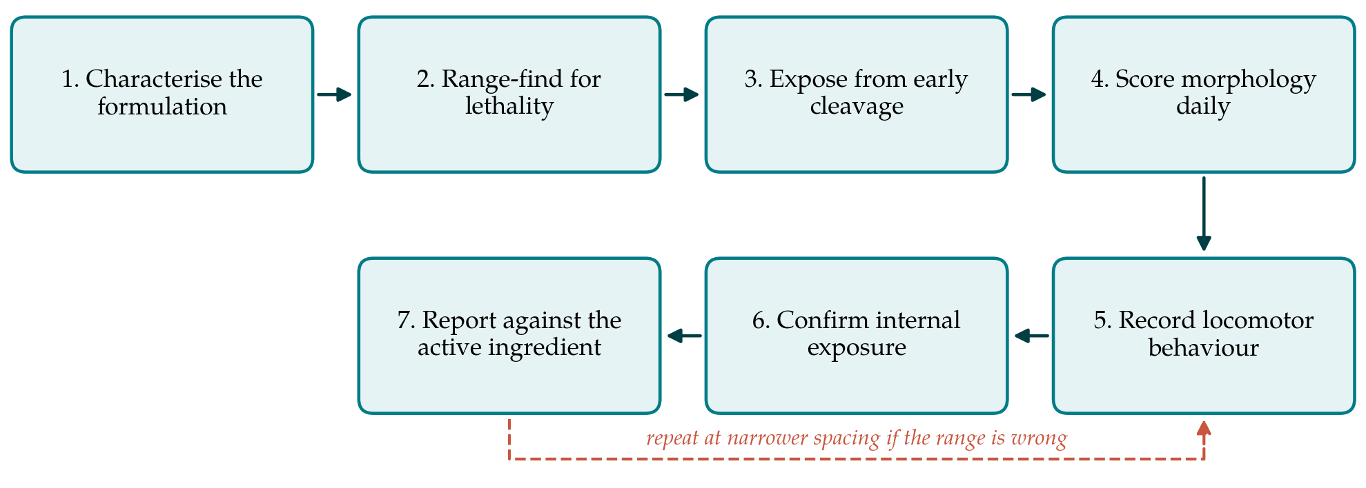
The protocol has seven steps. Figure 1 shows how they connect and where the procedure loops back on itself. The settings that differ most between published studies, and that therefore need to be fixed explicitly, are listed in Table 1 with the reason for each choice.
| Setting | Proposed choice | Reason |
|---|---|---|
| Test item | Formulation and active ingredient in parallel | The comparison is the purpose of the screen |
| Exposure start | Before the end of cleavage | Covers gastrulation and primary neurogenesis |
| Exposure end | Five days after fertilisation | Before independent feeding (Strähle et al., 2012) |
| Medium | Standard dilution water, daily renewal | Limits loss by degradation and sorption (OECD, 2013) |
| Vehicle | Water where possible, solvent kept minimal | Solvents alter uptake and baseline activity |
| Plate material | Glass or low-sorption plastic | Lipophilic actives and surfactants adsorb to polystyrene |
| Behaviour | Light and dark alternation on day five | Transitions give a robust response (Irons et al., 2010) |
3.1. Step 1: characterise the test item
A formulation is a defined commercial product, and the record should state the product name, the batch, the declared active ingredient and its nominal content, and whatever is disclosed about the co-formulants. The matched active ingredient of known purity is obtained for parallel testing. Both are checked for solubility and for visible phase separation in the dilution water at the highest intended concentration, since a formulation that disperses rather than dissolves will give concentrations that are not what they appear.
3.2. Step 2: range-find on the acute endpoints
A widely spaced concentration series is run first against the coagulation, somite, tail detachment and heartbeat endpoints of the fish embryo acute toxicity test (OECD, 2013). The purpose is to place the main experiment below overt lethality and gross malformation, because a behavioural deficit in a deformed or moribund embryo says nothing specific about the nervous system. The main study then uses closely spaced concentrations spanning the region below that threshold.
3.3. Step 3: expose from early cleavage
Freshly collected eggs are sorted under a stereomicroscope, unfertilised and irregularly cleaving embryos are discarded, and exposure begins before the end of the cleavage period so that gastrulation and the whole of primary neurogenesis occur under exposure (Kimmel et al., 1995). Embryos are held individually in wells with daily renewal of the medium, and plates are kept at the standard temperature on a fixed light cycle. Husbandry follows established laboratory practice (Westerfield, 2000).
3.4. Step 4: score morphology daily
Each embryo is examined every day against a fixed list: coagulation, somite formation, tail detachment, heartbeat, oedema, spinal curvature, swim bladder inflation, eye and head size, and pigment pattern. Daily scoring serves two purposes. It records the general toxicity against which any behavioural finding must be interpreted, and it identifies the day on which an effect first appears, which carries information about the developmental process affected.
3.5. Step 5: record locomotor behaviour
On day five the plate is moved to a tracking system and acclimatised in darkness before a sequence of alternating light and dark periods. Movement is recorded continuously and summarised for each period. The dark phase startle and the subsequent habituation are the features most consistently reported in the literature (Irons et al., 2010). Temperature, time of day and plate position influence the response, so the protocol fixes them and randomises treatment across the plate rather than filling it column by column.
3.6. Step 6: confirm internal exposure
A negative result is uninterpretable unless the embryo was exposed. The chorion is a partial barrier, and surfactants in a formulation may alter its permeability, which is itself part of what the screen is testing. Where analytical capacity exists, the active ingredient is measured in the medium at the start and end of a renewal interval and, if possible, in pooled embryos. Where it does not, the protocol requires that the limitation be stated rather than passed over. The broader argument for treating internal exposure as part of the assay has been made for environmental risk assessment generally (Scholz et al., 2008).
3.7. Step 7: report against the active ingredient
Results for the formulation are reported alongside those for the matched active ingredient at equivalent nominal active concentrations, so that a difference between them is visible. Reporting follows the principles of the ARRIVE guidelines as far as they apply to embryonic stages (Percie du Sert et al., 2020), with the number of embryos, the number of independent clutches, the exclusion rules and the statistical approach all stated in advance.
4. Fitting the Protocol to a Working Laboratory
The protocol assumes a breeding colony, a stereomicroscope, an incubator and a tracking system, which is the equipment of an ordinary zebrafish facility rather than a specialised one. The main costs are the analytical confirmation in step six and the use of independent clutches, both of which are tempting to omit and both of which determine whether the result means anything. Clutch is the largest single source of variation in this system, and treatment groups drawn from one spawning cannot be distinguished from clutch effects.
Scheduling is governed by the five-day window. One operator can handle a small number of plates per cohort, and a formulation with its matched active ingredient and controls fills a cohort. A screening programme is therefore a sequence of weekly cohorts rather than a single large experiment, which has the advantage that the concentration range can be adjusted between cohorts. Plate position effects are worth checking once at the outset, since edge wells differ in evaporation and in illumination during tracking.
5. Limitations and What Remains to Be Shown
This protocol is a proposal. It has not been run, and three things would have to be established before it could be recommended for screening. The first is reproducibility: whether the behavioural read-out gives consistent rankings across clutches, operators and laboratories, which is the weakness most often identified in zebrafish behavioural work. The second is concordance: whether compounds known to be developmental neurotoxicants in mammals are flagged, and whether compounds known not to be are not. The third is whether the formulation against active ingredient comparison behaves as intended, which requires products whose co-formulants are known.
Two limits are structural rather than empirical. A behavioural change at five days is a sensitive but unspecific indicator, and distinguishing a neurodevelopmental effect from altered muscle function or general malaise needs follow-up with neuroanatomical or transcriptional endpoints. And the zebrafish lacks the placental and lactational exposure routes that the mammalian study covers, so the screen addresses direct exposure of the embryo and nothing else. Broader discussions of what neurotoxicity testing in non-mammalian models can and cannot deliver make the same point (Legradi et al., 2018).
6. Conclusion
Formulated pesticides are what organisms encounter, and the developmental neurotoxicity data that regulators hold describe something else. The zebrafish embryo is a practical system for narrowing that gap, provided the protocol is specified tightly enough that results can be compared between laboratories. The seven steps set out here fix the choices that most often differ, pair every formulation with its active ingredient, and require that internal exposure be addressed rather than assumed. Whether the screen performs as intended is an empirical question, and the reproducibility and concordance work needed to answer it is the natural next step.
Conflict of interest
To be declared by the author(s) before publication.
Funding
To be declared by the author(s) before publication.