1. Introduction
Recurrent pregnancy loss, defined in the European guideline as the loss of two or more pregnancies, has many proposed causes and few proven ones (ESHRE Guideline Group, 2018; Rai and Regan, 2006). A structural chromosome rearrangement carried by one partner is among the proven ones. Surveys of couples with repeated losses have consistently found carriers of reciprocal or Robertsonian translocations and inversions at a frequency of a few percent, well above that in the general population (De Braekeleer and Dao, 1990). The carrier has a full genetic complement and no symptoms. The difficulty arises at meiosis, when the rearranged chromosomes and their normal homologs pair as a quadrivalent and may segregate so that a gamete receives too much of one segment and too little of another (Gardner and Amor, 2018).
Karyotyping of both partners is the classical investigation, and its yield and cost have been debated. Franssen et al. (2005) showed that carrier probability varies with maternal age at the second loss, the number of losses and the family history, and argued for selective testing. Less discussed is the opposite problem: the couple who are tested, found normal and reassured, when one of them carries a rearrangement below the resolution of the light microscope. Genome sequencing of couples with recurrent miscarriage has shown that such hidden carriers exist in appreciable numbers (Dong et al., 2019).
This article sets out a workflow for the laboratory and the genetics clinic that is aimed at that second group. It is a methodological proposal. It describes no patient and reports no application of the workflow, and it has not been validated here. Each step is drawn from published techniques or guidelines; what is offered is their arrangement into an ordered sequence with stated decision points.
2. Why Balanced Translocations Escape Detection
A G-banded karyotype at the level of resolution usual for constitutional studies distinguishes segments of roughly five to ten megabases. A reciprocal exchange is visible when it alters the length or banding pattern of the chromosomes involved. It is invisible, or cryptic, when the translocated segments are smaller than this, or when they are of similar size and staining so that each derivative chromosome looks like its normal counterpart. Exchanges between the pale-staining terminal bands of two chromosomes are the textbook example, and the search for them in children with unexplained developmental disorders led to the first subtelomeric probe sets (Knight et al., 1999).
Chromosomal microarray replaced the karyotype as the first-tier test for many indications because it detects gains and losses far below microscopic resolution (Miller et al., 2010). A truly balanced translocation involves no gain or loss, and the array of a carrier is normal. The array becomes informative one generation later: an unbalanced conceptus that inherits one derivative chromosome shows a terminal deletion of one chromosome together with a terminal duplication of another, a combination that points directly at a parental exchange. Table 1 compares the methods available and what each can and cannot show.
| Method | What it shows | Limit for cryptic balanced exchanges |
|---|---|---|
| G-banded karyotype | Whole genome at single-cell level; position of every chromosome | Misses small or similarly banded segments |
| Chromosomal microarray | Copy number gain and loss at high resolution | Blind to balanced change; useful on pregnancy tissue |
| Subtelomeric FISH panel | Position of each chromosome end on metaphases | Terminal exchanges only; labor-intensive |
| Low-pass genome sequencing with large inserts | Breakpoints genome-wide (Talkowski et al., 2011) | Weak in repeats; needs dedicated analysis |
| Optical genome mapping | Long-molecule maps of structural variants (Mantere et al., 2021) | Misses centromeric and acrocentric short-arm breakpoints |
| Targeted metaphase FISH | Direct view of the derivative chromosomes | Requires prior knowledge of the regions involved |
Two families of genome-wide methods can see a balanced exchange directly. Sequencing of large-insert or mate-pair libraries at low depth identifies read pairs whose ends map to different chromosomes, an approach shown by Talkowski et al. (2011) to localize balanced breakpoints for diagnostic use and later applied to large series of balanced rearrangements (Redin et al., 2017). Optical genome mapping images labeled DNA molecules hundreds of kilobases long and detects translocations from the label pattern; it has shown close agreement with standard cytogenetic tests for constitutional rearrangements (Mantere et al., 2021). Both methods lose power where the breakpoints lie in long repeats, and neither sees the whole-arm exchanges at centromeres or the short-arm fusions of Robertsonian translocations, which the karyotype detects easily. The methods are complementary, and the workflow uses them in that way.
3. The Proposed Workflow
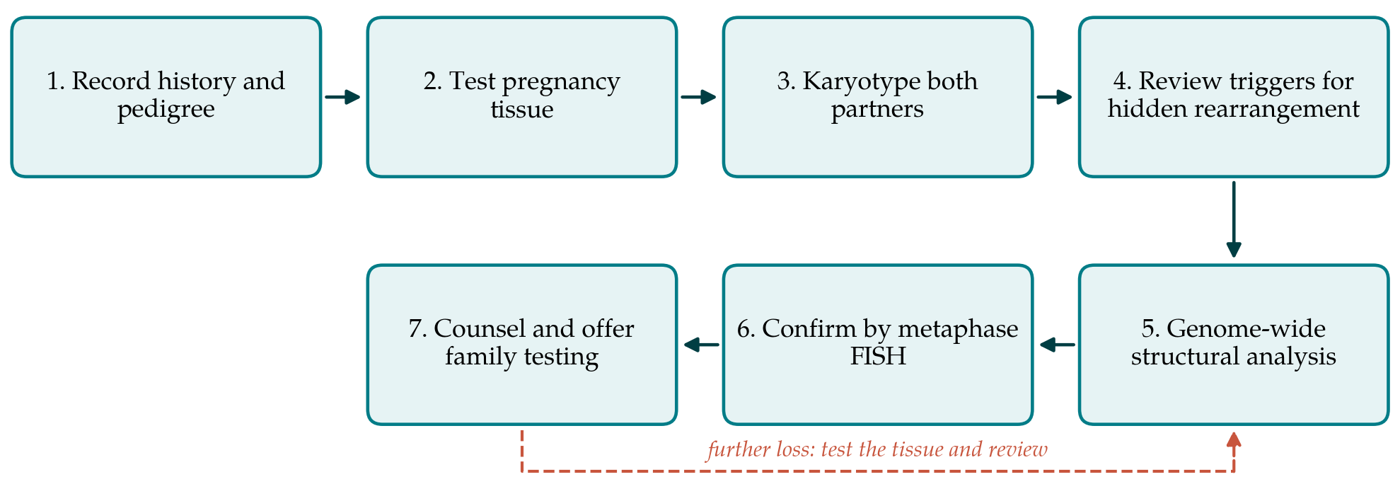
The workflow has seven steps, shown in Figure 1. The first three are part of existing practice in many centers. The fourth is the decision point that the proposal adds, and the last three follow only when that decision is positive.
3.1. Step 1: History and pedigree
The clinician records the number and gestational age of the losses, any pregnancy or child with congenital anomalies, and a three-generation pedigree for both partners that asks specifically about miscarriages, stillbirths and unexplained disability among siblings, parents, aunts and uncles. These are the factors that modify carrier probability in the selective testing model (Franssen et al., 2005), and they are also the factors that should keep suspicion alive when a first karyotype is normal.
3.2. Step 2: Cytogenomic analysis of pregnancy tissue
Whenever tissue from a loss is available it is examined by chromosomal microarray or an equivalent sequencing-based copy number method, with attention to maternal cell contamination. The European guideline regards such testing as optional and explanatory in purpose, and prefers array-based methods when it is done (ESHRE Guideline Group, 2018). Most abnormal results will be whole-chromosome aneuploidies that arose by chance and carry no implication for the parents. The result that matters for this workflow is the segmental one, above all a paired terminal deletion and duplication.
3.3. Step 3: Karyotyping of both partners
Peripheral blood from each partner is cultured and G-banded at a band level adequate for constitutional analysis, and the result is written in the current international nomenclature (McGowan-Jordan et al., 2020). If a rearrangement is seen, the couple proceeds straight to counseling. If the pregnancy tissue has already shown a segmental imbalance, the cytogeneticist is told which chromosome regions to scrutinize, since a directed look succeeds more often than a blind one.
3.4. Step 4: Review of triggers
With two apparently normal karyotypes in hand, the laboratory and clinician jointly ask whether any feature justifies a further search. Three triggers are proposed. The first is direct: pregnancy tissue with an unbalanced segmental rearrangement of the derivative type. The second is repetition, meaning that the same segmental imbalance has been found in more than one loss. The third is the pedigree: a pattern of losses or of children with anomalies in the relatives of one partner. A couple with none of these exits the workflow and continues with the general evaluation recommended by professional bodies (ASRM Practice Committee, 2012). The purpose of the step is restraint as much as detection, because indiscriminate genome-wide testing of all couples would generate variants of uncertain meaning at considerable cost.
3.5. Step 5: Genome-wide structural analysis
Couples who meet a trigger are offered one genome-wide method capable of detecting balanced change. The choice between low-pass large-insert sequencing and optical genome mapping should follow local availability and experience. Sequencing can use stored DNA. Optical mapping needs a fresh blood sample for the extraction of very long molecules. Where step 2 has identified the chromosome ends involved, a targeted fluorescence in situ hybridization study with probes for those ends is a legitimate and cheaper substitute. Analysis is restricted to structural variants; incidental sequence-level findings are not sought, and the consent discussion should say so.
3.6. Step 6: Confirmation on metaphase chromosomes
No rearrangement is reported on the strength of a genome-wide call alone. Probes flanking the predicted breakpoints, or subtelomeric probes for the arms concerned, are hybridized to metaphases of the suspected carrier to show the signals on the derivative chromosomes. This step confirms the finding by an independent principle, establishes that the exchange is present in the cells examined, and supplies the probe set that will be needed for testing relatives and for any later prenatal or preimplantation analysis.
3.7. Step 7: Counseling and family testing
The carrier and partner are counseled on the modes of segregation, on the likelihood that an unbalanced conception would miscarry or continue, and on the options open to them. The published outcome studies are relevant and on the whole encouraging. Carrier couples have more miscarriages than non-carrier couples, yet their chance of a healthy child through natural conception is good (Franssen et al., 2006; Stephenson and Sierra, 2006), although a less favorable course has been described for reciprocal translocation carriers in another series (Sugiura-Ogasawara et al., 2004). Preimplantation testing for structural rearrangements reduces the proportion of unbalanced embryos transferred (Munné et al., 2000), but the European guideline notes that evidence for a higher live birth rate is limited (ESHRE Guideline Group, 2018). Testing is offered to adult relatives at risk.
4. Fitting the Workflow to Local Practice
Centers differ in what they can do, and the workflow is meant to bend. Where pregnancy tissue is seldom collected, step 2 will rarely contribute and the pedigree carries more of the weight in step 4. Where neither sequencing nor optical mapping is available, a full subtelomeric probe panel on both partners is a workable form of step 5, with the understanding that it finds terminal exchanges only. Where a laboratory already uses a genome-wide structural method as its first-line test in place of the karyotype, steps 3 and 5 merge; conventional chromosome analysis should then be retained for the centromeric and Robertsonian rearrangements that such methods miss. In every variant, the feedback loop in Figure 1 applies: a further loss is an opportunity to test tissue and to revisit the triggers.
5. Limitations and What Remains to Be Shown
The workflow has not been tested, and its central assumptions are open to challenge. The triggers in step 4 are reasoned from the biology of segregation and from the selective karyotyping literature; their sensitivity and specificity for cryptic carriers are unknown. A prospective comparison would be needed in which all couples with normal karyotypes received genome-wide analysis and the triggers were then scored against the result. The proportion of hidden carriers reported from sequencing studies (Dong et al., 2019) may not hold in other populations or referral patterns, and the yield of the workflow depends on it.
A second uncertainty is clinical. Finding a cryptic translocation gives the couple an explanation and a basis for informed choices, but whether it changes the chance of a live birth is not established, for the same reasons that the benefit of testing for visible translocations remains debated. Genome-wide methods also uncover balanced rearrangements with breakpoints inside genes, whose significance for a healthy adult is usually nil but may be hard to explain (Redin et al., 2017). Laboratories adopting the workflow should decide in advance how such findings will be handled and reported.
6. Conclusion
A normal karyotype does not exclude a balanced translocation, and for a small number of couples with recurrent pregnancy loss the distinction matters. The workflow proposed here directs genome-wide structural testing to couples in whom pregnancy tissue, repetition or family history suggests a hidden rearrangement, and requires confirmation on metaphase chromosomes before any result is acted on. It draws only on established techniques. Whether its triggers select the right couples, and whether the diagnoses it yields improve outcomes, are questions for prospective study.
Conflict of interest
To be declared by the author(s) before publication.
Funding
To be declared by the author(s) before publication.