1. Introduction
Breast cancers that lack the estrogen receptor, the progesterone receptor and overexpression of HER2 are grouped as triple-negative. The label is one of exclusion, and the receptor definitions on which it rests come from consensus testing guidelines (Hammond et al., 2010). The tumors it covers are typically of high grade, arise more often in younger women and in carriers of BRCA1 mutations, and recur early when they recur at all (Foulkes et al., 2010). Gene expression studies have shown that the group is far from uniform and includes a subset with a strong immune signature (Lehmann et al., 2011).
For most patients with stage II or III disease, systemic treatment is now given before surgery. The preoperative setting has a property that adjuvant treatment lacks: the effect of the drugs can be read in the surgical specimen. Absence of invasive cancer in the breast and axillary nodes, termed pathological complete response, is followed by markedly better long-term outcome in triple-negative disease than in any other subtype, as the pooled CTNeoBC analysis showed (Cortazar et al., 2014). A marker available on the diagnostic core biopsy that anticipates this response would therefore be of practical value.
Pathologists have long noted that some breast carcinomas are heavily infiltrated by lymphocytes. Over the past fifteen years that observation has been turned into a quantitative variable and tested in thousands of archived trial specimens. This article reviews what those studies found about the density of tumor-infiltrating lymphocytes (TILs) and the response of triple-negative breast cancer to neoadjuvant chemotherapy, how firmly the findings stand, and what they do not yet justify.
2. Scope and Approach
This is a narrative review of the published literature. It draws on the consensus documents that standardized TIL scoring, on analyses of lymphocyte density in specimens from randomized neoadjuvant and adjuvant trials, on pooled analyses of those cohorts, and on earlier reviews of host immunity in breast cancer (Savas et al., 2016; Stanton and Disis, 2016). No formal search protocol was applied and no quantitative synthesis is attempted. Studies are discussed for what they contribute to four questions: how the marker is measured, how it relates to pathological complete response, what explains the relation, and whether the relation is ready to guide treatment.
3. Measuring Lymphocyte Density
The early studies scored lymphocytes in different ways, which made their results hard to compare. In 2014 an international group of pathologists and oncologists agreed a common method, published as the recommendations of the International TILs Working Group (Salgado et al., 2015). The method uses a single hematoxylin and eosin section. The reader estimates the proportion of the stromal area within the borders of the invasive tumor that is occupied by mononuclear inflammatory cells, and reports it as a continuous percentage. Lymphocytes in direct contact with carcinoma cells, called intratumoral, are less numerous and harder to see without immunohistochemistry, and the group advised that the stromal compartment be the principal measure.
Several rules protect the estimate from obvious distortions. Areas of necrosis, crush artifact, carcinoma in situ and normal lobules are excluded. The score is an average over the tumor stroma and is not taken from the densest focus. Granulocytes are not counted. Full sections are preferred, although core biopsies are accepted in the preoperative setting because nothing else is available before treatment. The same principles were later extended to other solid tumors (Hendry et al., 2017).
Agreement between trained readers is good at the extremes and weaker in the middle of the range, where most tumors fall and where infiltrates are often unevenly distributed. This matters whenever a cut-off is proposed. Image analysis of digitized slides offers a way to reduce reader variation, and the working group has set out what computational methods would need to demonstrate before they could replace visual scoring (Amgad et al., 2020). For the present, the evidence reviewed below rests almost entirely on visual estimates made by pathologists.
4. Lymphocyte Density and Pathological Complete Response
The studies that bear most directly on the question are summarized in Table 1. They fall into three groups: analyses of pretreatment biopsies from neoadjuvant trials, analyses of adjuvant trial specimens that address prognosis, and studies of the tissue that remains after treatment.
| Source | Material studied | Main observation on lymphocyte density |
|---|---|---|
| Denkert et al., 2010 | Pretreatment biopsies from two German neoadjuvant trials | Independent predictor of pathological complete response |
| Loi et al., 2013 | Adjuvant trial BIG 02-98, node-positive disease | Prognostic in triple-negative tumors, not in luminal tumors |
| Adams et al., 2014 | Two ECOG adjuvant trials, triple-negative cases | Prognostic value of stromal lymphocytes confirmed |
| Dieci et al., 2014 | Residual tumor after neoadjuvant chemotherapy | Infiltrate in residual disease linked to better survival |
| Denkert et al., 2018 | Pooled biopsies from six neoadjuvant trials | Graded rise in response rate with density in every subtype |
| Loi et al., 2019 | Pooled individual data, early triple-negative disease | Each increment in density adds prognostic information |
| Park et al., 2019 | Early triple-negative disease without chemotherapy | High density marks a group with favorable outcome |
4.1. Pretreatment biopsies from neoadjuvant trials
The first large study came from the German Breast Group. Denkert et al. (2010) examined pretreatment core biopsies from the GeparDuo trial and validated their findings in GeparTrio, both of which used anthracycline and taxane regimens. The proportion of lymphocytes in the tumor nests and in the stroma each predicted pathological complete response, and did so independently of age, grade, nodal status and receptor status. Tumors in which lymphocytes dominated the picture, which the authors called lymphocyte-predominant breast cancer, responded completely far more often than tumors with no infiltrate.
The same group later pooled biopsies from six of its randomized neoadjuvant trials, all scored under the working group method (Denkert et al., 2018). The analysis treated density as a continuous variable and also divided tumors into low, intermediate and high groups. In triple-negative disease the rate of complete response rose stepwise across the three groups, and the association held after adjustment for the standard clinical factors. No level was found below which lymphocytes ceased to matter or above which more made no difference. The relation looked like a gradient.
4.2. From response to survival
A predictor of response is of interest mainly because response predicts survival. The pooled German analysis reported that higher density was associated with longer disease-free and overall survival in triple-negative disease, whereas in luminal, HER2-negative tumors higher density was associated with shorter overall survival (Denkert et al., 2018). The contrast is a reminder that an infiltrate does not mean the same thing in every breast cancer.
Adjuvant trial cohorts, in which every patient had surgery first, point in the same direction. In BIG 02-98 each increment in lymphocyte density was associated with a lower risk of relapse and death among triple-negative cases only (Loi et al., 2013), and specimens from two ECOG trials confirmed the finding (Adams et al., 2014). A pooled analysis of individual patient data then established stromal density as a prognostic factor that adds to tumor size and nodal status (Loi et al., 2019). Because all of these patients received chemotherapy, the studies could not say whether lymphocytes mark a tumor with a good natural history or one that is sensitive to drugs. A cohort of patients who received no systemic treatment suggests that part of the effect is prognostic in the strict sense (Park et al., 2019).
4.3. Lymphocytes in residual disease
Most patients do not achieve a complete response, and for them the surgical specimen offers a second chance to assess the immune infiltrate. Dieci et al. (2014) found that a dense infiltrate in residual triple-negative tumor was associated with better survival than a sparse one. The extent of residual disease is itself graded by the residual cancer burden index (Symmans et al., 2007), and patients with residual invasive cancer are now offered further treatment such as capecitabine (Masuda et al., 2017). Whether the infiltrate in residual tumor refines the selection of patients for such treatment has not been tested prospectively.
5. Interpreting the Association
Two explanations are usually offered, and they are not exclusive. The first is that a dense infiltrate is the visible sign of an immune response already under way, which chemotherapy then assists. Anthracyclines and some other cytotoxic agents kill tumor cells in a manner that exposes signals recognized by dendritic cells, a process described as immunogenic cell death (Galluzzi et al., 2017). On this view the drugs do not act alone: part of their effect depends on T cells that were present before treatment began.
The second explanation is that lymphocytes are a marker of tumor biology. Triple-negative cancers with high proliferation, genomic instability and many altered proteins both attract lymphocytes and respond to DNA-damaging drugs, so the two could travel together without one causing the other. The immunomodulatory expression subtype described by Lehmann et al. (2011) overlaps with histologically infiltrated tumors, which fits either account. Observational tissue studies cannot separate the two, and the distinction has practical weight: only the first implies that strengthening the immune response should improve the effect of chemotherapy.
Density is also a crude summary. An infiltrate contains cytotoxic CD8-positive T cells, helper and regulatory T cells, B cells and plasma cells in varying proportions, and their arrangement in the tissue differs from tumor to tumor. Reviews of the field have argued that composition and spatial organization carry information beyond the percentage of stroma occupied (Savas et al., 2016). So far no refinement has displaced the simple stromal score, largely because nothing else has been validated in as many patients.
6. Neoadjuvant Immunotherapy and the Place of the Marker
The treatment against which the marker was studied has changed. In the KEYNOTE-522 trial the addition of the PD-1 antibody pembrolizumab to platinum-containing neoadjuvant chemotherapy raised the rate of pathological complete response in early triple-negative breast cancer (Schmid et al., 2020), and the IMpassion031 trial reported a similar gain with atezolizumab (Mittendorf et al., 2020). In both trials the benefit was seen whether or not the tumor expressed PD-L1. Chemotherapy with a checkpoint inhibitor has since become a standard preoperative approach for higher-risk disease.
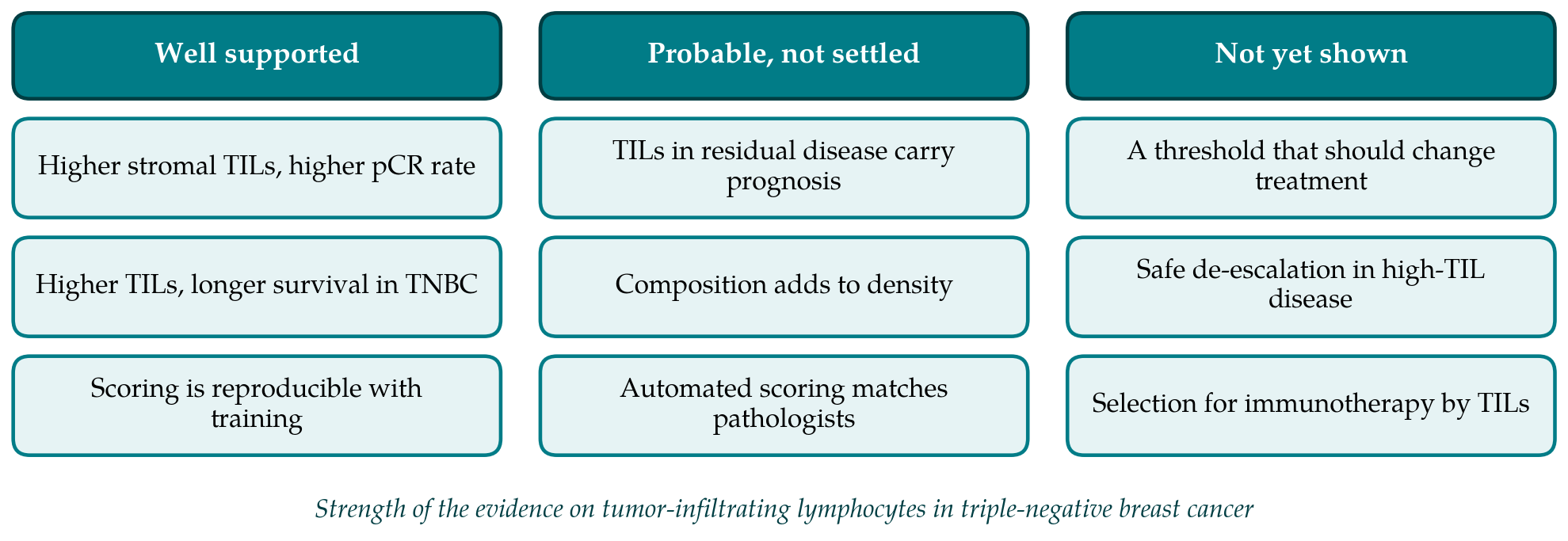
This shifts the question. It is no longer enough to ask whether lymphocyte density predicts response to chemotherapy. One must ask whether it identifies patients who gain little from the added antibody because they do well without it, or patients who gain most because the antibody has an existing response to release. The published trials were not designed to answer this, and exploratory biomarker analyses cannot settle it. Figure 1 sets out which statements about the marker are well supported, which are probable, and which have not been shown.
7. Limits of the Evidence and Open Questions
The body of work reviewed here is large and consistent, yet it shares several weaknesses. Every analysis was retrospective, performed on specimens collected for other purposes, and the cases with available tissue were a subset of those enrolled. The reporting recommendations for tumor marker studies, known as REMARK, were written with exactly these hazards in mind (McShane et al., 2005). The thresholds that divide low from high density were chosen in advance in some studies and by convention in others, and none was derived to support a treatment decision.
A core biopsy samples a small part of a tumor that may be unevenly infiltrated, and the reproducibility of scoring in the middle range is imperfect. Chemotherapy regimens in the source trials differed and mostly predate the routine use of platinum agents and checkpoint inhibitors. These points do not undermine the association. They limit how far it can be pushed.
Expert consensus reflects that position. The 2019 St Gallen panel accepted that lymphocyte density is a prognostic factor in triple-negative disease that may reasonably be reported, while declining to endorse it as a basis for giving more or less treatment (Burstein et al., 2019). The questions that would change this are specific. One is whether patients with small, node-negative, heavily infiltrated tumors can safely receive less chemotherapy. Another is whether density, alone or with other features, identifies patients for whom a checkpoint inhibitor adds nothing. A third is whether automated scoring can deliver a value stable enough to act on. Each requires a prospective design in which the marker is measured before treatment and the analysis is specified in advance.
8. Conclusion
Stromal lymphocyte density, scored on an ordinary stained section, is among the most thoroughly studied tissue markers in triple-negative breast cancer. Across trial cohorts and pooled analyses a denser infiltrate has gone with a higher rate of pathological complete response to neoadjuvant chemotherapy and with longer survival, and the relation is graded and independent of the usual clinical factors. The marker is inexpensive and can be reported today as prognostic information. It should not yet be used to select or withhold treatment. That step waits on prospective evidence, and on a clearer account of what the marker means when immunotherapy is part of the regimen.
Conflict of interest
To be declared by the author(s) before publication.
Funding
To be declared by the author(s) before publication.