1. Introduction
Inhibitors of intestinal α-glucosidases slow the final step of carbohydrate digestion and blunt the rise in blood glucose after a meal. Acarbose, the best-studied drug of the class, lowers glycated haemoglobin modestly and causes gastrointestinal side effects in many patients, as a Cochrane review of the trials concluded (Van de Laar et al., 2005). That combination of a validated target and an imperfect drug has encouraged a long search for inhibitors from natural sources (Tundis et al., 2010).
Seaweeds feature prominently in that search. Brown algae contain phlorotannins, oligomers of phloroglucinol found in no terrestrial plant, and extracts and purified compounds from genera such as Ecklonia and Ascophyllum inhibit α-glucosidase and α-amylase in vitro (Zhang et al., 2007; Lee and Jeon, 2013). Red algae yield bromophenols with similar activity (Kurihara et al., 1999; Kim et al., 2008). Screening collections of species has therefore become common, and the convenience of the plate assay has made it the default method. This note concerns how that assay should be set up and read when the samples are crude algal extracts.
2. The Standard Plate Assay and Its Assumptions
In the usual format, enzyme from Saccharomyces cerevisiae is incubated with the test sample in phosphate buffer near neutral pH, the chromogenic substrate p-nitrophenyl α-D-glucopyranoside is added, and after a fixed time the reaction is stopped with sodium carbonate, which also converts the released p-nitrophenol to its yellow anion. Absorbance is read at about 405 nm, and inhibition is calculated from the difference between wells with and without sample. Acarbose is included as a positive control.
Three assumptions are built in. The first is that the yeast enzyme predicts behaviour towards the mammalian brush-border enzymes. It often does not. Oki et al. (1999) showed that inhibitory activity depends strongly on the origin of the enzyme, with compounds active on the yeast enzyme proving weak on mammalian preparations and the reverse; acarbose itself is a poor inhibitor of yeast α-glucosidase. The second is that an artificial aryl glucoside stands in adequately for maltose and sucrose. The third is that a fall in absorbance means that the enzyme has been specifically inhibited. With seaweed extracts it is the third assumption that fails most often.
3. Sources of False Results With Seaweed Extracts
Crude algal extracts differ from the pure compounds for which the assay was designed in several ways, summarised in Table 1 with the control appropriate to each. Colour is the most obvious. Extracts made with organic solvents carry chlorophylls and carotenoids, among them fucoxanthin in brown algae, and oxidised phenolics are brown; all absorb in the violet and blue. Unless every sample has its own blank without enzyme, pigment absorbance is added to the product signal and inhibition is underestimated, or turbidity that develops on adding carbonate is misread.
| Property of the extract | Effect on the reading | Control |
|---|---|---|
| Pigments and brown oxidised phenolics | Absorbance near 405 nm unrelated to product | Sample blank without enzyme for every well |
| Phlorotannins | Non-selective binding and precipitation of protein | Repeat with added albumin; remove phenolics and retest |
| Alginate, fucoidan and other polysaccharides | Viscosity and enzyme binding slow the reaction | Fractionate by ethanol precipitation; test fractions separately |
| Residual sea salt | Ionic strength alters enzyme activity | Desalt; match salt in control wells |
| Colloidal aggregates | Enzyme adsorbed on particles | Repeat with non-ionic detergent (Feng and Shoichet, 2006) |
| Extraction solvent carried over | Solvent itself inhibits the enzyme | Solvent-only control at the same concentration |
Phlorotannins pose the deeper problem. Like tannins of land plants they bind and precipitate proteins, and an extract rich in them will inhibit almost any enzyme offered to it. The inhibition is real in the well and may even be relevant in the gut, where phlorotannins would meet digestive enzymes at high local concentration, but it is not selective, and it should not be presented as the discovery of an α-glucosidase inhibitor. Studies of phenolic-rich Ascophyllum nodosum fractions, and a comparison of extracts from Irish seaweeds, both found activity against α-amylase as well as α-glucosidase, which is what a general protein-binding mechanism would predict (Apostolidis and Lee, 2010; Lordan et al., 2013). A related artefact, well known from synthetic screening libraries, is inhibition by colloidal aggregates that adsorb enzyme; it is diagnosed by its disappearance when a little non-ionic detergent is added (Feng and Shoichet, 2006). Seaweed polysaccharides add viscosity, and extracts that have not been desalted bring variable ionic strength.
4. A Screening Sequence With Built-In Controls
These considerations suggest a sequence of five stages, each paired with the control that guards against the artefact most likely at that point (Figure 1). Extracts are prepared by a recorded method, dried, weighed and desalted where necessary, so that concentrations are expressed on a dry-mass basis and can be compared between species. The primary screen is run at a single concentration with a sample blank for every extract, a solvent control and acarbose on every plate, and plate quality is judged by a statistic such as the Z-factor of Zhang et al. (1999) before any well is interpreted.
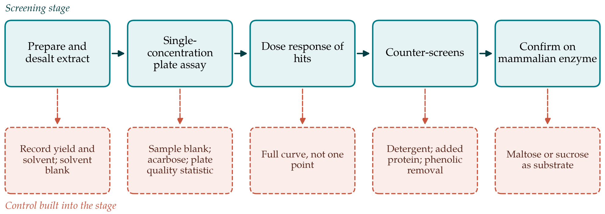
Extracts that pass a preset threshold are retested over a range of concentrations. A complete curve distinguishes graded inhibition from the abrupt, all-or-nothing behaviour typical of precipitation and aggregation, which a single concentration cannot do. Confirmed hits then go through counter-screens: the assay is repeated with detergent and with added albumin, and again after phenolics have been removed with an adsorbent such as polyvinylpolypyrrolidone. Activity that vanishes with the phenolics is assigned to them and described as such. Finally the surviving extracts are tested on a mammalian preparation, most simply rat intestinal acetone powder, with maltose or sucrose as substrate and glucose measured enzymatically. Only activity that persists to this stage justifies fractionation.
5. From a Screening Hit to a Credible Lead
Reporting conventions matter as much as bench controls. A potency value for a crude extract should state the enzyme source, substrate, extract concentration basis and the potency of acarbose in the same run; without these the figure cannot be compared with any other. Because acarbose is weak on the yeast enzyme, the statement that an extract is more potent than acarbose in a yeast assay carries little meaning (Oki et al., 1999). Species identity, collection site and season should be recorded and a voucher kept, since the phenolic content of seaweeds varies with all three.
Caution is also needed in moving from the plate to claims about health. In vitro inhibition establishes a mechanism worth testing and nothing more. Effects on post-prandial glucose in people have to be shown in controlled trials, and seaweed preparations raise safety questions of their own, notably iodine and inorganic arsenic content, which are subject to regulatory limits in several jurisdictions (Holdt and Kraan, 2011).
6. Conclusion
The yeast enzyme plate assay remains a sensible first filter for seaweed extracts provided that it is treated as a filter. Sample blanks, a plate quality statistic, dose-response confirmation, counter-screens for non-selective mechanisms and a final test on a mammalian enzyme with a natural substrate add little time, and they remove most of the results that would not survive further study. Screens arranged in this way will report fewer active species, and the ones they do report will be worth the effort of isolation.
Conflict of interest
To be declared by the author(s) before publication.
Funding
To be declared by the author(s) before publication.