1. Introduction
The anterior surface of the tibia lies directly under the skin, and the distal third of the leg carries tendons where the proximal leg carries muscle bellies. A road traffic collision, a fall from height or a crush injury can therefore expose fracture, fixation hardware and tendon in a wound that will not close by itself and cannot be grafted. The classification of Gustilo and Anderson (1976), refined for the most severe injuries by Gustilo et al. (1984), defines the type IIIB open fracture precisely by this problem: periosteal stripping and bone exposure that require a flap.
The outcome that matters is not flap survival alone. The Lower Extremity Assessment Project followed patients with limb-threatening injuries and found that functional outcome at two years was similar after reconstruction and after amputation, and was poor in many patients in both groups (Bosse et al., 2002). Salvage is thus a long undertaking whose soft-tissue stage should add as little morbidity as possible. That consideration, more than any change in survival rates, explains the movement from muscle flaps toward flaps that spare muscle.
This review asks how the published evidence should inform the selection of a perforator flap for a traumatic defect of the leg, and when another kind of flap remains the better choice. It reports no new patients and offers no pooled estimates.
2. Scope and Approach
The article is a narrative review of the published literature. It draws on the anatomical studies that defined perforator flaps, the consensus statements that fixed their terminology, the principal clinical series and comparative studies of soft-tissue cover in open tibial fractures, and national guidance on open fracture care. Sources were chosen for their influence on current practice and for the clarity with which they bear on the choice of flap. No formal search protocol was applied, so the selection reflects judgement and cannot claim to be complete. Reconstruction of the foot sole, replantation and bone transport are outside its scope.
3. Anatomical Basis of Perforator Flaps
Three bodies of anatomical work underlie present practice. The first is the classification of muscle blood supply by Mathes and Nahai (1981), which made muscle and musculocutaneous flaps predictable and established them as the workhorses of leg reconstruction. In the same year Pontén (1981) showed that long skin flaps on the lower leg survive when the deep fascia is raised with them, which drew attention to the vessels running at that level. The second is the angiosome concept of Taylor and Palmer (1987), who mapped the body into three-dimensional territories supplied by named source arteries and linked to one another by choke vessels, an account that explains why skin can be carried on a vessel belonging to the neighbouring territory.
The third is the demonstration that a flap can live on a single perforating vessel dissected through muscle. Koshima and Soeda (1989) raised abdominal skin on a perforator of the inferior epigastric artery and left the rectus abdominis in place, and the principle was soon carried to other donor sites. The history of the idea, and the argument over which flaps deserve the name, is recounted by Hallock (2003). Terminology was settled in large part by the Gent consensus, which defined a perforator flap as skin and subcutaneous fat supplied by vessels that pass through or between deep tissues, and named each flap after its source artery (Blondeel et al., 2003).
More recent injection studies refined the picture. Saint-Cyr et al. (2009) described the territory of an individual perforator, the perforasome, and showed that adjacent perforasomes are joined by direct and indirect linking vessels. In the limbs, flow from a perforator spreads chiefly along the axis of the limb. Two practical consequences follow. A flap can be designed freely around any adequate perforator, and its long axis in the leg should be longitudinal.
4. Muscle or Fasciocutaneous Cover Over a Fracture
The case for muscle rested on the belief that it brings a richer blood supply to a contaminated fracture, obliterates dead space and resists infection better than skin. Experimental work in animals supports some of these claims, particularly an advantage for muscle in early fracture healing. The clinical literature has not shown a corresponding difference. Yazar et al. (2006) compared free muscle with free fasciocutaneous flaps for open fractures of the distal third of the tibia and the ankle and found no significant difference in flap survival, infection or bone union. A structured review of the question by Chan et al. (2012) reached a measured conclusion: laboratory data favour muscle, clinical data show equivalence, and no adequately designed trial exists.
Where the two do differ is in what follows. A fasciocutaneous flap can be lifted again for bone grafting or revision of fixation, tolerates secondary thinning, and glides over tendons at the ankle. A muscle flap with a skin graft atrophies to a good contour over the tibial crest but is harder to re-elevate and sacrifices a functional unit, which is not a trivial loss in a patient who will depend on the upper body and the remaining limb during rehabilitation. These are differences of handling and donor morbidity. They are well described in the series cited here, but they have seldom been measured with patient-reported instruments.
5. Free Perforator Flaps in Common Use
Among free flaps, the anterolateral thigh flap dominates the literature. It was described by Song et al. (1984) as a flap based on a septocutaneous vessel, and the large experience reported by Wei et al. (2002) established that it could be raised reliably even though most of its perforators take a course through the vastus lateralis. Its attractions for the injured leg are a long pedicle of good calibre, a skin paddle large enough for extensive degloving, the option of carrying fascia lata or a segment of muscle on the same pedicle, and a donor site on the same limb that allows a second team to work at the same time. Table 1 sets it beside the main alternatives, and Figure 1 arranges the options as they present themselves at the planning stage.
| Flap | Source vessel | Usual role in the leg | Main limits |
|---|---|---|---|
| Anterolateral thigh (Wei et al., 2002) | Descending branch, lateral circumflex femoral | Large or composite defects at any level | Bulk in a thick thigh; variable perforator course |
| Medial sural artery perforator (Cavadas et al., 2001) | Medial sural artery | Thin cover for ankle, heel cord and dorsum of foot | Limited width for direct donor closure |
| Thoracodorsal artery perforator (Angrigiani et al., 1995) | Thoracodorsal artery | Broad, thin cover; latissimus dorsi preserved | Lateral positioning; distant donor site |
| Superficial circumflex iliac artery perforator (Koshima et al., 2004) | Superficial circumflex iliac artery | Thin, pliable cover with a concealed scar | Short pedicle and small vessels |
| Propeller flap (Pignatti et al., 2011) | Posterior tibial or peroneal perforator | Small and moderate defects of the distal third | Venous congestion; pedicle near the injury |
| Free muscle with skin graft (Mathes and Nahai, 1981) | Named pedicle of gracilis or latissimus dorsi | Deep cavities and irregular dead space | Loss of a muscle; harder to re-elevate |
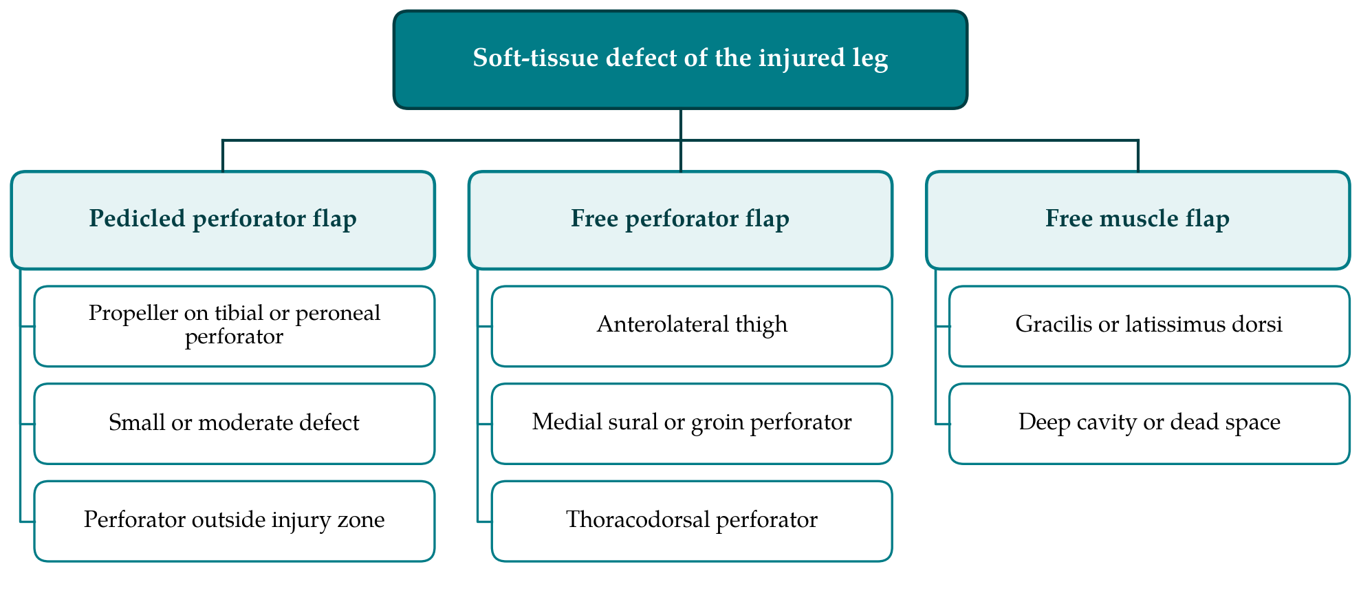
The thinner free flaps answer the main criticism of the thigh flap, which is bulk around the ankle and foot. The medial sural artery perforator flap, raised from the calf over the medial head of gastrocnemius, provides thin skin and a long pedicle (Cavadas et al., 2001). The thoracodorsal artery perforator flap gives the surface area of a latissimus dorsi flap without the muscle (Angrigiani et al., 1995). The superficial circumflex iliac artery perforator flap, developed for limb defects by Koshima et al. (2004), is thinner still and hides its scar in the groin, at the price of a short pedicle and small vessels that demand refined technique. None of these has been compared with the others in a controlled study. The reports are single-centre series from units with a particular interest in the flap they describe.
6. Pedicled Perforator and Propeller Flaps
A local flap avoids microvascular anastomosis and replaces lost skin with skin from the same leg. The propeller design, introduced by Hyakusoku et al. (1991) and developed for the limb by Teo (2010), is an island of skin rotated about a skeletonised perforator by up to a half turn. The Tokyo consensus fixed its definition and classification (Pignatti et al., 2011). In the leg the flap is usually based on a perforator of the posterior tibial or peroneal artery, found with a handheld Doppler probe and confirmed by exploration through one margin of the planned flap before the island is committed.
The systematic review by Gir et al. (2012) gathered the published propeller flaps of the lower extremity and found that most survived, with partial flap loss and venous congestion as the leading complications. Two reservations apply to trauma. The pooled cases mix defects of many causes, and traumatic wounds are a minority studied without separate analysis in many reports. More fundamentally, the perforators that would carry a propeller flap lie in the same leg that absorbed the injury. A perforator that pulses under the Doppler probe may still sit in bruised, degloved tissue whose venous drainage is compromised. The literature therefore supports propeller flaps for small and moderate defects after lower-energy injury, or for defects whose surrounding skin is demonstrably healthy, and offers much less assurance after a severe crush.
7. Injury Zone, Timing and Recipient Vessels
Selection of a flap cannot be separated from the condition of the leg that receives it. The zone of injury extends beyond the visible wound, and vessels within it are friable and prone to thrombosis. Classical teaching places the anastomosis proximal to that zone on a vessel of normal appearance, which in turn demands a flap with a long pedicle and favours the anterolateral thigh and thoracodorsal donor sites. An alternative is to use a perforator close to the defect as the recipient vessel, a strategy reported for the knee region by Hong and Koshima (2010). It shortens the dissection and spares the major arteries of a limb that may have only one or two patent, but it depends on small-vessel technique and on a perforator that has itself escaped injury.
Timing has been debated since Godina (1986) reported fewer flap failures and less infection when free tissue transfer followed radical debridement within the first seventy-two hours. Current guidance in England asks for definitive soft-tissue cover at the time of debridement or within seventy-two hours of injury (NICE, 2016). Later reconstruction is plainly possible and is often unavoidable in patients with multiple injuries, but the tissues are then oedematous and fibrotic, which strengthens the argument for a flap with a long pedicle and for recipient vessels well away from the wound.
These constraints are the reason the reconstructive ladder, with its rule of trying the simplest method first, has given way in trauma to the reconstructive elevator of Gottlieb and Krieger (1994): the surgeon goes directly to the method that best fits the defect, which after high-energy injury is often free tissue transfer.
8. Gaps in the Evidence
Almost everything known about flap choice in the injured leg comes from retrospective series. They come from centres that perform many such operations, they report flap survival more consistently than function, and surgeons choose the flap with knowledge of the wound, which confounds any comparison. The end points that would discriminate between flaps, such as time to union, deep infection, ability to wear ordinary footwear, return to work and the patient's own assessment of the limb and the donor site, are reported unevenly and by differing definitions.
Several specific questions remain open. Whether the experimental advantage of muscle for fracture healing has any clinical counterpart in the most contaminated injuries has not been tested in a prospective comparison. The safe extent of injury beyond which a propeller flap should not be attempted has no agreed definition. The contribution of preoperative imaging of perforators to outcome in trauma, as distinct from elective surgery, is largely assumed. Agreement on a core set of outcomes for open fracture reconstruction, and registries that record the flap used alongside them, would do more to settle these matters than further single-flap series.
9. Conclusion
The literature justifies treating perforator flaps as a first-line option for soft-tissue cover of the traumatised leg. Comparative data show flap survival and bone healing similar to those achieved with muscle, and the practical advantages of sparing muscle and of easy re-elevation are well described. The same literature gives no ground for a universal choice. The anterolateral thigh flap suits large and composite defects, thinner free flaps suit the ankle and foot, propeller flaps suit limited defects surrounded by healthy skin, and muscle retains a place where a deep cavity must be filled. Extent of the injury zone, the time since injury and the state of the recipient vessels should decide among them.
Conflict of interest
To be declared by the author(s) before publication.
Funding
To be declared by the author(s) before publication.