1. Introduction
The primary cilium is a single, non-motile projection found on most vertebrate cells. Its membrane concentrates receptors and its interior is a compartment in which several developmental signals are processed, the best studied being Hedgehog (Goetz and Anderson, 2010). Inherited defects in ciliary proteins cause a group of conditions known as ciliopathies, which include Joubert syndrome, Bardet-Biedl syndrome, Meckel syndrome and nephronophthisis and which affect the brain, kidney, retina and skeleton in overlapping combinations (Hildebrandt et al., 2011). The list of genes involved has grown steadily, and with it the recognition that cilia can be disturbed secondarily in disorders whose primary lesion lies elsewhere (Reiter and Leroux, 2017).
Organoids have made it possible to look for such disturbances in human tissue. Cerebral organoids reproduce features of early cortical development and were used from the outset to model a genetic form of microcephaly (Lancaster et al., 2013). Kidney organoids contain segmented nephrons (Takasato et al., 2015), and organoids carrying engineered mutations in polycystic kidney disease genes form cysts (Freedman et al., 2015). An investigator who stains such cultures for ciliary markers while studying an unrelated rare disorder may see an unexpected difference between patient-derived and control organoids.
What should be done next is the subject of this article. It proposes a framework for testing whether an apparent ciliary defect in an organoid model is real, attributable to the genotype, specific to the cilium and functionally meaningful. The framework is a proposal. It describes a procedure and its rationale, and reports no application to any particular disorder, cell line or patient.
2. Why Cilia Are Difficult to Judge in Organoids
Three features of the system make a naive comparison unreliable. Each corresponds to a class of alternative explanation that the framework is designed to exclude.
2.1. Ciliation depends on cell state
A primary cilium grows from the mother centriole after it has docked at the plasma membrane as a basal body, and it is built and maintained by intraflagellar transport (Ishikawa and Marshall, 2011). Because the centrioles are also needed for the mitotic spindle, the cilium is assembled in quiescent or G1 cells and resorbed before mitosis (Plotnikova et al., 2009). The proportion of ciliated cells in a population therefore falls when more cells are cycling, and cilium length differs between cell types. A mutation that alters proliferation or differentiation will change the number of cilia observed without acting on the cilium at all.
2.2. Organoids are variable
Organoids self-organise, and no two are identical. Cell-type composition can differ between batches and between individual organoids, although protocols that guide differentiation more tightly reduce this (Velasco et al., 2019). Induced pluripotent stem cell lines add a further source of variation, since lines from different donors, and different clones from one donor, differ in genetic background and in their propensity to differentiate (Volpato and Webber, 2020). A comparison of one patient line with one unrelated control line confounds the mutation of interest with everything else that distinguishes two people and two clones.
2.3. Measurement is not neutral
Cilia are usually identified by immunofluorescence for a protein of the axoneme, such as acetylated tubulin, or of the ciliary membrane, such as ARL13B, together with a marker of the basal body. These markers are themselves subject to regulation. ARL13B is a ciliary protein whose loss alters axoneme structure and Hedgehog signalling (Caspary et al., 2007), and its level in the cilium can change when ciliary composition changes. A cilium that is present but poorly stained will be scored as absent. Antibody penetration into a thick specimen, the angle at which a cilium lies relative to the optical section, and the judgement of the person counting all affect the result.
3. The Proposed Framework
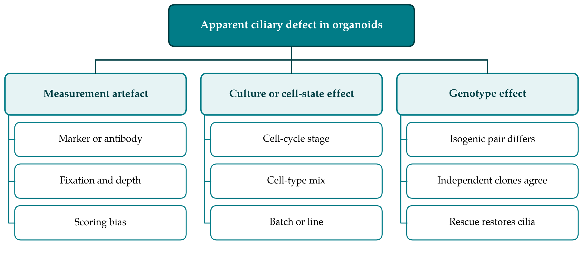
The framework asks four questions in a fixed order. The order matters because each later question is expensive to answer and meaningless if an earlier one fails. Figure 1 shows the three classes of explanation for an apparent defect and the kinds of evidence that separate them, and Table 1 sets out the questions with their controls and assays.
| Question | Controls and assays | Outcome needed to proceed |
|---|---|---|
| 1. Is the measurement sound? | Two ciliary markers plus basal body marker; blinded or automated scoring; electron microscopy | Difference seen with each method |
| 2. Does it follow the genotype? | Isogenic pairs; independent clones; repeated differentiations; rescue | Difference tracks the variant in every pair |
| 3. Cilium or cell state? | Proliferation and cell-type markers; analysis within matched cell types | Difference persists in matched cells |
| 4. Is there a functional consequence? | Response to pathway agonist; ciliary localisation of pathway proteins | Signalling response is altered |
| Throughout | Pre-specified measures; organoid or batch as unit of analysis | Conclusions stated at the level tested |
3.1. Question 1: Is the measurement sound?
The first task is to show that the difference survives a change of method. Cilia should be labelled with at least two independent markers, one for the axoneme and one for the membrane, always together with a basal body marker so that a cilium is counted only when it arises from a centriole. Organoids of the groups being compared are fixed, sectioned or cleared, stained and imaged together, with identical settings. Images are coded so that the person scoring does not know the genotype, or scoring is done by a segmentation routine whose parameters are fixed before the comparison is made. The quantities to record are the fraction of cells bearing a cilium and the length of each cilium measured in three dimensions. Where a defect in structure is suspected, electron microscopy of the axoneme and the transition zone provides confirmation that does not depend on any antibody.
3.2. Question 2: Does the difference follow the genotype?
If the measurement holds, the next step is to separate the mutation from the genetic background. The most direct design uses isogenic pairs. The variant is corrected in the patient-derived line, or introduced into a well characterised control line, by genome editing with CRISPR-Cas9 (Ran et al., 2013), so that the lines compared differ at one site. The reprogramming method that produced the patient line (Takahashi et al., 2007) and the editing step can each introduce changes of their own, and so at least two independently derived clones of each genotype should be examined and should agree. Lines from additional unrelated patients, where they exist, add weight. The strongest evidence is rescue: restoring the normal gene product restores the cilia.
Differentiation should be repeated on separate occasions, and the unit of analysis should be the independent differentiation or the organoid and never the single cell. Thousands of cilia measured in two organoids are two observations. Plotting each replicate's mean together with the underlying measurements makes the structure of the data visible to the reader (Lord et al., 2020).
3.3. Question 3: Is it a property of the cilium or of the cell state?
A difference that follows the genotype may still be indirect. Organoids of the two genotypes should be compared for the proportion of cycling cells, using a proliferation marker, and for cell-type composition, using lineage markers or single-cell RNA sequencing. Ciliation is then compared within matched populations: progenitors with progenitors, post-mitotic cells with post-mitotic cells, at matched stages of differentiation. If the difference disappears within matched cell types, the mutation affects what the cells become and the change in cilia is a consequence. If it persists, the cilium itself is implicated. A complementary test is to examine the undifferentiated stem cells or a simple derivative induced to enter quiescence, where composition is uniform. Agreement between the simple and the complex system strengthens the conclusion, and a defect seen only in organoids suggests dependence on cell type.
3.4. Question 4: Is there a functional consequence?
Structure and function do not always go together. Cilia of normal appearance can signal poorly when the transition zone, which controls what enters the compartment, is defective (Garcia-Gonzalo and Reiter, 2017), and short cilia can signal adequately. Hedgehog signalling is the most accessible readout, because vertebrate cells require intraflagellar transport to respond to it (Huangfu et al., 2003) and because the pathway component Smoothened moves into the cilium on activation (Corbit et al., 2005). The proposed test is to stimulate organoids or their dissociated cells with a pathway agonist and to measure both the accumulation of Smoothened in cilia and the induction of the target genes GLI1 and PTCH1. A blunted response in mutant cells, corrected in the isogenic control, connects the structural finding to a mechanism by which development could be disturbed.
4. Reporting and Interpretation
A finding that passes all four questions supports a specific statement: that the variant impairs cilia in the cell types and under the conditions examined. Reports should give the origin, clone identity and quality control of every line, the differentiation protocol and the age of the organoids, the markers, the scoring procedure and the number of independent differentiations. Negative and partial results deserve reporting too, since a defect confined to one cell type is informative.
Caution is needed in moving from the dish to the patient. Organoids model early stages of organ formation and lack vasculature, flow and many mature cell types. A ciliary defect in vitro does not show that cilia are abnormal in the affected tissues of a person, nor that the person's condition should be reclassified. Functional studies can contribute to the assessment of whether a variant is pathogenic, and the criteria for giving them weight are set out in consensus guidelines (Richards et al., 2015), which require well established assays. An exploratory finding in organoids does not meet that standard by itself, and nothing in this framework should be read as guidance on diagnosis or treatment.
5. Limitations and What Remains to Be Shown
The framework has not been tested. It is a reasoned ordering of controls that are individually established, and whether the order is efficient, and whether the four questions are sufficient, can only be learned by applying it. Known ciliopathy genes offer an obvious positive control: organoids carrying a well characterised mutation in a transition zone or intraflagellar transport gene should pass every step, and a framework that failed them would need revision. A negative control is equally necessary, for instance a mutation known to change proliferation without acting at the cilium, which should be caught at the third question.
Practical limits are also evident. Isogenic lines take months to make and validate. Matching cell types across genotypes is difficult when the mutation alters differentiation profoundly. Motile cilia, which matter in airway and ependymal tissue, require different assays and are outside the scope of this proposal. For very rare disorders only one patient line may ever be available, and in that situation the introduction of the variant into a control line becomes the principal test.
6. Conclusion
An unexpected ciliary phenotype in an organoid model is a lead, and it should be treated as one. The framework proposed here converts the lead into a sequence of tests that deal in turn with measurement, genotype, cell state and function, and it specifies the evidence required at each stage before the next is attempted. It awaits application and validation. Used with that understanding, it may help investigators distinguish a new link between a rare disorder and the cilium from the ordinary variability of a complex culture system.
Conflict of interest
To be declared by the author(s) before publication.
Funding
To be declared by the author(s) before publication.