1. Introduction
Mitochondria were once described in stem cell biology chiefly by their absence. Quiescent adult stem cells and pluripotent cells in culture rely heavily on glycolysis, their mitochondria are few and structurally simple, and oxidative metabolism rises as cells commit to a lineage. Haematopoietic stem cells, for instance, have a metabolic profile suited to the low-oxygen environment of their niche (Simsek et al., 2010), and somatic cells that are reprogrammed to pluripotency shift from oxidative phosphorylation to glycolysis before they acquire the markers of the new state (Folmes et al., 2011). From observations of this kind it was natural to conclude that stem cells keep their mitochondria switched off and out of the way.
That conclusion has not survived closer study. Stem cells whose mitochondria are damaged, or that cannot dispose of damaged mitochondria, lose the capacity to renew themselves even when their supply of ATP is adequate. The organelle supplies intermediates for biosynthesis and for the enzymes that modify chromatin, produces reactive oxygen species that act as signals, buffers calcium, and reports its own stress to the nucleus (Shyh-Chang et al., 2013; Chandel et al., 2016). A stem cell therefore has reason to keep a small population of mitochondria in very good order.
The machinery that does so is collectively called mitochondrial quality control. This review asks how far the published evidence supports the claim that it takes part in fate decisions, as distinct from merely keeping cells alive. It first states how the literature was approached, then takes the levels of quality control in turn, and ends with the weaknesses of the evidence and the work that would address them.
2. Approach to the Literature
The review is narrative. It is built around primary studies that manipulated a defined component of mitochondrial quality control in a defined stem cell population and measured an outcome related to fate, such as long-term reconstitution after transplantation, the balance of self-renewal and differentiation in culture, or the onset of senescence. Reviews are cited for background on mechanism. Most of the relevant work has been done in the mouse and in cultured human cells, and the conclusions should be read with that limit in mind. No formal search protocol was used and the coverage is selective.
3. The Layers of Mitochondrial Quality Control
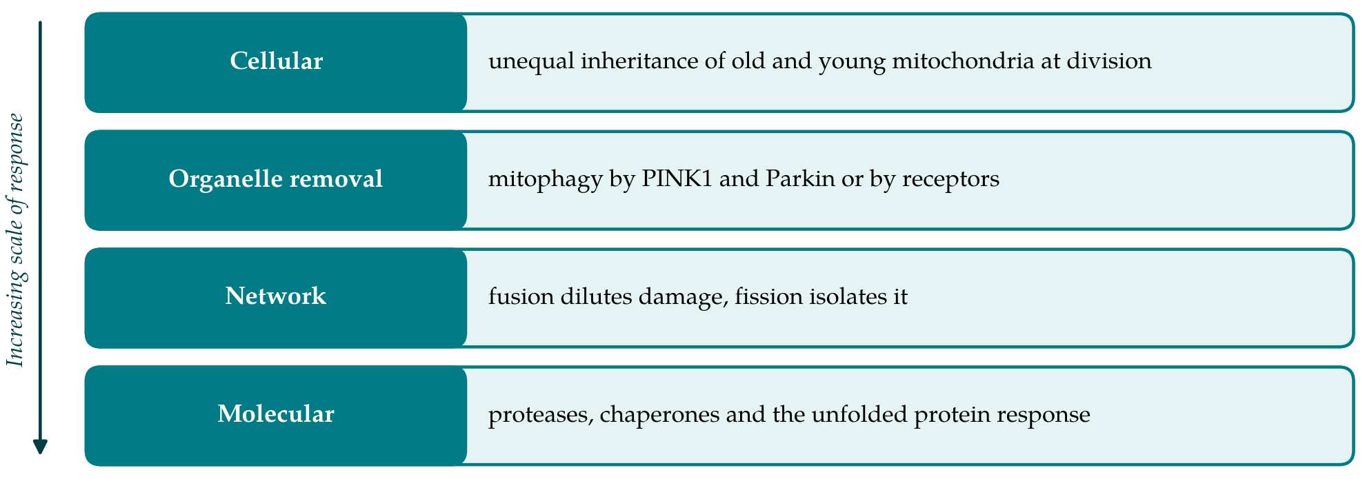
Quality control operates at several scales, from single proteins to the whole population of organelles in a cell (Pickles et al., 2018). Figure 1 arranges them as four layers. The layers are connected, since damage that is not repaired at one level is passed to the next, and a fault in any one of them tends to raise the load on the others.
3.1. Proteostasis and stress signalling
Mitochondrial proteins are encoded by two genomes, and the complexes of the respiratory chain are assembled from subunits of both. An imbalance between the two sets, or the accumulation of misfolded protein in the matrix, activates a transcriptional programme known as the mitochondrial unfolded protein response, which raises the expression of chaperones and proteases and adjusts metabolism (Quirós et al., 2016). The response is one of several routes by which the organelle communicates with the nucleus, and it is the first line of defence in the sense that it acts before an organelle is damaged beyond repair.
3.2. Fusion and fission
Mitochondria form a network that is continually remodelled. Fusion of the outer membrane depends on the mitofusins MFN1 and MFN2 and fusion of the inner membrane on OPA1, while fission is carried out by the dynamin-related protein DRP1. Fusion allows the contents of organelles to mix, so that a mitochondrion with a defective component can be complemented by its neighbours. Mice lacking either mitofusin die during embryonic development, which shows how much the process matters (Chen et al., 2003). Fission does the opposite work of sorting. Twig et al. (2008) observed that a division often yields one daughter organelle with a lower membrane potential than the other, and that the depolarised daughter is less likely to rejoin the network and more likely to be removed by autophagy.
3.3. Mitophagy
The selective autophagy of mitochondria is the best characterised layer. In the pathway that depends on PINK1 and Parkin, the kinase PINK1 is normally imported into healthy mitochondria and degraded. When membrane potential is lost it accumulates on the outer membrane, and Parkin, a ubiquitin ligase, is recruited from the cytosol to the damaged organelle (Narendra et al., 2008). Ubiquitin chains on outer membrane proteins, phosphorylated by PINK1, then recruit autophagy receptors that link the organelle to the forming autophagosome (Lazarou et al., 2015). A second group of pathways uses receptors that reside in the outer membrane and bind the autophagy machinery directly. The receptor NIX, for example, is required for the programmed clearance of mitochondria from maturing red blood cells (Sandoval et al., 2008). The mechanisms are reviewed by Youle and Narendra (2011).
3.4. Unequal inheritance
A dividing cell has one further option, which is to give its two daughters mitochondria of different quality. Katajisto et al. (2015) labelled mitochondria according to their age in cultured human mammary cells with stem-like properties and found that divisions were often asymmetric in this respect. The daughter that received fewer old mitochondria retained stem cell traits, and interfering with fission abolished both the asymmetry and the difference in fate. The finding implies that the quality of the inherited organelles is itself a determinant of what a daughter cell becomes.
4. Evidence Linking Quality Control to Fate
The studies that tie these mechanisms to stem cell behaviour are summarised in Table 1 and discussed below by tissue. They share a design: a quality control component is removed or enhanced, and the consequences for self-renewal or differentiation are followed.
| Layer | Key components | Reported effect on stem cells | Representative sources |
|---|---|---|---|
| Proteostasis | SIRT7, NRF1, chaperones, prohibitins | Restrains proliferation under stress; declines with age | Mohrin et al., 2015; Zhang et al., 2016 |
| Fusion | MFN1, MFN2, OPA1 | Supports self-renewal of neural and lymphoid-competent blood stem cells | Khacho et al., 2016; Luchsinger et al., 2016 |
| Fission | DRP1 | Needed for sorting of aged mitochondria at division | Katajisto et al., 2015 |
| Mitophagy | PINK1, Parkin, autophagy genes | Required for expansion of blood stem cells; prevents senescence in muscle | Ito et al., 2016; García-Prat et al., 2016 |
| Bulk autophagy | ATG12 and core machinery | Keeps blood stem cells metabolically quiet | Ho et al., 2017 |
| Remodelling in reprogramming | Mitochondrial clearance and rejuvenation | Accompanies return to pluripotency | Prigione et al., 2010 |
4.1. Haematopoietic stem cells
The blood system offers the most stringent test of stem cell function, because a single cell can be asked to rebuild haematopoiesis in a transplanted animal. Several findings point the same way. The fraction of haematopoietic stem cells with low mitochondrial membrane potential has the greater capacity for long-term reconstitution, and lowering the potential chemically favours self-renewal in culture (Vannini et al., 2016). Ito et al. (2016) found that a highly purified population of these cells depends on mitophagy for its self-renewing expansion and that silencing the genes for Parkin or PINK1 impaired it. The general metabolic requirements of the cells are reviewed by Ito and Suda (2014).
Autophagy more broadly has a related function. When Ho et al. (2017) deleted an essential autophagy gene in the blood system of mice, stem cells accumulated mitochondria, became metabolically active, and showed impaired self-renewal with a bias towards myeloid output, a pattern that resembles ageing. The same study reported that old stem cells differ among themselves in autophagic activity and that those which keep it high retain better function. Fusion matters as well, in a lineage-specific way: Luchsinger et al. (2016) showed that mitofusin 2 is needed to maintain stem cells with strong lymphoid potential, an effect they traced to the handling of calcium and not to energy supply. Finally, Mohrin et al. (2015) described a checkpoint in which the sirtuin SIRT7 limits mitochondrial protein-folding stress; its expression falls in aged stem cells, and restoring it improved their regenerative capacity.
4.2. Neural and muscle stem cells
In the developing and adult mouse brain, Khacho et al. (2016) found that the shape of the mitochondrial network changes as neural stem cells commit, and that deleting fusion proteins in these cells reduced self-renewal and led to depletion of the stem cell pool. The effect was mediated by a rise in reactive oxygen species that altered a nuclear transcriptional programme, so that the mitochondrial change preceded and caused the change in identity. Reactive oxygen species of mitochondrial origin are also required for differentiation in other settings: human mesenchymal stem cells need those generated at complex III of the respiratory chain in order to form adipocytes (Tormos et al., 2011). The signal is thus used in both directions, and its meaning depends on the cell type and the dose.
Muscle stem cells, or satellite cells, spend most of adult life in quiescence. García-Prat et al. (2016) showed that their autophagic activity fails in very old mice, that the failure leads to mitochondrial dysfunction and oxidative stress, and that the cells then enter senescence; restoring autophagy reversed the process. Zhang et al. (2016) reached a compatible result from another direction. Supplying a precursor of NAD+ to aged mice activated the mitochondrial unfolded protein response in muscle stem cells and delayed their senescence.
4.3. Pluripotent cells
Pluripotent stem cells provide the clearest example of mitochondria being remodelled as fate changes. When fibroblasts are reprogrammed to induced pluripotent stem cells their mitochondria return to an immature form with poorly developed cristae, and markers of mitochondrial oxidative stress fall (Prigione et al., 2010). The change is reversed on differentiation. Because the metabolic shift is detectable before pluripotency genes are fully expressed (Folmes et al., 2011), it is unlikely to be a passive consequence of the new state.
5. Limits of the Evidence and Future Work
The case that mitochondrial quality control contributes to fate decisions is strong in outline. Its weaknesses are of four kinds. First, most experiments remove a gene, and the proteins involved have other functions. Mitofusin 2 tethers mitochondria to the endoplasmic reticulum, autophagy genes act on many substrates, and Parkin has targets outside mitochondria. An effect on fate after deletion does not by itself show that mitochondrial quality was the cause. Second, measurements of mitochondrial mass and membrane potential in stem cells rely on fluorescent dyes whose retention is affected by the efflux pumps that these cells express at high levels, and this has led to conflicting reports. Third, the evidence comes from a few tissues and largely from the mouse. Fourth, the signalling step between an altered organelle and an altered nucleus is identified in only some of the studies.
Better tools exist for several of these problems. Mice that express the pH-sensitive reporter mt-Keima allow mitophagy to be measured in tissues without dyes (Sun et al., 2015), and methods that tag mitochondria by age can test whether unequal inheritance occurs in vivo. Experiments that rescue a phenotype by correcting a defined downstream signal, as was done for reactive oxygen species and for calcium in the studies cited above, are more persuasive than deletion alone. Whether enhancing quality control can preserve stem cell function in human tissues is unknown. The animal studies justify the question, and they do not answer it.
6. Conclusion
The published record shows that stem cells depend on active maintenance of their mitochondria and that failures of maintenance alter fate in recognisable ways: loss of self-renewal, skewed differentiation and premature senescence. Proteostasis, network dynamics, mitophagy and unequal inheritance each contribute, and they act on the nucleus through metabolites, reactive oxygen species, calcium and stress signalling. What remains to be shown is how general these relationships are across tissues and species, and whether the mitochondrial change is the initiating event or one link in a longer chain. The answer will come from experiments that follow the quality of the organelles and the fate of the cell in the same lineage over time.
Conflict of interest
To be declared by the author(s) before publication.
Funding
To be declared by the author(s) before publication.