1. Introduction
The situation is one every stroke team will recognise. A young adult arrives at an emergency department with sudden weakness of one arm and leg. The ambulance crew has given advance notice of a stroke, the team is waiting, and the clock that governs reperfusion treatment is running. In nearly every such call the cause lies in the brain or is one of the familiar mimics: seizure, migraine, hypoglycaemia, a functional disorder. Very occasionally it lies in the cervical spinal canal, where a haematoma in the epidural space is compressing one half of the cord.
Spontaneous spinal epidural haematoma is uncommon enough that most emergency physicians and many neurologists will never have seen a case. The estimate usually quoted, from Holtås et al. (1996), is in the order of one case per million people per year. Its hemiparetic presentation has been reported repeatedly in the case literature, often with the observation that stroke treatment was started or seriously considered before the diagnosis was made. A rare condition does not justify a new investigation for every patient. It may justify a small change in what is asked and looked at, provided the change costs almost nothing in the common case.
This article proposes such a change in the form of a stepwise diagnostic pathway for emergency and stroke teams. It is written with younger adults in mind, because in them the prior probability of atherosclerotic stroke is lower and unusual causes take a larger share. The pathway is assembled from published evidence and has not been tested in practice here.
2. Clinical Background
Three things about the condition shape the design: how it arises and presents, why stroke assessment tends to miss it, and what is lost when it is missed.
2.1. The condition
A spinal epidural haematoma is called spontaneous when it occurs without significant trauma or an invasive procedure. In their survey of 613 published cases of spinal haematoma, Kreppel et al. (2003) found that no cause could be identified in a substantial share, and that anticoagulant treatment and vascular malformations were the most frequent associations among the rest. The source of bleeding is debated. Groen and Ponssen (1990) argued for rupture of the internal vertebral venous plexus, perhaps after a rise in pressure from straining or coughing, whereas Beatty and Winston (1984) held that the speed of cord compression in the cervical region pointed to an arterial origin. The clot usually lies posterior or posterolateral to the cord and spans several segments, with the cervicothoracic junction a common site.
The typical story is of sudden, severe pain in the neck or between the shoulder blades, sometimes radiating into an arm, followed within minutes to hours by weakness and sensory change below the level of the lesion. When the haematoma is central the result is paraparesis or tetraparesis. When it is lateral the corticospinal tract on one side takes most of the pressure and the weakness is in the arm and leg of that side.
2.2. Why it imitates stroke
A hemiparesis of sudden onset is the core of every stroke recognition tool. The Recognition of Stroke in the Emergency Room scale scores face, arm and leg weakness, speech disturbance and visual field defect (Nor et al., 2005), and the National Institutes of Health Stroke Scale quantifies the same domains in more detail (Brott et al., 1989). Neither asks about pain and neither tests for a sensory level on the trunk. A patient with unilateral arm and leg weakness from a cervical lesion will score positively on both. In the Brain Attack Study, close to a third of suspected strokes assessed at the bedside proved to be mimics (Hand et al., 2006), so stroke teams are used to mimics, but the common ones are medical conditions for which a short delay or even inadvertent thrombolysis carries little risk.
There is a further trap. Neck pain with hemiparesis in a young adult rightly suggests cervical artery dissection, a leading cause of stroke in this age group (Debette and Leys, 2009). The clinician who thinks of dissection orders vessel imaging, finds the arteries normal, and may then return to a diagnosis of stroke of undetermined cause without having considered the canal that the same images pass through.
2.3. What is at stake
Guidelines for acute ischaemic stroke recommend intravenous thrombolysis within four and a half hours of onset in eligible patients and stress that treatment should not be delayed for investigations that are not essential (Powers et al., 2019). Thrombolysis given to patients who turn out to have a mimic is generally safe, with a low rate of symptomatic haemorrhage in cohort studies (Zinkstok et al., 2013; Tsivgoulis et al., 2015). An epidural haematoma is the exception to that reassurance, since a fibrinolytic or antithrombotic drug can be expected to promote further bleeding in a closed space.
Delay matters for a second reason. Outcome after surgical evacuation depends on the severity of the deficit before operation (Foo and Rossier, 1981) and on the interval between onset and decompression. Lawton et al. (1995) found better recovery when surgery was performed within twelve hours, and Groen and van Alphen (1996) concluded that patients with complete deficits did best when operated on within thirty-six hours and those with incomplete deficits within forty-eight. A more recent analysis reached similar conclusions about the weight of preoperative status (Bakker et al., 2015). Hours spent on a stroke unit under the wrong diagnosis are taken from that interval.
3. The Proposed Pathway
The pathway has seven steps, shown in Figure 1. The first two apply to every patient assessed for suspected stroke and are meant to add well under a minute. The remainder apply only when those steps raise suspicion. Table 1 lists the features that should do so, set against what is usual in a hemispheric stroke.
| Feature | Usual in hemispheric stroke | Suggests cervical epidural haematoma |
|---|---|---|
| Pain at onset | Absent or mild headache | Sudden severe neck or interscapular pain, may radiate to arm |
| Face | Lower face often weak on the affected side | Spared |
| Language and vision | Aphasia, neglect or field defect may be present | Normal |
| Sensory pattern | Loss on the same side as the weakness | Crossed pain and temperature loss, or a level on the trunk |
| Other limbs | Normal | Subtle weakness may appear and progress |
| Course in first hours | Maximal at onset or fluctuating | Often progressive after the pain; occasionally resolving |
| Bladder | Usually unaffected early | Retention may develop |
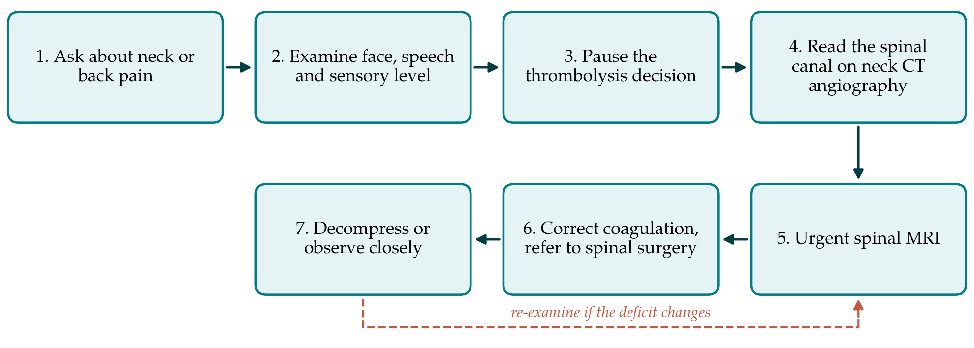
3.1. Step 1: Ask about pain
Every patient with sudden limb weakness is asked one question: whether there was pain in the neck, shoulders or upper back at or just before the onset. Acute ischaemic stroke is usually painless. Severe axial pain at onset should prompt the clinician to think of dissection, of aortic dissection, and of a spinal cause.
3.2. Step 2: A targeted examination
Four findings are sought. The first is whether the face is involved, since a lesion below the brainstem spares it. The second is whether language, attention and visual fields are normal despite a dense weakness of the dominant side. The third is sensation: loss of pain and temperature on the side opposite the weakness, or a sensory level on the trunk, indicates the cord. The fourth is any weakness, however slight, of the supposedly unaffected limbs. The International Standards for Neurological Classification of Spinal Cord Injury provide a common vocabulary for recording these findings (Kirshblum et al., 2011).
3.3. Step 3: Pause the thrombolysis decision
If severe axial pain at onset is combined with any one of the cord features in Table 1, the pathway asks the team to withhold thrombolytic and antithrombotic drugs until the spinal canal has been looked at. This is a pause and not a refusal. It is justified by the asymmetry of harms: a short delay in a patient who does have a stroke against a serious risk in a patient who does not.
3.4. Step 4: Read the canal on the images already taken
Most stroke protocols include computed tomographic angiography from the aortic arch to the vertex. The cervical canal is in the field of view. An acute epidural haematoma may be visible on these images as a dense collection behind or beside the cord, and the pathway asks the radiologist, when alerted by the clinical team, to review the canal on soft tissue windows before reporting the study as normal. This step adds no radiation, no contrast and no time in the scanner.
3.5. Step 5: Urgent magnetic resonance imaging
Magnetic resonance imaging of the spine is the definitive investigation. Holtås et al. (1996) described the appearances and their change with time: in the first day the haematoma is typically close to the cord in signal on T1-weighted images and bright or mixed on T2-weighted images, and it becomes bright on T1-weighted images later. Where a service performs magnetic resonance imaging of the brain for suspected stroke, sagittal sequences through the cervical spine can be added in the same session. If suspicion is high, a negative review of the canal at Step 4 should not prevent Step 5.
3.6. Step 6: Correct coagulation and refer
Once a haematoma is confirmed, anticoagulant and antiplatelet drugs are stopped and any coagulopathy is corrected according to local protocols and haematology advice. The patient is referred immediately to the spinal surgical service, with the time of onset, the examination findings and their trend stated clearly, since these determine urgency.
3.7. Step 7: Decompress or observe
Surgical evacuation, usually by laminectomy or hemilaminectomy, is the established treatment for patients with significant or progressing deficits (Kreppel et al., 2003). Conservative management has been reported with good results in patients whose deficits are mild or are already improving within the first hours, and Groen (2004) has reviewed that experience. The decision belongs to the surgical team. The pathway asks only that it be made early, with repeated examination in the interval so that deterioration is detected at once.
4. Fitting the Pathway to a Stroke Service
The pathway is meant to be added to an existing stroke protocol as a prompt and not to stand as a separate document. In practice this means one line on the assessment form for pain at onset, one for facial involvement and crossed sensory signs, and a standing agreement with radiology about the cervical canal. It asks nothing of ambulance crews, whose prehospital tools are rightly kept simple. Training can be brief, because the examination uses skills every clinician already has. The points most likely to meet resistance are the pause at Step 3, which runs against the habit of speed that stroke services have worked hard to build, and access to spinal magnetic resonance imaging out of hours. A service adopting the pathway should agree in advance who authorises the pause and how the scan is obtained.
5. Limitations and What Remains to Be Shown
The pathway has not been validated. Its red flags are drawn from case reports and series of a rare condition, which describe the patients who were eventually diagnosed and say little about how often the same features occur in people with stroke. Neck pain accompanies dissection, and sensory findings in an acutely unwell patient are unreliable. The specificity of the Step 3 trigger is therefore unknown, and if it is low the pathway could delay treatment for patients with stroke more often than it protects a patient with a haematoma.
Two measurements would address this. A stroke service could record, for consecutive calls, how often each red flag is present and what the final diagnosis was, which would give the frequency of triggers and the delay they cause. Review of the cervical canal on archived angiograms from known cases of epidural haematoma would show how often the lesion is visible on that modality. Neither study requires the pathway to be in use. Given the rarity of the condition, sensitivity can probably be estimated only through collaboration between centres or a registry.
6. Conclusion
A cervical epidural haematoma that presents as hemiparesis is a rare mimic of stroke with a specific danger, because the treatment for the suspected condition may worsen the real one. The pathway proposed here relies on one question, a few examination findings and a second look at images already acquired to bring the diagnosis to mind in time. It is offered as a reasoned proposal for stroke teams to examine, adapt and test, and its effect on diagnosis and on treatment times remains to be measured.
Conflict of interest
To be declared by the author(s) before publication.
Funding
To be declared by the author(s) before publication.