1. Introduction
Nontuberculous mycobacteria (NTM) are environmental organisms. They live in soil, in natural water and in engineered water systems, where biofilms and tolerance of disinfectants allow them to persist (Falkinham, 2009). Most people inhale or swallow them without consequence. Clinical disease is usually confined to one organ: slowly progressive lung disease in people with bronchiectasis or emphysema, cervical lymphadenitis in young children, or skin and soft tissue infection after injury or a procedure. Management of those syndromes is set out in international guidance (Daley et al., 2020).
Disseminated disease belongs to another category. When an NTM species is cultured from blood or bone marrow, or from several anatomically separate sites, the organism has crossed defences that ordinarily contain it, and the clinician should assume a defect in cell-mediated immunity until one has been looked for. Before effective antiretroviral therapy, disseminated Mycobacterium avium complex infection was a defining illness of advanced human immunodeficiency virus (HIV) infection and its relationship to the CD4 count was well described (Horsburgh, 1991). An adult with the same syndrome and a negative HIV test presents a harder problem.
That problem now has a recognised answer in a substantial proportion of cases. Adults, most of them of East or South East Asian origin, have been described with disseminated NTM and other intracellular infections caused by neutralising autoantibodies against interferon-gamma, a condition sometimes called adult-onset immunodeficiency (Browne et al., 2012). Other possibilities include late-presenting inborn errors of the interleukin-12 and interferon-gamma circuit, idiopathic CD4 lymphocytopenia, haematological malignancy and the effects of immunosuppressive drugs. This article proposes an ordered pathway for working through them. It is a proposal drawn from published series and guidelines, and it has not been validated in practice.
2. Background to the Pathway
Control of mycobacteria depends on a conversation between infected macrophages and T cells. Macrophages produce interleukin-12, which drives T cells and natural killer cells to secrete interferon-gamma, which in turn activates the macrophage to restrict intracellular growth. Genetic lesions at almost every node of this circuit, in the receptors for interleukin-12 and interferon-gamma, in STAT1, in IRF8 and in the NEMO regulator, cause Mendelian susceptibility to mycobacterial disease, in which weakly virulent environmental mycobacteria and Bacille Calmette-Guerin cause severe infection in otherwise healthy people (Casanova and Abel, 2002; Bustamante et al., 2014). The classification of these and other inborn errors of immunity is maintained by an expert committee and is the reference point for genetic work-up (Tangye et al., 2022).
The same circuit can be interrupted without a mutation. High-titre neutralising autoantibodies to interferon-gamma block signalling and produce a phenocopy of the genetic defects in adults who were previously well (Browne and Holland, 2010). The large comparative study that defined the syndrome described patients in Thailand and Taiwan with disseminated NTM, salmonellosis, penicilliosis and varicella zoster, almost all of them with these autoantibodies and none with an identified genetic cause (Browne et al., 2012). Susceptibility is associated with particular HLA class II alleles, which fits the geographical concentration of reported cases (Chi et al., 2013). Follow-up of such patients has shown a chronic, relapsing course in which the autoantibody titre tends to persist (Chi et al., 2016).
Two further diagnoses complete the main differential. Idiopathic CD4 lymphocytopenia, defined as persistently low CD4 T cells without HIV infection or another cause, was characterised in a national survey when such patients came to attention during the HIV epidemic, and it predisposes to the same opportunistic organisms (Smith et al., 1993). Iatrogenic immunosuppression is probably now the commonest explanation of all: tumour necrosis factor antagonists in particular carry a recognised risk of mycobacterial disease (Winthrop et al., 2009), and corticosteroids, calcineurin inhibitors, cytotoxic chemotherapy and transplantation all contribute.
3. The Proposed Pathway
The pathway has seven steps. Figure 1 shows how they follow one another, and Table 1 gives the purpose of each step and the result that moves the patient on. The order is deliberate: inexpensive and widely available tests come before specialised ones, and the tests that change immediate management come before those that explain the mechanism.
| Step | Purpose | Finding that directs the next step |
|---|---|---|
| 1. Confirm dissemination | Establish that infection is not confined to one organ | Growth from blood, marrow or multiple sites |
| 2. Identify the species | Guide therapy and gauge virulence (Daley et al., 2020) | A weakly virulent species raises suspicion |
| 3. Exclude HIV and drugs | Find the common causes first (Horsburgh, 1991) | Negative serology and no immunosuppressive drug |
| 4. Lymphocyte subsets | Detect CD4 lymphocytopenia (Smith et al., 1993) | Repeatedly low CD4 count with no other cause |
| 5. Anti-interferon-gamma antibodies | Test for the acquired phenocopy (Browne et al., 2012) | High-titre neutralising autoantibody |
| 6. Functional and genetic study | Look for an inborn error (Bustamante et al., 2014) | Impaired signalling or a causative variant |
| 7. Joint review | Agree treatment and duration of follow-up | A plan recorded by both services |
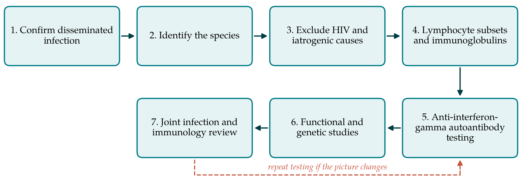
3.1. Step 1: Confirm that the infection is disseminated
Dissemination should be documented and not assumed. Mycobacterial blood cultures and, where there are cytopenias or unexplained fever, bone marrow aspirate and trephine with culture and histology are the core investigations. Tissue from an accessible site, a lymph node or a skin lesion, should be sent for both histology and mycobacterial culture rather than histology alone. Cross-sectional imaging defines the extent of disease. A single positive respiratory culture does not meet the definition, because airway colonisation is common and the diagnostic criteria for pulmonary disease require repeated isolation with compatible radiology (Daley et al., 2020).
3.2. Step 2: Identify the species
Species identification by a reference laboratory, using line probe assays, mass spectrometry or sequencing, serves two purposes. It determines treatment, since regimens for Mycobacterium avium complex, Mycobacterium kansasii and the rapidly growing species differ. It also carries diagnostic weight: the weakly virulent species that rarely trouble anyone with intact immunity are the ones most associated with defects of the interferon-gamma axis. Susceptibility testing should follow the methods and the drug-organism combinations recommended in guidance, since in vitro results correlate with outcome for only some agents.
3.3. Step 3: Exclude HIV infection and iatrogenic causes
A fourth-generation HIV antigen and antibody assay is mandatory whatever the stated risk, with nucleic acid testing if infection is strongly suspected despite a negative result. A careful drug history is equally important and is easily done poorly. It should cover biologic agents, particularly tumour necrosis factor antagonists and newer agents acting on cytokine pathways (Winthrop et al., 2009), systemic corticosteroids including intermittent courses, transplant immunosuppression, recent chemotherapy and non-prescribed preparations. Diabetes mellitus, malnutrition, chronic kidney disease and alcohol use disorder modify risk without accounting on their own for disseminated disease.
3.4. Step 4: Lymphocyte subsets and immunoglobulins
Flow cytometric enumeration of T, B and natural killer cells, with serum immunoglobulin concentrations and serum protein electrophoresis, is the next layer. Persistently reduced CD4 counts in the absence of HIV infection, confirmed on a separate occasion, support idiopathic CD4 lymphocytopenia (Smith et al., 1993). Lymphopenia with a paraprotein or marrow infiltration points to haematological disease, which is why the marrow examination in step one earns its place. Normal subsets do not end the evaluation; the autoantibody syndrome characteristically leaves routine immunological tests unremarkable.
3.5. Step 5: Test for anti-interferon-gamma autoantibodies
Where disseminated NTM disease occurs in an adult with normal lymphocyte subsets and no drug or viral cause, testing for anti-interferon-gamma autoantibodies should be considered early rather than last, especially in patients of East or South East Asian origin and in those with concurrent salmonellosis, disseminated Talaromyces marneffei infection or severe zoster. Detection combines an immunoassay for binding antibody with a functional assay showing that patient plasma blocks interferon-gamma-induced STAT1 phosphorylation or downstream gene expression in normal cells (Browne and Holland, 2010; Browne et al., 2012). Both are performed in specialist laboratories, so samples and transport should be arranged in advance. The association with particular HLA class II alleles may support the diagnosis but does not establish it (Chi et al., 2013).
3.6. Step 6: Functional and genetic study of the cytokine axis
If no autoantibody is found, the question becomes whether an inborn error has presented late. Functional testing measures interferon-gamma production by stimulated whole blood and the cellular response to interferon-gamma, which localises the defect to the production or the response arm of the circuit. Targeted sequencing of the known genes, or whole exome sequencing where the panel is negative and suspicion remains, follows (Bustamante et al., 2014; Tangye et al., 2022). Hypomorphic variants may produce an adult presentation, so an adult age at onset does not exclude a genetic cause. Variants should be interpreted against standard criteria and with functional corroboration, since the clinical consequences of a wrong attribution are considerable.
3.7. Step 7: Joint infection and immunology review
The pathway ends with a joint consultation at which the microbiological diagnosis, the immunological findings and the treatment plan are brought together. Antimycobacterial therapy for disseminated disease is prolonged and combination based, and in patients whose immune defect persists it may need to continue long after cultures are negative. Decisions about immunomodulatory treatment in the autoantibody syndrome, about prophylaxis against other opportunistic organisms and about interferon-gamma replacement in proven receptor-side defects belong to specialists and are outside the scope of this pathway. The review should set the interval for reassessment, because relapse is common and the underlying defect may not resolve (Chi et al., 2016).
4. Fitting the Pathway to Different Settings
Few hospitals can perform every test. The pathway is arranged so that each step is useful in isolation and so that the first four can be completed in any centre with a diagnostic laboratory and flow cytometry. Steps five and six depend on referral, and the practical requirement is a named referral route and an agreed sample protocol rather than local capability. In settings where tuberculosis is common, the first consideration is often whether the organism is Mycobacterium tuberculosis at all, since the therapeutic consequences differ entirely and misidentification is a recognised hazard. Where resources are limited, the greatest return comes from the early steps: confirming dissemination, getting the species right and excluding HIV infection and drug-induced immunosuppression.
5. Limitations and What Remains to Be Shown
This article proposes a sequence and does not evaluate it. Three questions would have to be answered before it could be recommended without qualification. The first is yield: in consecutive adults presenting with disseminated NTM infection, how often each step produces the explanation, and whether the order chosen here finds it sooner than an unstructured approach. The second is timing, since a pathway that reaches the right answer after many months of untreated infection has limited value; the interval from presentation to specific diagnosis would be the natural measure. The third is transferability, as the relative frequency of the underlying causes differs by region and the pathway may need reordering where the autoantibody syndrome is rare.
Two cautions apply to the pathway itself. A negative evaluation does not exclude an immune defect, and patients in whom no cause is found should be followed rather than discharged, because new phenotypes continue to be described. And the pathway addresses the cause of susceptibility, not the treatment of the infection, which follows the relevant guidance and the identified species.
6. Conclusion
Disseminated nontuberculous mycobacterial infection in an adult is a signal that the interleukin-12 and interferon-gamma axis or the T cell compartment has failed, and the cause can usually be identified if it is sought in an orderly way. The seven-step pathway proposed here moves from confirming dissemination and identifying the species, through the common acquired causes, to the autoantibody syndrome and the inborn errors, and it ends with a plan agreed between infection and immunology services. Its value in practice remains to be measured, and the measurements described above are within reach of centres that see these patients.
Conflict of interest
To be declared by the author(s) before publication.
Funding
To be declared by the author(s) before publication.