1. Introduction
A model that assigns a probability of 0.8 to malignancy is calibrated if, among all the images to which it assigns 0.8, about four in five are malignant. Discrimination, the ability to rank diseased cases above healthy ones, is a different property, and a model can have one without the other. For decisions that depend on thresholds, such as whether to refer, to biopsy or to reassure, the probability itself is the input, and a miscalibrated probability can mislead even when ranking is excellent. Van Calster et al. (2019) called calibration the Achilles heel of predictive analytics for this reason, and the TRIPOD reporting guideline asks that it be reported for every clinical prediction model (Collins et al., 2015).
Deep image classifiers present a particular difficulty. Guo et al. (2017) showed that modern networks tend to be overconfident and that a one-parameter rescaling of the logits, temperature scaling, corrects much of the error on data like the training set. Many alternatives followed. What has not followed is a common way of testing them in medical imaging. Two papers that both report an expected calibration error for a chest radiograph classifier may have used different bins, different splits and different rules about what the recalibration step was allowed to see.
This article proposes a protocol for benchmarking calibration in medical image classifiers so that results from different groups can be set side by side. The protocol is assembled from established statistical practice and published machine learning methods. It is offered as a proposal and has not been applied to any dataset in this article; no results are reported.
2. Background
The design rests on three bodies of prior work: how calibration is measured, how it is repaired, and why medical imaging strains both.
2.1. Measuring calibration
The oldest measure is the Brier score, the mean squared difference between forecast probability and outcome (Brier, 1950). Murphy (1973) showed that it separates into a reliability term, which is the calibration component, a resolution term and an uncertainty term fixed by the data. The Brier score and the negative log-likelihood are proper scoring rules: their expected value is optimised only by reporting the true probabilities (Gneiting and Raftery, 2007). They cannot be gamed, but they mix calibration with discrimination in a single figure.
The expected calibration error isolates calibration. Predictions are sorted into bins by confidence and the gap between mean confidence and observed accuracy is averaged across bins, weighted by bin size (Naeini et al., 2015). The measure is intuitive and pairs naturally with a reliability diagram. It is also fragile. Its value depends on the number of bins and on whether they have equal width or equal mass; in multi-class problems it usually examines only the top predicted class; and Nixon et al. (2019) documented how these choices alter the ranking of methods. Kumar et al. (2019) proved that the binned estimate can understate the true error of a continuous recalibration method and proposed a debiased estimator.
2.2. Repairing calibration
Post-hoc methods adjust the outputs of a trained model using held-out data. Platt scaling fits a logistic function to the scores (Platt, 1999); isotonic regression fits a monotone step function (Zadrozny and Elkan, 2002); and the two were compared across classical learners by Niculescu-Mizil and Caruana (2005). Temperature scaling is the single-parameter relative of Platt scaling for multi-class networks (Guo et al., 2017). Other methods act during training: label smoothing softens the targets (Müller et al., 2019), and averaging several independently trained networks, a deep ensemble, improves both accuracy and uncertainty estimates (Lakshminarayanan et al., 2017).
2.3. Why medical imaging strains both
Three features of clinical image data matter. Datasets are small and classes are unbalanced, so bins at high confidence for a rare disease may hold very few positive cases and any binned estimate is noisy. One patient often contributes several images, and if images from the same patient fall on both sides of a split the test is contaminated. And the deployment population differs from the development population in scanner, protocol and disease prevalence. Ovadia et al. (2019) found that calibration deteriorates as data move away from the training distribution and that post-hoc scaling tuned on in-distribution data is among the methods that degrade, a finding with obvious weight for a model carried from one hospital to another.
3. The Proposed Protocol
The protocol consists of seven steps. Table 1 lists what is fixed at each step and the record that should be published with the results, and Figure 1 shows the order in which the steps run. The steps are then described one at a time.
| Step | What is fixed in advance | Record to publish |
|---|---|---|
| 1. Task and splits | Label definition; patient-level train, validation, calibration and test splits | Split files with patient identifiers hashed |
| 2. Training | Architecture, preprocessing, augmentation, stopping rule, number of seeds | Configuration files and seeds |
| 3. Recalibration | Methods compared; fitted on the calibration split only | Fitted parameters per model and seed |
| 4. Metrics | Proper scores, binned and class-wise error, binning scheme | Metric code and reliability diagrams |
| 5. Uncertainty | Bootstrap by patient; spread across seeds | Intervals for every reported value |
| 6. Shift and subgroups | External test set; subgroups named before analysis | Results per site and subgroup |
| 7. Release | Licence and location of artefacts | Code, splits, per-image predictions |
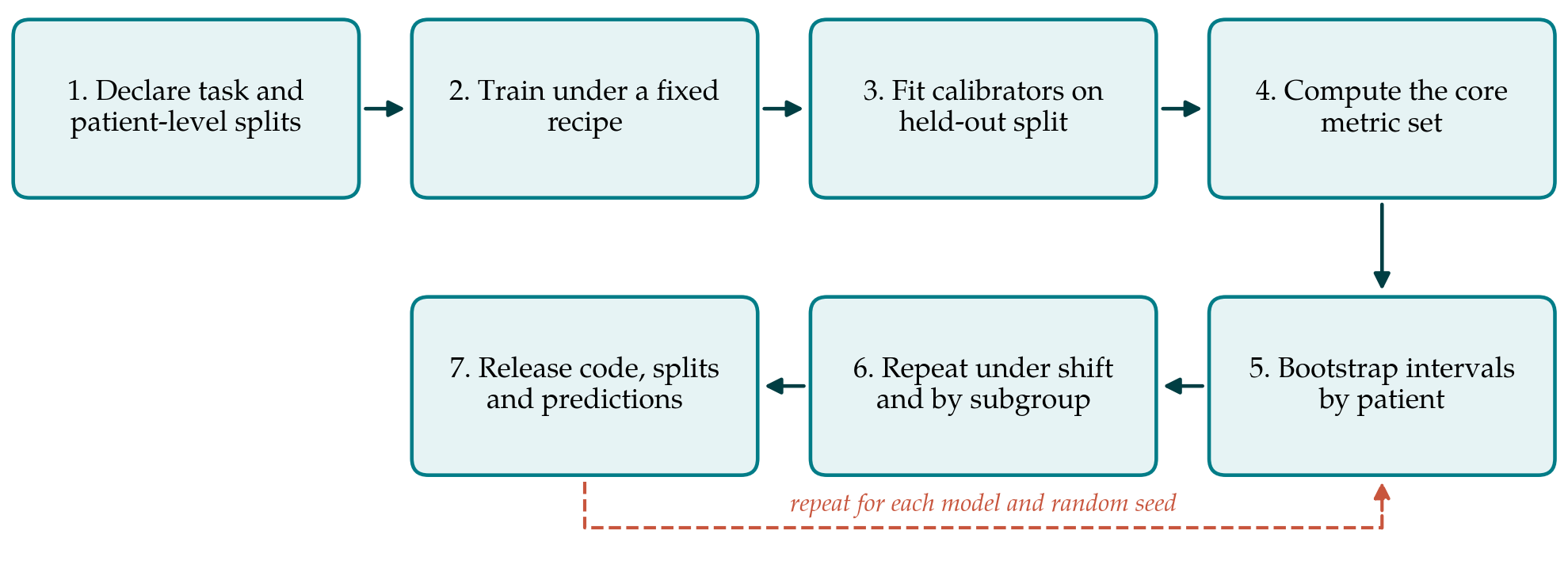
3.1. Step 1: Declare the task and fix the splits
The benchmark begins with a written definition of the prediction target, including how labels were obtained and whether the problem is binary, multi-class or multi-label. Data are then divided at the level of the patient into four parts: training, validation for model selection, a calibration split used only to fit post-hoc methods, and a test split that nothing is fitted on. The fourth split is the one most often missing in published work. When recalibration parameters are tuned on the validation data that also chose the model, or on the test set itself, the reported calibration is optimistic. Split files are published so that others evaluate on identical cases.
3.2. Step 2: Train under a declared recipe
Each model is trained with a fixed and published configuration, and training is repeated with several random seeds. Calibration is sensitive to choices that are rarely reported, among them the stopping criterion, weight decay and augmentation, so these belong in the record. Repetition matters because the difference between two calibration methods can be smaller than the variation between two runs of the same method.
3.3. Step 3: Recalibrate on the reserved split
Post-hoc calibrators are fitted on the calibration split and frozen. The protocol asks for at least the uncalibrated model, temperature scaling and one non-parametric method such as isotonic regression, so that every benchmark shares a common set of reference points. Training-time methods and ensembles are entered as separate models and pass through the same later steps.
3.4. Step 4: Compute a core set of metrics
No single number is adequate, so the protocol fixes a small set. The Brier score and negative log-likelihood are reported as proper scores. Expected calibration error is reported with equal-mass bins, with the number of bins stated, together with a class-wise variant of the kind described by Nixon et al. (2019), because top-label error hides miscalibration in rare classes. A reliability diagram accompanies each model. Discrimination is reported alongside, following the framework of Steyerberg et al. (2010), who treat overall performance, discrimination and calibration as separate questions. Where the intended clinical decision is known, net benefit from decision curve analysis may be added (Vickers and Elkin, 2006).
3.5. Step 5: Quantify uncertainty
Every reported value carries an interval. The protocol specifies a nonparametric bootstrap of the test set (Efron and Tibshirani, 1993) in which patients, not images, are resampled, so that correlated images from one person stay together. Variation across training seeds is reported separately, since it answers a different question: the bootstrap describes uncertainty about this model's calibration, and the seeds describe how much the training procedure itself varies.
3.6. Step 6: Evaluate under shift and within subgroups
The metric set is computed again on at least one test set drawn from a different institution or acquisition device, without refitting the calibrators. Results are also broken down by subgroups that were named before the analysis, typically sex, age band and scanner type where the data allow. A model that is calibrated on average may be overconfident for one group and underconfident for another, and the averages conceal it. Small subgroups will have wide intervals, and the protocol asks that these be shown and not suppressed.
3.7. Step 7: Release the artefacts
The benchmark is reproducible only if its parts are available. Code for training and for the metrics, the split files, fitted calibration parameters and the per-image predicted probabilities for every model and seed are released under a stated licence. Per-image predictions are the most valuable of these, because they allow a later reader to compute a metric that the original authors did not.
4. Fitting the Protocol to Practice
The protocol adds cost in two places. Reserving a calibration split takes cases away from training, which hurts when data are scarce; cross-fitting, in which calibrators are fitted on folds held out in turn, is an acceptable substitute provided the test split remains untouched. Training with several seeds multiplies computation, and for large models a smaller number of seeds with the count stated plainly is better than a single unqualified run.
Some data cannot be shared. Where patient images are restricted, the protocol can still be followed in part by releasing code, hashed split identifiers and predictions, none of which contain images. Multi-label tasks, common in chest radiography, are handled by treating each finding as its own binary problem and reporting the metric set per finding before any average.
5. Limitations and What Remains to Be Shown
The protocol has not been tested. It is not yet known whether following it narrows the disagreement between independent groups evaluating the same method, which is its whole purpose, and that would need to be demonstrated by having several teams apply it to a shared dataset. Nor is it established that the chosen metric set is the smallest that suffices, or that equal-mass binning is the best default across the range of prevalence found in practice.
There are limits of principle as well. Calibration is assessed against labels, and in medical imaging labels carry their own uncertainty from disagreement between readers; a model may look miscalibrated against one reader and not against a consensus. The calibration measured here is what Van Calster et al. (2019) describe as moderate calibration at best, agreement on average among cases given the same predicted risk, and it says nothing certain about any individual patient. A good benchmark result is evidence about a model on particular datasets. It does not establish that the model is safe to use in a clinic, which requires prospective evaluation in the setting concerned.
6. Conclusion
Calibration results for medical image classifiers will remain hard to compare for as long as each study chooses its own splits, bins and rules of access. The seven-step protocol proposed in this article fixes those choices in advance, reserves data for recalibration, reports proper scores with patient-level intervals, looks beyond the development distribution, and makes the predictions public. Whether it delivers the comparability it aims for is an open empirical matter, and the article invites its testing.
Conflict of interest
To be declared by the author(s) before publication.
Funding
To be declared by the author(s) before publication.