1. Introduction
Bronchopulmonary sequestration accounts for a small proportion of congenital lung malformations, and most radiologists see few cases in a career. The defining features have been stable since the term was introduced by Pryce (1946): a portion of lung parenchyma that does not communicate normally with the bronchial tree and is supplied by an anomalous systemic artery. Two forms are distinguished. Extralobar sequestration is invested in its own pleura and is typically found in fetal life or infancy, often with other anomalies. Intralobar sequestration lies within the pleura of an otherwise normal lobe and may remain silent for decades (Savic et al., 1979; Corbett and Humphrey, 2004).
The setting of diagnosis has changed. Older series were dominated by patients with recurrent lower-lobe pneumonia investigated by bronchography and aortography. Computed tomography (CT) is now performed in young adults for trauma, suspected pulmonary embolism, persistent cough and many other indications, and unexpected findings in the chest are common enough to have prompted formal guidance on their handling (Munden et al., 2018). A sequestration found in this way poses two questions that the classical literature answers only in part: how confidently the diagnosis can be made from an examination that was not designed to look for it, and what should be done about a lesion that has caused no symptoms.
This review addresses those questions for the young adult. It presents no new case and draws only on published work.
2. Approach to the Literature
The review is narrative. It relies on the foundational pathological descriptions, on the radiologic-pathologic correlation papers and imaging reviews that established the modern appearances, and on the larger surgical series and literature reviews that describe presentation and outcome. Much of the primary literature on sequestration consists of single case reports, which are numerous and unrepresentative: unusual presentations are published and ordinary ones are not. Reports of that kind were not used as evidence of how often anything occurs. Where frequencies are mentioned they come from collected series and are given in approximate terms, because the series differ in era, in case mix and in how the diagnosis was confirmed. No systematic search or pooled analysis was undertaken.
3. Anatomy, Classification and Origins
Table 1 compares the two forms, and Figure 1 places them within the wider group of related anomalies. In the collected series of Savic et al. (1979), and in the much larger review of the Chinese literature by Wei and Li (2011), intralobar lesions outnumbered extralobar ones by a wide margin, most lay in a lower lobe, usually in the posterior basal segment, and the left side was affected more often than the right. The feeding artery arose from the descending thoracic aorta in roughly three quarters of cases and otherwise from the abdominal aorta or one of its upper branches, the coeliac axis among them, or from an intercostal artery. A minority of lesions had more than one feeding vessel.
| Feature | Intralobar sequestration | Extralobar sequestration |
|---|---|---|
| Pleural covering | Within the visceral pleura of the affected lobe | Separate pleural investment |
| Relative frequency | The commoner form in collected series (Savic et al., 1979) | The less common form |
| Usual age at diagnosis | Later childhood or adult life | Fetal life or infancy |
| Typical site | Posterior basal segment of a lower lobe, more often left | Between lower lobe and diaphragm, usually left; occasionally below it |
| Arterial supply | Thoracic or abdominal aorta or a branch | Thoracic or abdominal aorta or a branch |
| Venous drainage | Usually pulmonary veins | Usually systemic veins: azygos, hemiazygos, inferior vena cava |
| Associated anomalies | Uncommon | Frequent, including diaphragmatic hernia (Rosado-de-Christenson et al., 1993) |
| Usual presentation | Recurrent infection, haemoptysis or an incidental finding | Mass on prenatal or neonatal imaging; respiratory distress |
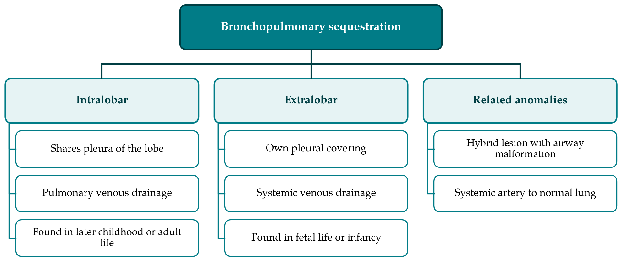
Venous drainage is the more reliable discriminator between the forms. Intralobar sequestration nearly always drains to the pulmonary veins, so that systemic arterial blood returns to the left atrium. Extralobar sequestration usually drains to systemic veins, the azygos or hemiazygos system or the inferior vena cava, though exceptions occur in both directions (Frazier et al., 1997; Rosado-de-Christenson et al., 1993). The extralobar form may lie anywhere between the neck and the upper abdomen; most are found between the lower lobe and the diaphragm on the left.
The origin of the intralobar form has been argued over for decades. Extralobar sequestration is accepted as congenital, arising from a supernumerary lung bud. For the intralobar form, Stocker (1986) proposed an acquired mechanism: chronic bronchial obstruction and infection in a lower lobe, with enlargement of small systemic arteries that normally run in the pulmonary ligament. The rarity of intralobar lesions in neonatal autopsies and the lack of associated anomalies supported the idea. Against it stand the detection of intralobar sequestration before birth and the existence of hybrid lesions that combine a systemic artery with the histology of congenital pulmonary airway malformation (Langston, 2003; Biyyam et al., 2010). Many authors now treat sequestration as part of a spectrum of bronchopulmonary-vascular malformations in which airway, parenchyma, arterial supply and venous drainage may each be abnormal independently (Sade et al., 1974; Clements and Warner, 1987).
4. How Sequestration Comes to Light in Adults
The classical adult presentation is recurrent infection in the same lower-lobe segment. Although the sequestered lung has no normal bronchial connection, it is aerated through collateral channels from adjacent lung, and organisms reach it by the same route; drainage is poor, and infection recurs. Cough, sputum, fever and chest pain are the usual complaints, and haemoptysis occurs in a proportion (Gezer et al., 2007; Halkic et al., 1998). Rarer reported events include massive haemoptysis, haemothorax, and superinfection with fungi or mycobacteria. Because the feeding vessel is an elastic artery carrying systemic pressure, flow through a large lesion can be considerable, though heart failure from the shunt is essentially a problem of infancy (Corbett and Humphrey, 2004).
A proportion of adults have no symptoms at diagnosis. In surgical series the proportion is modest, but surgical series select for patients who were unwell enough to be referred, and the true share of silent lesions is unknown (Petersen et al., 2003; Wei and Li, 2011). The incidental lesion in a young adult is commonly a basal opacity or cystic area noticed on a scan done for another purpose, or a persistent abnormality on a chest radiograph taken for employment or after minor trauma. The task of the radiologist in that situation is to think of the diagnosis, since the finding is otherwise liable to be reported as pneumonia, bronchiectasis or an indeterminate mass and followed without resolution.
5. Imaging Appearances
The parenchymal abnormality and the vascular anatomy have to be considered separately, because the first is variable and the second is what makes the diagnosis.
5.1. Chest radiography
The radiograph shows a homogeneous or cystic opacity in the posterior basal region, sometimes with an air-fluid level if infection has opened a communication with the airway. The appearance is non-specific. What should raise suspicion is persistence: an opacity in the same medial basal position on films separated by months or years, in someone too young for most acquired causes (Zylak et al., 2002).
5.2. Contrast-enhanced computed tomography
On CT the parenchymal component takes several forms: a solid mass or area of consolidation, a single cyst or a multicystic lesion with or without fluid levels, a region of hyperlucent, emphysema-like lung with sparse vessels, or mixtures of these. Mucus-filled bronchi may be seen within it. Hyperlucency at the margin of the lesion, attributed to air trapping in lung ventilated only by collateral drift, is a helpful sign when present (Frazier et al., 1997; Walker et al., 2014).
None of these appearances is diagnostic. The diagnosis rests on showing the systemic artery, and multidetector CT angiography with multiplanar and three-dimensional reformats does this well enough to have replaced catheter aortography for diagnosis (Ko et al., 2000; Lee et al., 2008). The artery is usually a single vessel of some size leaving the aorta near the diaphragm and passing through the pulmonary ligament to the lesion. The veins should be traced with equal care, since drainage determines the type and occasionally reveals an associated anomaly.
A report of a suspected sequestration is most useful when it records the items on which later decisions turn: the lobe and segments involved and the form of the parenchymal change; the number of systemic arteries, the level and vessel of origin of each, and their calibre and course; the route of venous drainage; whether the lesion appears to share the pleura of the lobe; signs of current or past infection; and any associated anomaly of the diaphragm, airway or pulmonary vessels. Stating plainly what the examination could not show, for instance that the aorta was poorly opacified or that the upper abdomen was not included, is as valuable as the positive findings, because it tells the referrer whether further imaging is needed before a surgical opinion is sought.
Whether the artery is seen on an incidental examination depends on how that examination was performed. A routine arterial or portal-venous phase chest CT opacifies the aorta and usually shows the vessel if the reader looks for it. CT pulmonary angiography is timed for the pulmonary arteries; the aorta may be only faintly opacified, and a small systemic branch can be invisible. Thick-section reconstructions hide it further, and a scan that stops at the lung bases may miss an artery arising below the diaphragm. An unenhanced scan cannot confirm the diagnosis at all. When a basal lesion of the kinds described is found on such a study, the appropriate step is to review thin sections around the lower descending aorta and, if doubt remains, to recommend a dedicated angiographic acquisition that includes the upper abdomen (Walker et al., 2014).
5.3. Magnetic resonance imaging and catheter angiography
Magnetic resonance angiography can show the feeding artery and draining veins without ionising radiation, an advantage in young patients and in pregnancy, and is established in the assessment of congenital lung lesions in children (Ko et al., 2000; Biyyam et al., 2010). Its depiction of the parenchyma is inferior to that of CT. Catheter angiography is now reserved for cases in which embolisation is planned.
6. Differential Diagnosis and Pitfalls
The differential diagnosis depends on which form the parenchymal abnormality takes. A solid basal lesion in a young adult suggests pneumonia, rounded atelectasis or a tumour; a cystic one suggests abscess, bronchiectasis, congenital pulmonary airway malformation or an infected bronchogenic cyst; a hyperlucent one with a central mucus plug suggests bronchial atresia (Berrocal et al., 2004; Zylak et al., 2002). In each case the systemic artery settles the matter, with one qualification. Chronic inflammatory lung disease of any cause recruits enlarged bronchial and other systemic collaterals, and these can be mistaken for a sequestration artery. Collaterals are typically multiple, small and tortuous, whereas the artery of a sequestration is a discrete vessel of substantial calibre.
Two vascular anomalies deserve separate mention. Systemic arterial supply to otherwise normal lung, in which the basal segments have normal bronchi but receive an aortic branch in place of pulmonary arterial flow, was the first of the variants described by Pryce (1946) and is not a sequestration in the strict sense. The scimitar, or hypogenetic lung, syndrome combines a small right lung with anomalous venous drainage to the inferior vena cava and often a systemic artery to the lower lobe (Clements and Warner, 1987). An extralobar sequestration below the diaphragm may be taken for an adrenal or other retroperitoneal mass (Rosado-de-Christenson et al., 1993).
7. Management After an Incidental Finding
For symptomatic sequestration the literature is consistent: resection is recommended, because infection recurs and haemorrhage is a risk, and the results of surgery in collected series are good (Halkic et al., 1998; Gezer et al., 2007). Lobectomy has been the usual operation for intralobar lesions, since chronic infection often leaves no clear plane, with segmental or wedge resection where the anatomy allows; thoracoscopic approaches are now widely reported. The principal surgical hazard is the feeding artery. It is thin-walled, atheromatous change in it has been described, and it can retract below the diaphragm if divided unrecognised. Preoperative imaging that states the number, origin and course of the arteries is therefore part of safe treatment and not an optional refinement (Savic et al., 1979; Lee et al., 2008).
For the asymptomatic adult the position is less settled. The argument for elective resection is that most lesions eventually become infected, that operation is easier before they do, and that rare complications such as major haemorrhage are unpredictable. The argument for observation is that these statements come from surgical series, in which the denominator of silent, never-diagnosed lesions is missing, and that some adults reach middle or old age with a sequestration that has caused no trouble (Petersen et al., 2003). The nearest body of comparative evidence concerns congenital cystic lesions detected before birth, where a meta-analysis found elective surgery to carry fewer complications than surgery after symptoms had developed (Stanton et al., 2009). How far that applies to an adult whose lesion has been silent for twenty or thirty years is uncertain.
Endovascular embolisation of the feeding artery has been reported as an alternative to resection or as a preliminary to it, mainly in children (Cho et al., 2012). It is less invasive, but the parenchyma remains, and long-term results in adults are not well documented. In practice the decision is made case by case in a multidisciplinary setting, taking account of the size and character of the lesion, any evidence of past infection, the calibre of the artery, and the preference of the person concerned after an explanation of what is and is not known.
8. Gaps and Future Work
The gaps in knowledge are mostly epidemiological. The prevalence of sequestration among adults undergoing chest CT has not been established, and nor has the natural history of lesions that are found by chance and left alone. Both could be addressed by registries that record incidentally detected cases, their imaging features and their course, whether treated or not. Such data would allow the advice given to an asymptomatic young adult to rest on something firmer than extrapolation from operated patients.
On the imaging side, the practical need is for recognition. Published guidance on incidental thoracic findings concentrates on common mediastinal and cardiovascular problems (Munden et al., 2018), and sequestration is too rare to justify a rule of its own. A simple habit serves instead: any persistent or unexplained focal abnormality in a medial basal segment of a young patient should prompt a deliberate look at the lower descending aorta and the pulmonary ligament. Whether spectral CT or newer magnetic resonance techniques add anything to conventional angiographic CT for this diagnosis has not been examined in any systematic way.
9. Conclusion
Intralobar pulmonary sequestration can stay hidden until adult life, and cross-sectional imaging performed for other reasons now uncovers some of these lesions before they cause illness. The parenchymal appearances are varied and imitate commoner diseases. The diagnosis depends on demonstrating a systemic feeding artery, which contrast-enhanced CT does reliably when the aorta is opacified, thin sections are reviewed and the scan reaches the upper abdomen, and which the same examination can miss when timed and reconstructed for another purpose. Once the artery and the venous drainage are defined, symptomatic lesions are resected with good results. For lesions found by chance in young adults who are well, the published evidence does not decide between early operation and observation, and that uncertainty should be shared openly with the person affected.
Conflict of interest
To be declared by the author(s) before publication.
Funding
To be declared by the author(s) before publication.