1. Introduction
Fusarium wilt, or Panama disease, destroyed the trade in Gros Michel bananas during the first half of the twentieth century and forced the industry to move to resistant Cavendish cultivars (Ploetz, 2015). The causal fungus, Fusarium oxysporum f. sp. cubense, enters through the roots, colonises the xylem and kills the plant by blocking water transport. It persists in soil for decades as chlamydospores, moves with planting material, soil, water and tools, and is not controlled by any fungicide in practical use (Ploetz, 2015). A strain able to attack Cavendish, tropical race 4, has since spread from Southeast Asia to other continents (Ordóñez et al., 2015), and was reported from Colombia in 2019 (García-Bastidas et al., 2020).
With no cure and few resistant cultivars acceptable to markets, management depends on exclusion and on rapid containment of new foci (Dita et al., 2018). Both require diagnosis. External symptoms, yellowing and collapse of older leaves and splitting of the pseudostem base, resemble those of bacterial wilts and of water stress, and the races of the pathogen cannot be told apart by eye. A laboratory test based on the polymerase chain reaction is available for tropical race 4 (Dita et al., 2010), but in many producing areas the nearest laboratory able to run it is far from the field, and the plant in question stays in the ground while samples travel.
This article proposes a protocol for running a loop-mediated isothermal amplification (LAMP) test beside the plantation, so that a presumptive answer is available the same day. It describes a procedure and the reasoning for each step. It does not report an application of the protocol, and it does not supply new primers: the protocol is written around primer sets already published and tested by their authors.
2. Background
Three matters need to be settled before a field protocol can be written: what exactly is to be detected, why LAMP suits the setting, and what published assays offer.
2.1. The diagnostic target
The name F. oxysporum f. sp. cubense covers several distinct lineages that share the ability to wilt banana (Ploetz, 2006). Races are defined by the cultivars attacked: race 1 affects Gros Michel and several other dessert types, race 2 affects cooking bananas of the Bluggoe type, and race 4 affects Cavendish as well. Tropical race 4 corresponds to one vegetative compatibility group and has been described as a separate species, Fusarium odoratissimum, in a revision that is not universally followed (Maryani et al., 2019). A field test must therefore state whether it detects the pathogen in the broad sense or tropical race 4 specifically. For quarantine purposes the second is usually what matters, and the protocol below assumes a race-specific primer set.
2.2. Why LAMP suits field use
LAMP, introduced by Notomi et al. (2000), amplifies DNA at a single temperature of about 60 to 65 °C using a strand-displacing DNA polymerase and four primers that recognise six regions of the target; two further loop primers shorten the reaction time (Nagamine et al., 2002). No thermal cycler is required. The reaction produces so much DNA that it can be seen without instruments, through turbidity, through a metal indicator such as hydroxynaphthol blue added before the run (Goto et al., 2009), or through a pH indicator in a weakly buffered mix (Tanner et al., 2015). Visual detection methods are reviewed with a working protocol by Tomita et al. (2008).
Two further properties count in the field. The polymerase is less affected than Taq polymerase by substances carried over from crude samples, and the reaction tolerates moderate variation in temperature and pH (Francois et al., 2011). This makes it possible to dispense with DNA purification. Tomlinson et al. (2010) showed for Phytophthora on woody hosts that a very short extraction followed by isothermal amplification could be carried out at the sampling site, and the same logic is applied here.
2.3. Published assays for the pathogen
LAMP assays directed at race 4 of the banana wilt pathogen have been described. One used conventional end-point detection (Li et al., 2013), and another used real-time fluorescence to detect and quantify tropical race 4 in soil (Zhang et al., 2013). Both were developed and tested under laboratory conditions. What they do not provide is a complete field procedure, with sampling, crude extraction, controls, reading rules and biosecurity treated as parts of one method. That is the gap this protocol is intended to fill.
3. The Proposed Protocol
The protocol has seven steps, listed in Table 1 with the safeguard attached to each and shown in sequence in Figure 1. A two-person team is assumed, one handling plants and one handling reactions, so that hands contaminated with plant sap never touch reaction tubes.
| Step | Action | Safeguard or record |
|---|---|---|
| 1. Select and record | Choose plants with early symptoms; note position, cultivar and symptoms | Field form with coordinates and photograph |
| 2. Sample | Cut a small window in the pseudostem and remove discoloured strands | New blade per plant; wound sealed; waste bagged |
| 3. Extract | Macerate tissue in alkaline lysis reagent, then dilute | Sample tube labelled before tissue is added |
| 4. Set up | Add diluted lysate to pre-dispensed reaction tubes and close them | Positive, negative and extraction controls in every run |
| 5. Incubate | Hold at the validated temperature for the validated time | Temperature checked with an independent thermometer |
| 6. Read | Compare colour with controls in daylight | Tubes stay closed; photograph kept |
| 7. Report | Record result as presumptive and send duplicate tissue to a laboratory | Chain-of-custody form; plant marked and isolated |
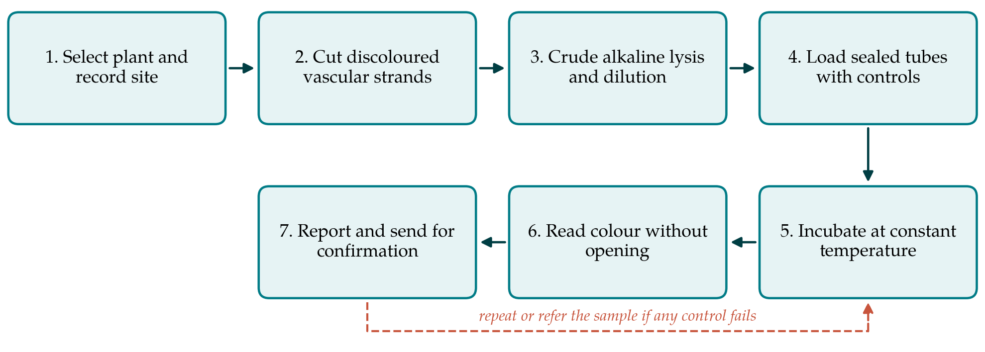
3.1. Step 1: Select the plant and record the site
Plants showing early symptoms are preferred to collapsed ones, because rotting tissue carries secondary organisms and more inhibitors. For each plant the team records the location, the cultivar, the symptoms seen and a photograph before any cut is made. A neighbouring plant without symptoms is sampled in the same way to act as a local comparison.
3.2. Step 2: Take vascular tissue
The pathogen is found in the xylem, which turns reddish brown in infected plants. A small window is cut in the outer leaf sheaths of the pseudostem, and a few discoloured vascular strands are lifted out with forceps. Each plant is sampled with a new disposable blade, or with tools flamed or disinfected between plants. The wound is closed, cut material is bagged for destruction, and footwear is cleaned before the team leaves the plot. These measures are part of the method. A diagnostic visit that carries infested soil or sap to the next field does more harm than a late diagnosis (Dita et al., 2018).
3.3. Step 3: Crude extraction
The strands are crushed in a small volume of alkaline polyethylene glycol reagent of the kind described by Chomczynski and Rymaszewski (2006), which lyses cells and yields a solution that can be added to an amplification reaction without neutralisation. The lysate is then diluted in water. Dilution lowers sensitivity slightly but reduces the phenolic compounds and polysaccharides in which banana tissue is rich, and it prevents the alkaline extract from shifting the colour of a pH-based indicator.
3.4. Step 4: Set up sealed reactions with controls
Reaction mix containing polymerase, primers and indicator is dispensed into tubes beforehand in a clean room, preferably as a dried or stabilised preparation that does not need a cold chain. In the field the operator only adds sample and closes the lid. Every run includes a positive control containing target DNA, a no-template control containing water, and an extraction control made from the symptomless plant. The positive control tube is prepared and sealed before the team travels and is never opened in the field.
3.5. Step 5: Incubate
Tubes are held at the temperature and for the time fixed when the assay was optimised. A battery-powered dry block is the simplest heat source; an insulated flask of water checked with a thermometer is a workable substitute. The time limit should be respected in both directions, since prolonged incubation increases the risk of non-specific amplification in negative tubes.
3.6. Step 6: Read the result without opening the tubes
With hydroxynaphthol blue, a positive reaction changes from violet to sky blue (Goto et al., 2009); with a phenol red system, from pink to yellow (Tanner et al., 2015). The run is valid only if the positive control has changed and the no-template and extraction controls have not. Sample tubes are read against the controls in daylight and photographed. Tubes are never opened after amplification. LAMP products are abundant and stable, and a single opened tube can contaminate equipment so thoroughly that every later run gives false positives. Used tubes are sealed in a bag and disposed of away from the working area.
3.7. Step 7: Report and refer
A positive field result is reported as presumptive. The plant is marked, access to it is restricted, and a duplicate tissue sample is sent to a reference laboratory for confirmation by the established polymerase chain reaction test (Dita et al., 2010) and, where possible, by isolation of the fungus. A negative result from a plant with clear symptoms is also referred, since the pathogen is unevenly distributed in the pseudostem and a single sample can miss it.
4. Fitting the Protocol to Field Conditions
Three practical constraints shaped the design. Heat and humidity degrade liquid enzyme mixes, which is why pre-dispensed, stabilised reagents are specified and why the number of liquid transfers in the field is kept to two. Operators will often be extension or plantation staff and not molecular biologists, which is why the readout is a colour compared with controls and not a curve to be interpreted. And the consequences of a wrong answer are asymmetric. A false positive may lead to needless destruction of plants and loss of market access; a false negative leaves an infectious focus in place. The protocol therefore treats the field test as a screen that directs laboratory confirmation and containment, and never as the final word.
Where the pathogen is a regulated quarantine organism, the national plant protection organisation decides who may test, how positive material is handled and how results are notified. The protocol should be adopted only within that framework. Positive control DNA should be a synthetic fragment or non-viable extract, so that no living culture of the pathogen is carried into production areas.
5. Limitations and What Remains to Be Shown
The protocol is a proposal and has not been validated in this article. Before it could support official decisions, its performance would have to be measured against the criteria that diagnostic standards lay down for plant pest tests: analytical sensitivity, analytical specificity against other races and against the non-pathogenic F. oxysporum strains common in banana soils, selectivity across cultivars and tissues, repeatability, and reproducibility between operators and sites (EPPO, 2014).
Several specific uncertainties deserve mention. The effect of crude banana extract on the chosen primer set and indicator is unknown until tested, and may differ between cultivars. The specificity of published primers has been established against the isolates their authors held; the diversity of the pathogen in its centre of origin is large (Maryani et al., 2019), and new variants could escape detection or cross-react. Colour judgement varies between observers and with lighting. Finally, the test detects DNA and not viability, which is acceptable in a living plant but would complicate any extension of the method to soil or water.
6. Conclusion
A same-day presumptive test for banana Fusarium wilt at the field edge is technically within reach. The chemistry is established, race-specific primer sets have been published, and crude extraction and colour readout remove the need for laboratory instruments. The seven-step protocol proposed here joins these elements with the controls, contamination precautions and biosecurity measures that field use demands. Its diagnostic performance has yet to be measured, and until it has been, results obtained with it should prompt confirmation and precaution, not regulatory action.
Conflict of interest
To be declared by the author(s) before publication.
Funding
To be declared by the author(s) before publication.